![]()
![]()
![]()
Use LEFT and RIGHT arrow keys to navigate between flashcards;
Use UP and DOWN arrow keys to flip the card;
H to show hint;
A reads text to speech;
37 Cards in this Set
- Front
- Back
|
Give 4 differences between the pelves of men and women
|
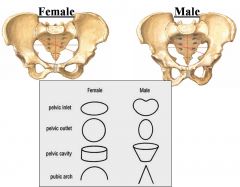
- Sub-pubic angle 120 in females, 60 in males = similar for thyroid cartilage angles
|
|
|
Describe the anatomy of the pelvic inlet
|
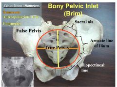
Pelvic inlet boundaries:
- Anteriorly by pubic crest - Laterally by arcuate and pectineal lines - Posteriorly by ala of sacrum and sacral promontory |
|
|
Describe the boundaries of the true and false pelves
|
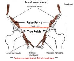
False Pelvis
- the expanded portion of the cavity situated above and in front of the pelvic brim - It supports the intestines (specifically, the ileum and sigmoid colon), and transmits part of their weight to the anterior wall of the abdomen. - Bounded laterally by the ilial ala - Posterior by lower lumbar vertebrae - Anteriorly by lower rectus abdominis True pelvis - that part of the pelvic cavity which is situated below and behind the pelvic brim - Bounded anterior by the pubic symphysis and the superior rami of the pubes - Posteriorly by the pelvic surfaces of the sacrum and coccyx - Laterally, by the inner surfaces of the body and superior ramus of the ischium, and that part of the ilium which is below the arcuate line. |
|
|
What is the pelvic outlet?
|
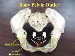
It has the following boundaries:
- Anteriorly: the pubic arch - Laterally: the ischial tuberosities - Posterolaterally: the inferior margin of the sacrotuberous ligament - Posteriorly: the tip of the coccyx |
|
|
Describe the pelvic diaphragm
|
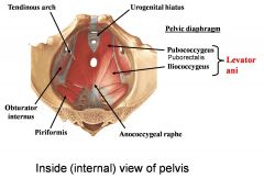
- Composed of the levator ani muscles
1. Pubococcygeus 2. Puborectalis 3. Ileococcygeus - In males it is pierced by 2 structures = rectum and ureters - In females it is pierced by 3 structures = rectum, ureter and vagina - The tendinous arch forms a common point of muscle attachment for these muscles - Inferiorly to the urogenital hiatus in females (urorectal in males) is the urogenital membrane, forming the sphincter urethrae |
|
|
Describe congenital abnormalities of the uterus and vagina
|
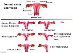
|
|
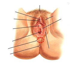
Label this diagram
|

|
|
|
What is the vestibule and why is it clinically significant?
|
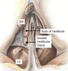
- Space between labia minora
containing urethra, vagina & greater vestibular glands. (Bartholin’s gland ) - Greater vestibular glands open either side of vagina. - Lesser glands between urethral & vaginal orifices. All secrete mucus into vestibule during sexual arousal. - Bulb of vestibule is female homologue of the bulb of the penis. Consists of erectile tissue - Bartholin’s gland is site or origin of vulval adenocarcinomas. Also prone to infection & cysts. |
|
|
What is the broad ligament?
|
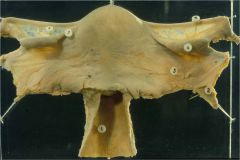
- The wide fold of peritoneum that connects the sides of the uterus to the walls and floor of the pelvis
- Like a flat sheet that is folded over the uterus - The contents of the broad ligament include the following: • Reproductive: 1. Fallopian tube 2. ovary • Vessels 1. ovarian artery (is a suspensory ligament) 2. uterine artery • Ligaments 1. ovarian ligament 2. round ligament of uterus 3. suspensory ligament of the ovary (Some sources consider consider it a part of the broad ligament while other sources just consider it a "termination" of the ligament.) |
|
|
Describe the position of the uterus
|
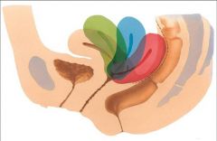
- Uterus position varies with fullness of bladder and rectum
- Normal position anteverted (tipped anterosuperiorly relative to the axis of vagina) and anteflexed (flexed anteriorly relative to the cervix) - Proper position can be key to ability to carry foetus to term or clinical problems associated with pregnancy |
|
|
Describe the vaginal fornices
|
- The vaginal fornix = the recess around the cervix
- Has anterior, posterior and lateral parts - The posterior fornix is the deepest past and is closely related to the rectouterine pouch → danger area as a botched abortion can pierce into the peritoneal cavity herem and it may hide all sorts of implanted devices e.g. old tampons |
|
|
Describe the vaginal fornices
|
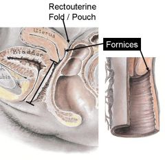
- The vaginal fornix = the recess around the cervix
- Has anterior, posterior and lateral parts - The posterior fornix is the deepest past and is closely related to the rectouterine pouch → danger area as a botched abortion can pierce into the peritoneal cavity herem and it may hide all sorts of implanted devices e.g. old tampons |
|
|
Describe the role of the pudendal nerve in the innervation of the external genitalia
|
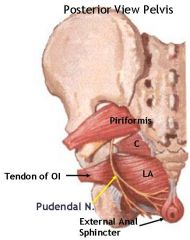
- Course
• Exits the pelvis via the greater sciatic foramen • Enters the lesser sciatic foramen around the brachial spine to pass through the ischioanal fossa to reach all the perineum - Somatic motor innervation = continence • External anal sphincter • Muscles assisting with erectile tissues • Muscles of the deep perineal pouch (including sphincter urethrae) - Somatic sensory innervation • Anus • Scrotum/labia • Penis/clitoris • Lower 1/4 of vagina - Consequently, to anaesthetise the whole of the area of skin around the perineum put anaesthetic into the pudendal nerve |
|
|
Describe the features of the cervix
|
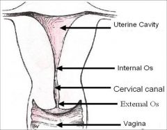
- Cervix connects uterine cavity to vagina
- Consists of an internal os, cervical canal and external os which protrudes into the vagina |
|
|
How would you examine the vagina and cervix?
|

|
|
|
Describe the blood supply to the uterus
|
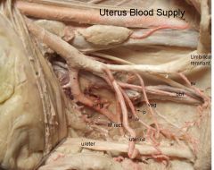
- Blood supply to the uterus derives mainly from the uterine arteries, which come off the internal iliac arteries. It may also have a supply from the ovarian arteries which run off the abdominal aorta (as different embryological origins)
|
|
|
Describe the examination of the uterus
|
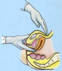
- The bimanual exam
- Two fingers placed in the vagina. Other hand used to palpate the pelvic organs through the abdominal wall - Size / position / mobility / consistency of uterus can be examined |
|
|
Describe the relations of the vagina and uterus
|
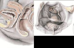
Vagina:
- Anteriorly: urethra, base of bladder - Laterally: levator ani, pelvic fascia and ureter - Posteriorly: anal canal, rectum and rectouterine pouch Uterus: - Anteriorly: vesicouterine pouch, superior surface of rectum - Posteriorly: rectouterine pouch, anterior surface rectum - Laterally: broad and cardinal ligaments, ureters |
|
|
Describe the support and ligaments of the cervix and upper vagina
|
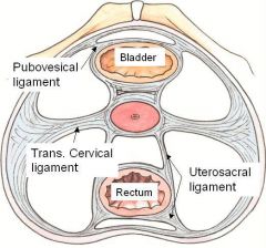
- Main supporting mechanism = pelvic fascia
- Visceral layer ensheathes pelvic organs - Two become continuous where organs penetrate the pelvic floor - At junction thickening of the fascia forms tendinous arch of pelvic fascia from pubis to sacrum - Anterior part of this forms pubovesicular ligament - Posterior part forms uterosacral ligament - Lateral extension of fascia around cervix forms transverse cervical ligaments (aka lateral cervical, cardinal ligaments) |
|
|
Describe the support and ligaments of the uterus
|
- The main support of the uterus is derived from the tone of the pelvic floor muscles
- Other supporting structures called ligaments: • Broad ligament = double layer of peritoneum that surrounds the uterus with the uterine tubes forming its superior border. It doesn't support the uterus • Round ligament of the uterus = from the uterus body to the labia majora through the inguinal canal (remnant of lower sections of the gubernaculum). Holds the uterus in the anteverted position and is a minor support • Uterosacral ligaments = from the cervix either side of the rectum to piriformis muscle over the sacrum. Supports the uterus • Transverse cervical ligaments = from the cervix and superior vagina to the lateral pelvic walls. Supports the uterus • Pubocervical ligaments = from the pubis to the cervix. Do not support the uterus |
|
|
Describe the neurovascular supply to the ovary and uterine tube
|
- Arterial = ovarian arteries arise from the abdominal aorta and descend along the posterior abdominal wall. Ascending branches of the uterine arteries help supply the medial aspects of the ovaries and the uterine tubes and anastomose with the the ovarian branches
- Venous = Veins draining the ovary form a pampiniform plexus of veins in the broad ligament near the ovary and uterine tube. These usually fuse to form a ovarian vein, which ascends to enter the inferior vena cava on the left, and the right drains into the right renal vein - Lymph = Lymphatic vessels from the ovary join those from the uterine tubes and fundus and follow the ovarian blood vessels to ascend to the right and left lumbar lymph nodes - Nerves = Sympathetic from the ovarian plexus. Parasympathetics from the uterine and inferior hypogastric plexi and the plexic splanchnic nerves |
|
|
Describe the pouches of the periotneum
|
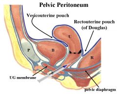
- Vesicouterine pouch between the bladder and the uterus. Lost when the bladder fills
- Rectouterine pouch of Douglas between the uterus and the rectum. Very deep and anaesthetics are given here |
|
|
Describe the functions of the pelvic floor
|
1. Support internal organs against gravity
2. Allow increases of intra-abdominal pressure 3. Allow passage and function of urethra, vagina and rectum |
|
|
Describe the structures that make up the anterior pelvic floor
|
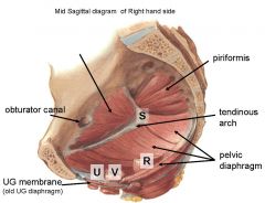
- Urogenital membrane:
• striated membrane between fascia • stretched between pubic arches • surrounds sphincter urethrae (and vagina) → continence mechanism - Deep transverse perineal muscle = inserts into the perineal body/vagina |
|
|
Describe the structures of the posterior pelvic floor
|
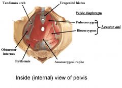
- Funnel shaped muscular floor made up of levator ani = 3 components
1, Iliococcygeus 2. Pubococcygeus 3. Puborectalis (sling around rectum) - Also coccygeus muscle - All muscles are striated and meet in the median plane along raphe (and the perineal body) - The muscles attach posteriorly to the ischial spines, the pubic bones anteriorly, and to the pelvic tendinous arch (thickening in the obturator fascia) in between, forming a basin effect |
|
|
Describe the blood supply and innervation to the pelvic floor
|
- Blood supply = branches of the anterior trunk of the internal iliac artery
• Pudendal artery • Vaginal artery • Inferior rectal artery - Innervation by pudendal nerve = S2,3,4 |
|
|
What is the perineal body?
|
- AKA central perineal tendon
- Condensation of connective tissue - Contains fibres from levator ani and urogenital membrane - Reinforces area between vagina and rectum - Centreal fulcrum for pelvic support |
|
|
Describe 7 factors that can increase the risk of pelvic floor dysfunction
|
1. Strenuous work e.g. heavy labour
2. Straining, chronic cough 3. Pelvo-abdominal mass, obesity 4. Smoking, aging, tissue damage 5. Childbirth, multiple pregnancies 6. Poor repair of obstetric injuries 7. Female = much more likely than male (3 opening in pelvic diaphragm so inherently weaker) |
|
|
Describe the effects of pregnancy on the pelvic floor
|
- Musculo-fascial supports are weakened and stretched
- Perineal body is attenuated by thinning (reduced in strength) - Levator hiatus is widened and never really closes - Tears and lacerations - Stretch of pudendal nerve → neuropraxia and muscle weakness |
|
|
What is the most common pelvic floor dysfunction and what are the types?
|
- Prolapse:
• Cystocoele = bladder • Urethrocoele = urethra • Rectocoele = rectum • Uterine prolapse • Procidentia (complete prolapse so bladder and uterus completely external) • Combinations of the above |
|
|
Describe how prolapses can be prevented
|
1. Reduce the risk factors
- weight control - smoking - lifting properly etc 2. Pelvic floor exercises 3. Episiotomy usage = shouldn't be routinely carried out 4. Identify and repair all obstetric damage |
|
|
What are the possible treatments for pelvic floor dysfunction?
|
- Varies with type of defect = some less severe than others
- Varies with patient, age, lifestyle and medical condition - Surgical procedure = most involve hysterectomy - Support techniques e.g. pessaries to keep internal organs internal |
|
|
Describe the histology of the ovary
|
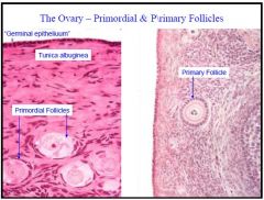
- Ovary has 3 components:
1. Surface = simple cuboidal epithelium called the germinal epithelium (nothing to do with germ cells) 2. Cortex = connecting tissue stroma supporting thousands of follicles - preovulatory, postovulatory and degenerating 3. Medulla = composed of supporting stroma, it contains a rich network of vessels and nerves that enter the ovary from the mesovarium |
|
|
Describe the histology of the uterine tubes
|

- The uterine tubes are lined by ciliated and secretory colmnar cells that waft the oocyte towards the uterus and supply it with nutrients
- Two layers of spiral muscle surround this lining and help move the oocyte and sperm by peristalsis. - These muscles are sensitive to sex steroids, so that motility is most rapid when sex steroids are highest |
|
|
Describe the histology of the uterus
|
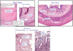
The body and fundus are composed of 3 tissue layers:
1. Serosa (Perimetrium)= peritoneal covering 2. Myometrium = thick smooth muscle layer that is sensitive to hormones e.g. oxytocin 3. Endometrium = inner lining of the uterus that varies through the menstral cycle. Sensitive to hormones e.g. oestrogen and progesterone - The endometrium can also be subdivided into a further 2 layers: • Deep basal layer = changes little during the menstral cycle • Superficial functional layer = hormone-sensitive layer that proliferates in response to oestrogen and becomes secretory in response to progesterone. It is shed at the end of the menstral cycle and regenerates from cells in the basal layer - The superficial functional layer has outer compact and inner spongy layers - The arterioles of the superficial endometrial layer lie alongside the glands of the endometrium. They have a spiral appearance, unlike the straight arterioles of the basal layer. |
|
|
Describe the histology of the cervix
|
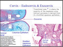
- The cervix consists mainly of collagen and small amounts of smooth muscle
- The columnar epithelium lining the endocervical canal secretes mucous that changes in consistency during the menstral cycle - The cervix is divided into 2 sections 1. Endocervix = superiorly - related to the uterus body and has columnar endometrial epitherlium 2. Ectocervix = inferior, related to the vagina and has stratified squamous vaginal epithelium - Note that the anatomical division is usually located higher than the histological division - During pregnancy and puberty high oestrogen levels cause the simple columnar epithelium of the cervix to extend beyond the external os into the vagina → cervical ectropy. In the vagina, the acidic pH induces squamous metaplasia of the columnar cells - The cells that change are in an area called the transitional zone and they are susceptible to dysplasia |
|
|
Describe the histology of the vagina
|
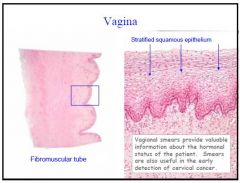
- Walls of the vagina are composed of 4 layers:
1. Stratified squamous epithelial lining for protection 2. Elastic lamina propria 3. Fibromuscular layer (2 layers of smooth muscle) 4. Fibroelastic adventitia - The wall contains very few sensory fibres and no glands and the lining is therefore lubricated by cervical mucous |

