Reading...
![]()
Play button
![]()
Play button
![]()
Use LEFT and RIGHT arrow keys to navigate between flashcards;
Use UP and DOWN arrow keys to flip the card;
H to show hint;
A reads text to speech;
52 Cards in this Set
- Front
- Back
- 3rd side (hint)
|
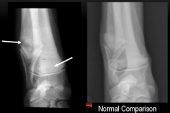
Osteochondrosis
Osteochondritis Dissecans |
• Usually around 6-9 months old
• Large breed, rapidly growing dogs • Often BILATERAL • Most common in caudal humeral head • Also femoral condyle, talus |
• Flattening or concave defect of articular bone surface, surrounding sclerosis
• Adjacent mineral body in joint (OCD)- osteochondral fragment |
|
|
Fragmented Medial Coronoid Process
|
• #1 in young, large breed forelimb
|
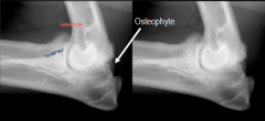
• Cranial margin of ulna indistinct close to the joint space
• Osteophytes • May see fragment • May see osteochondrosis of humeral condyle o Concave defect o Sclerosis of bone adjacent to ulnar notch |
|
|
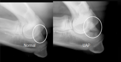
Ununited Anconeal Process
|
• Should be united by 6 months
|
• See broken process
|
|
|
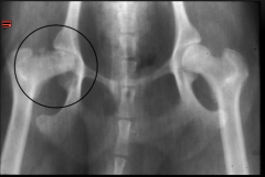
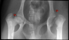
Avascular Necrosis of the Femoral Head
|
• Adolescent toy and small breed dogs
• Damaged blood supply to femoral head causes bone necrosis • Usually unilateral, can be bilateral |
• Small, misshapen
• Heterogenous opacity- necrosis and remodeling • May have mineralized chip- “joint mouse” |
|
|
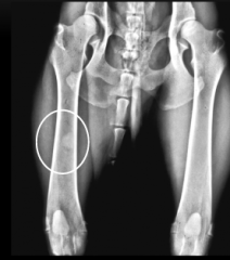
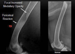
Canine Panosteitis
|
• Large breed, especially German Shepherds and Basset Hounds
• Long bones, usually big ones |
• Circumscribed areas of increased opacity in diaphyseal medullary cavity
• Often near nutrient foramen; progresses from around this area • Smooth new periosteal bone |
|
|
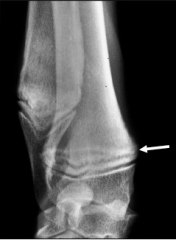
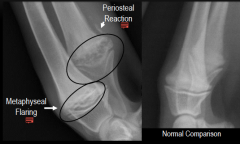
Hypertrophic Osteodystrophy (HOD)
|
• Systemic disease
• Large and giant breeds • Onset around 4 months old • Self-limiting; no specific Tx • Good examples: distal radius and ulna • Almost always BILATERAL |
• Hallmark sign: transverse radiolucent lines in metaphysis
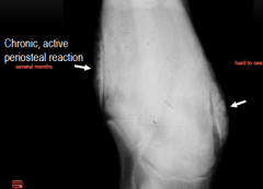
• Metaphysis flaring or irregular new bone • Periosteum lifting off of bone- probably necrosis and pus underneath • When chronic- harder to see lines, heal on own, just hope no limb angular deformities |
|
|
Craniomandibular Osteopathy
|
• Terriers, bulldogs
• 3-8 months old • Heritable component • Self-limiting, ends at maturity • Painful, disfiguring |
• Irregular osseous proliferation on mandible, TMJ, bullae, occasionally calvarium
|
|
|
Hyperparathyroidism/ Metabolic Bone Disease
|
• Typically primary- adenoma of parathyroid gland in old dogs
• Can be nutritional or renal |
• Generalized increase in bone opacity
• Thin bone cortices • Loss of lamina dura around teeth roots • Pathologic fractures • Spinal deformities |
|
|
OC/OCD
|
failure of endochondral ossification
-articular cartilage and subchondral bone |
|
|
|
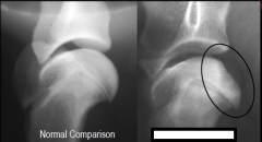
Osteochondrosis
|
flattening/defect and subchondral lucency
|
|
|
OCD caudal humeral head
|
flattening and thin fragment
|
|
|
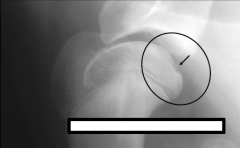
OCD caudal humeral head
|
flattening and thin fragment
|
|
|
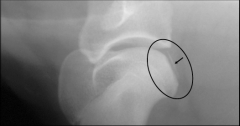
OC Caudal humeral head
|
subchondral lucency
|
|
|
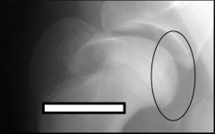
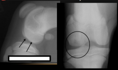
OC Femoral Condyle
|
Flattening of condyle
subchondral lucency |
|
|
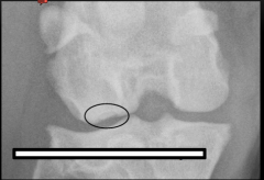
OC femoral condyle
|
subchondral defect with ovoid fragment
|
|
|
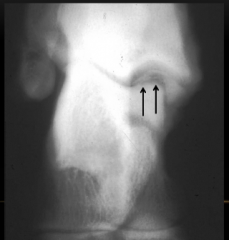
OCD talus
|
medial trochlear ridge
|
|

|
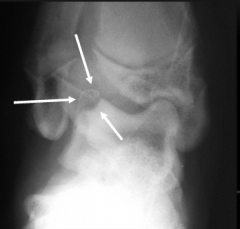
OCD talus
|
widening jt space
fragment |
|
|
OCD talus
______ view |
flexed DorsoPalmar View
|
|
|
Developmental condition caused by joint incongruity and/or osteochondrosis
|
elbow dysplasia
|
|
|
|
elbow dysplasia
|
one or more of
ununited anconeal process fragmented medial coronoid process OCD of medial aspect of humeral condyle |
results in early OA/DJD
|
|
|
Elbow dysplasia
____ view |
flexed lateral view
|
|
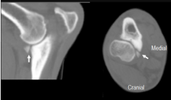
CT -
|
Fragmented Medial Coronoid Process
|
|
|
|
fragmented medial coronoid process
|
|
|
|
fragmented medial coronoid process
|
|
|
|
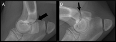
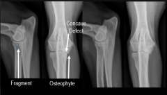
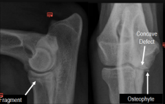
FMCP + Osteochondrosis of Humeral Condyle
|
|
|
|
FMCP + Osteochondrosis of Humeral Condyle
|
sclerosis of ulnar notch
OC of distal humerus small triangular fragment cranial margin of coronoid process |
|
|
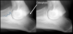
osteochondrosis of humeral condyle
|
coronoid process disease
"lazy" oblique |
|
|
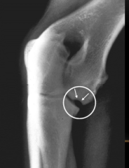
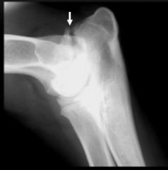
ununited anconeal process
|
united by 6 months
|
|
|
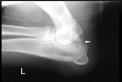
ununited anconeal process
view: |
flexed lateral projection
|
|
|
ununited anconeal process
|
|
|
|
Orthopedic foundation for animals
|
flexed lateral view >24 months
Grade I Grade II Grade III |
Grade I: <3 mm
Grade II: 3-5 mm Grade III: >5 mm |
|
|
Avascular Necrosis
|
|
|
|
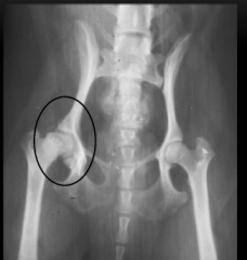
avascular necrosis
|
|
|
|
avascular necrosis
|
jt space too wide
femoral head small and misshapen chronic, femoral head gone from necrosis |
|
|
canine panosteitis
|
|
|
|
canine panosteitis
|
progress to diffuse medullary increased opacity
subtle smoothly marginated periosteal rxn |
|
|
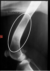
canine panosteitis
|
diffuse medullary increased opacity
smooth new periosteal bone |
|
|
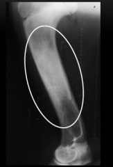
canine panosteitis
|
hard to distinguish cortex from medullary cavity
trx with nsaids can be any long bones (tibia femur radius ulna humerus) |
|
|
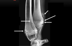
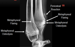
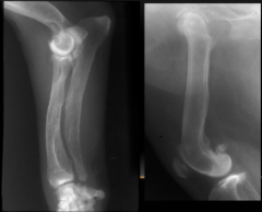
hypertrophic osteodystrophy
|
transverse radiolucent lines in metaphysis (double physis sign)
metaphyseal flaring or irregular new bone very painful |
|
|
hypertrophic osteodystrophy
|
|
|
|
hypertrophic osteodystrophy
|
between periosteum and bone
inflammatory response purulent exudate |
|
|
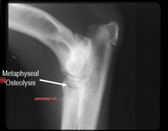
hypertrophic osteodystrophy
|
chronically affected
**metaphyseal osteolysis **both legs |
|
|
hypertrophic osteodystrophy
|
|
|
|
hypertrophic osteodystrophy
|
|
|
|
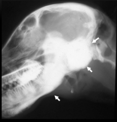
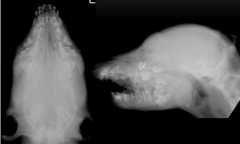
craniomadibular osteopathy
|
irregular osseous proliferation on mandible, TMJ, bullae
occasionally calvarium smooth margins |
|
|
craniomandibular osteopathy
|
proliferative new bone, formation on tympanic bulla
difficulty eating and opening mouth painful |
|
|
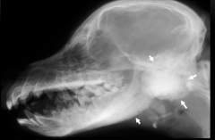
craniomandibular osteopathy
|
mandible proliferation
disfiguring, painful to pressure ddx: bone tumor |
|
|
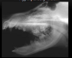
Hyperparathyroidism/ Metabolic Bone Disease
|
most common
farily common in old dogs with hypercalcemia -PU/PD, muscle weakness, lethargic |
|
|

|
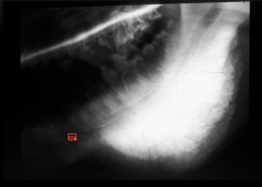
hyperparathyroidism
absence of lamina dura opacity like soft tissue = demineralization |
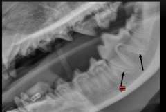
normal lamina dura
|
|
|
hyperparathyroidism
|
|
|
|
hyperparathyroidism
|
|
|
|
hyperparathyroidism
thin bone cortices decreased bone opacity |
|