![]()
![]()
![]()
Use LEFT and RIGHT arrow keys to navigate between flashcards;
Use UP and DOWN arrow keys to flip the card;
H to show hint;
A reads text to speech;
24 Cards in this Set
- Front
- Back
|
Ribozymes? Proteins? Cofactors? General formula of amino-acid? Draw---> |
All known enzymes are proteins except Ribozymes. Ribozymes are self cleaving and selfsplicing RNA molecules!
Proteins are macromolecules made from amino acids which arejoined together by a covalent bond known as a peptide bond. Afunctional protein usually have a specific 3-dimensional structure Proteins are the most abundant biological macromolecules occurringin all cells and all parts of cells Proteins are the molecular instruments through which the geneticinformation is expressed All the proteins are made from the same set of 20 amino acids, the body can only produce 10 out of the 20 amino acids. The group of 20 amino acids may be regarded as the alphabet inwhich the language of protein structure is written Many enzymes require the presence of other compounds - cofactors - before their catalytic activity canbe exerted. |
|
|
Structure of enzyme? held together by? structures of proteins?(folding)? bonds in the structures? Binding site? Allosteric site? |
They are all proteins( globular proteins). They are long chains of amino acid units heldtogether by peptide bonds, looped and folded into secondary and tertiary (andoften quaternary) structures by disulfide bonds, hydrophobic interactions andsalt bridges. The enzyme has anactive site, that is the substrate binding site with the catalytic site and iswhere the catalyzation takes place. It has a high specificity. There can also be anallosteric site for where the regulating molecule can attach. The enzyme isalso composed of other binding sites. |
|
|
Simple enzymes? Holoenzymes? |
Simple enzymes are only proteins Holoenzymes They have two parts: Apoenzyme(the protein part) Coenzyme |
|
|
Experiment1 Optimal temperature for the amylase action? Denaturation? Temperature? Starch-Iodine complex? Boiled tube? Unboiled tube? How pH effects amylase? |
Amylase catalyses the break down of starch into its monomers. Denaturation is a process in which proteins or nucleic acids loose their structure (Quart,Tert,Secondary) If amylase is exposed to temperature >70C atoms starts to vibrate so much that atoms smash into each other causing irreversible damage to the molecular structure. Iodine angles the starch molecule into a spiral which forms the Starch-Iodine complex! This can then be used as a marker for the presence of starch, cause if starch is present it will give a dark blue color Boiled tube - Dark blue color cause amylase enzyme is destroyed Unboiled tube - No color since amylase enzyme is working properly and catalyzing the breakdown of starch Effective pH range of Amylase=6,8-7,0 Ionic charge creates the functional shape of the enzyme When pH changes the ionic charges changes and the structure of the enzyme is altered making it stop function or work with severely reduced capacity! |
|
|
Net charge of pH 0-1? Around 3? Around 6-8? above 11? Zwitter ion? pI? Polyelectrolytes Formation of peptide bonds? |
+1 0 -1 -2 Zwitter ion - No overall charge but contains parts that have + or - charge Isoelectric Ph = Where a molecule carries no electric charge Netcharge changes with pH and concentration Formation of peptide bonds is followed by net loss of 1 positive and 1 negative charge per peptide bond formed. |
|
|
Action of amylase?(Draw and explain) Structures of protein?(Draw and explain) Induced fit model?(Draw and explain) Key lock principle?(Draw and explain) Basic structure of proteins Basic structure of peptide bond |
(Draw and explain) ON PAPER :) |
|
|
Enzymes are Three distinctive features Enzymes work by |
biological catalysts 1) catalytic power, specificity, and regulation 2) Mainly globular proteins, but a smaller number of ribonucleic acids, calledribozymes 3) lowering the activation energy and thus acceleratingreaction rates as much as 10^21 over uncatalyzed levels In dilute aqueous solutions under mild conditions of temperature and pH |
|
|
Activity of enzymes Rate of reaction?(see picture) Michaelis constant High Km Low Km |
The rate of the reaction increases as thesubstrate concentration increases until reachesVmax were reaction attains maximal velocity at that point a increase in substrate concentration does notincrease reaction rate because of saturation Substrate concentration that causes thereaction to proceed at its half maximal velocity(1/2 Vmax) Km High Km – low affinity to the substrate Low Km – high affinity to the substrate |
|
|
Classes of enzymes 1)One way 2)Both ways |
1) Hydrolases - Creating 2 separate molecules and addition of H2O Ligase - ATP involved Lyase - Clevage of C-C, C-O, C-N, Generating double bonds 2) Isomerase - Geometric or structural changes Oxireductase - (C-H forming C=O) Transferase - Transfer of any group (NH3, CH3, PO4) |
|
|
Oxidoreductases: Transferases: Hydrolases: Lyases: Isomerases: Ligases: |
Oxidoreductases: Exampes: -dehydrogenases -oxidases -peroxidases -hydroxylases -oxygenases Transferases: Exampes: -transases (such as: transaminase, transkelotase, transaldolase, transmethylase.) Hydrolases: Exampes: -esterases -amidases -peptidases -phosphatases -glycosidases Lyases: Exampes: -decarboxylases -aldolases -synthases -cleavage enzymes -hydrases or hydratases or dehydratases -deaminases Isomerases: Exampes: -isomerases -racemases -epimerases -mutases Ligases: Exampes: -synthetases |
|
|
Amyloclastic force |
Amyloclasticforce that is the volume of the 0,1 % starch (stivelse) solution in millilitersthat is hydrolyzed by 1 ml saliva(spytt) at 38°C during 30 min. |
|
|
1. Absolute specificity 2. Bond specificity 3. Group specificity 4. Stereospecificity Draw reaction for each group |
1) enzyme can bind only one substrate and catalize only one reaction a) Lactase, b) Sucrase c) Maltased) Urinasee) L-glutamate dehydrogenase 2) enzyme will recognize a specific type of chemical bond in the substrate molecule but does not care about the surroundings a. Amylase, which acts on α 1-4glycosidic bonds in starch, dextrin and glycogen. b.Lipase that hydrolyzes ester bonds in different triglycerides 3) enzyme will recognize not only a specific chemical bond but also the structure surrounding the bond Ex. pepsin – peptide bond + Tyr, Phe, Trp 4) enzyme can recognize only a specific stereoisomer a) L amino acid oxidase acts onlyon L amino acids. b)α- glycosidase acts only on α- glycosidic bonds |
|
|
Quantitative regulation of enzyme activity? Qualitative regulation? |
Quantitative regulation of enzyme activity? Regulation of the expression of the gene! Ex. LAC operon, Post-translation regulation Qualitative regulation? Activators and inhibitors |
|
|
Inhibition and what bonds that form? |
Irreversible inhibitors form covalent bonds; reversible ones use weaker modes of bonding: Hydrogen bonds Electrostatic interactions (salt bridges) Lipophilic interactions |
|
|
Enzyme activation Allosteric regulation, ------Homotropic effector ------Heterotropic effector proteolytical cleavage, covalent modification ------Phosphorylation ------protein kinases ------protein phosphatases |
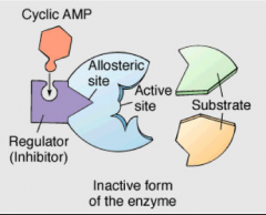
allosteric regulation, An allosteric enzyme is a protein that contains one or more binding sites called allosteric sites – usually separate from its active site Substances that bind to allosteric sites are called effectors If the effector is a substrate, it is described as homotropic Homotropic effector: the substrate itself serves as an effector (in most cases as positiveeffector) Heterotropic effector : the effector is different from the substrate example: fructose 2,6-biphosphate -> activates phosphofructokinase 1 -> increases the rate of glycolysis in response to the hormon glucagon proteolytical cleavage, Zymogens – inactive precursors of enzymes Common for enzymes involved in digestion The zymogens are activated only after they have entered the stomach or intestine In this way the molecules they encounter along the way are protected from digestion example: Pepsinogen --> Pepsin (In stomach) covalent modification Can completely turn on an inactive form of an enzyme or shut down an activeenzyme Phosphorylation is a common example of covalent modification |
|
|
Enzyme Inhibition |
Competitive (reversible, non-reversible) Uncompetitive Non-competitive Allosteric effect Covalent modification Proteolytic cleavage |
|
|
Competitive (reversible, non-reversible) |
REVERSIBLE Compete with substrate for binding to the same active site Usually similar in structure to substrate High concentrations of inhibitor can eliminate the binding of substrate High concentrations of substrate can block the binding of inhibitor E+S---->ES E+I----->EI IRRIVERSIBLE (Toxins, Ex. Sarin) Sarin acts on cholinesterase by forming a covalent bond with the particular serine residue at the active site. Fluoride is the leaving group, and the resulting phosphoester is robust and biologically inactive. A build-up of acetylcholine in the synaptic cleft, due to the inhibition of cholinesterase, means the neurotransmitter continues to act on the muscle fibre, so that any nerve impulses are effectively continually transmitted. |
|
|
Uncompetitive |
Binds reversibly to enzyme-substrate complex, but are unable to bind free enzyme E+I--->no reaction ES+I--->ESI |
|
|
Non-competitive |
Binds reversibly to both free enzyme and enzyme-substrate complex Usually binding of inhibitor is not at the active site Usually not similar in structure to substrate Inhibitor binds to ES-complex E+I--->EI ES+I--->ESI Substrate also binds to EI complex EI+S--->ESI
|
|
|
Creatinekinase |
Use in medicine: It can be used to diagnose and monitor disorders in which all types of muscle are damaged. Which organ: Heart and muscles Normal level in blood: 50-330 U/L adult male 40-175 U/L adult female |
|
|
Lactate dehydrogenase |
1. Lactate Dehydrogenase Lactate dehydrogenase is an enzyme found in most human cells and catalyzes the production of lactate from pyruvate Measurement of LDH isoenzymes helps determine the location of tissue damage. Which organ: Heart, muscles, lungs, pancreas, liver, brain Normal levels in blood: 140-280 U/L |
|
|
Aspartate Aminotransferase (AST) |
Use in medicine: The amount of AST in the blood is directly related to the extent of the tissue damage in certain organs Which organ: Heart and liver Normal levels in blood: 14-20 U/L adult males 10-36 U/L adult females |
|
|
Alanine aminotransferase (ALT) |
Use in medicine: ALT is measured to see if the liver is damaged or diseased. Which organ: Liver Normal levels in blood: 10-40 U/L adult male 7-35 U/L adult female |
|
|
Feehlings reagent |
Cu2+ Ions give the orange color Fehlings A Solution is blue Fehlings B Colorless Presence of carbohydrates in the solution gives a orange color when mixed in together in the final solution. |