Reading...
![]()
Play button
![]()
Play button
![]()
Use LEFT and RIGHT arrow keys to navigate between flashcards;
Use UP and DOWN arrow keys to flip the card;
H to show hint;
A reads text to speech;
69 Cards in this Set
- Front
- Back
|
List the 4 diseases classified under Blistering (Bullous) Diseases
|
1. Pemphigus Vulgaris
2. Bullous Pemphigoid 3. Dermatitis Herpetiformis 4. Epidermolysis Bullosa |
|
|
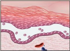
Subcorneal = beneath the Stratum Corneum layer
|
What type of blister is this?
|
|
|
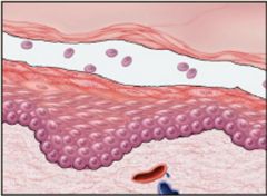
Suprabasal = roof & base of the blister are Keratinocytes
|
What type of blister is this?
|
|
|
Subepidermal = base is connective tissue, roof is basal layer of keratinocytes
|
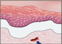
What type of blister is this?
|
|
|
Define Pemphigus Vulgaris
|
Autoimmune bullous disease mediated by cytotoxic antibody (IgG) to Desmoglein (protein in desmosomes) that results in dyshesion
|
|
|
What type of blister does Pemphigus Vulgaris cause?
|

Intra-epidermal blister = Suprabasal
|
|
|
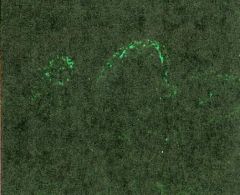
What does the DIF look like in Pemphigus Vulgaris?
|
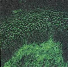
lace-like pericellular IgG in epidermis
|
|
|
What does the DIF look like in Pemphigus Vulgaris?
|
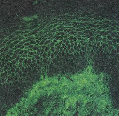
lace-like pericellular IgG in epidermis
|
|
|
Pemphigus Vulgaris
-suprabasal acantholytic blister |
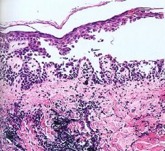
What blistering disease is this?
|
|
|
Pemphigus Vulgaris
-IgG & complement encircling epidermal cells |

What blistering disease is this from?
|
|
|
Pemphigus Vulgaris:
1. most common age? 2. Where do blisters commonly occur 3. Treatment? |
1. 30-50 years of age
2. scalp, mucous membrane, periumbilical, intertriginous areas 3. Prednisone + Immunosuppressive agents |
|
|
Define Bullous Pemphigoid
|
autoimmune bullous disease mediated by antibodies to 2 basement membrane glycoproteins in the Lamina Lucida (IgG ab's to hemidesmosomes)
|
|
|
What type of blister does Bullous Pemphigoid produce?
|
Epidermal-dermal interface blister = SUBEPIDERMAL
|
|
|
What does the DIF look like in Bullous Pemphigoid?
|
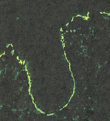
Linear deposition of IgG & C3 along the dermal-epidermal jxn
|
|
|
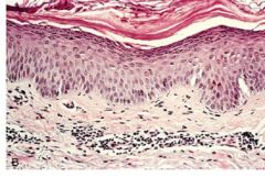
Bullous Pemphigoid
-subepidermal bullae -perivascular infiltrate of |
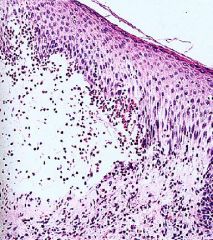
What blistering disease is this?
|
|
|
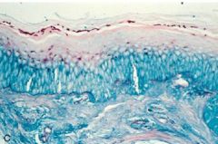
Bullous Pemphigoid
-linear band of complement & IgG deposition along BM |
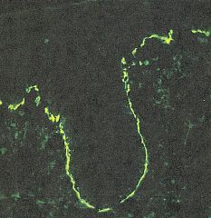
What blistering disease is this?
|
|
|
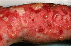
What are the clinical features of Bullous Pemphigoid?
|
MC affects men > 60 years old
Large & tense blisters Blisters on medial thighs & flexor aspect of the forearm |
|
|
A 51-year-old man presents with painful blisters over his entire body. He notes that he currently has several blisters in his mouth as well. A blister appears when you rub his skin with your finger. Skin biopsy demonstrates acantholysis.
|
Pemphigus Vulgaris
|
|
|
What type of hypersensitivity reaction are Bullous Pemphigoid & Pemphigus Vulgaris
|
Type II HS = Cytotoxic
|
|
|
What is the treatment for Bullous Pemphigoid?
|
Corticosteroids
|
|
|
Bullous Pemphigoid
|
What disease?
|
|
|
Bullous Pemphigoid
-subepidermal bullae -perivascular infiltrate of eosinophils & lymphocytes |
What blistering disease?
|
|
|
Blistering disease strongly associated with Gluten sensitivity (Celiac Disease) in individuals w/ HLA-B8 & HLA-DRw3
|
Dermatitis Herpetiformis
|
|
|
Blistering disease in which IgA to Gliadin cross-react or these immune complexes deposit at the tips of Dermal Papilla
|
Dermatitis Herpetiformis
|
|
|
Dermatitis Herpetiformis
-fibrin & neutrophils accumulate selectively at the tips of dermal papillae forming microabscesses |
What blistering disease is this?
|
|
|
Dermatitis Herpetiformis
-granular deposits of IgA localized in the tips of Dermal Papillae |
What blistering disease is this?
|
|
|
Dermatitis Herpetiformis
Celiac disease |
What blistering disease? What is it associated with?
|
|
|
Dermatitis Herpetiformis
|
Intensely pruritic cutaneous eruption
Urticaria-like plaques & small vesicles over the extensor surfaces of the body |
|
|
What is the treatment for Dermatitis Herpetiformis?
|
Gluten-free diet
|
|
|
What is Epidermolytic (simplex) Epidermolysis Bullosa?
|
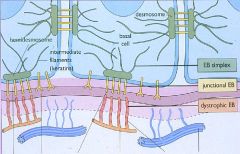
Gene mutations in Keratins 5 & 14 in the basal keratinocytes
|
|
|
What is Junctional Epidermolysis Bullosa?
|

Gene mutation in Laminin in the Basement Membrane (Lamina Lucida)
|
|
|
What is Dermolytic (Dystrophic) Epidermolysis Bullosa due to?
|

Gene mutation in Collagen Type 7 (beneath the lamina densa)
|
|
|
Epidermolysis Bullosa
1. Where do blisters tend to form? 2. When is this disease usually noted? |
1. at sites of minor trauma
2. at birth or shortly thereafter |
|
|
Epidermolysis Bullosa
|
What is this?
|
|
|
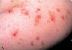
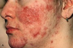
This disorder of epidermal appendages is a result of phsiologic hormonal variations & alterations in hair follicle maturation
|
Acne vulgaris
|
|
|
Describe the pathogenesis of Acne Vulgaris
|
Bacterial lipases of Propionibacterium acnes -> liberating highly irritating fatty acids -> expanding mass of lipid & keratin within the mid portion of the hair follicle -> distended follicle = Comedone
|
|
|
Describe the 2 types of Acne Vulgaris
|
1. Noninflammatory
-Obstructive-closed comedos = whiteheads -Open comedos = Blackheads 2. Inflammatory -follicular rupture -> extensive acute & chronic inflammation -> scarring |
|
|
Acne vulgaris
|
What is this?
|
|
|
Acne Vulgaris
|
What is this?
|
|
|
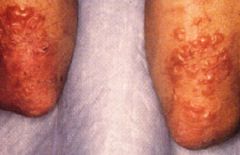
What skin disease is characterized as a Panniculitis (inflammation of subcutaneous fat)?
|
Erythema Nodosum
|
|
|
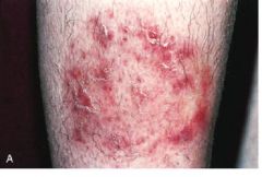
Erythema Nodosum
|
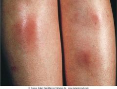
Self-limited, tender and erythematous nodules usually on the anterior portion of the shins
|
|
|
Inflammation of the fibrous septa of the subcutaneous tissue with Giant cells at the interface between the septa & adipose tissue
|
Erythema Nodosum
|
|
|
What triggers Erythema Nodosum?
|
Drugs or Microorganisms = exogenous agents
|
|
|
Erythema Nodosum
-Inflammation in the fibrous septa of the subcutaneous tissue, and giant cells at the interface between the septa and adipose tissue |
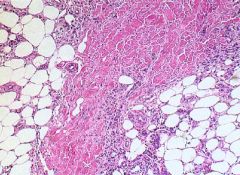
What is this showing?
|
|
|
What is the cause of Verrucae?
|
Human Papilloma Virus (HPV)
|
|
|
What is the cause of Verruca Vulgaris?
|
HPV 1, 2, 3, 4
|
|
|
Verruca vulgaris caused by HPV 1-4
|
What is this?
|
|
|
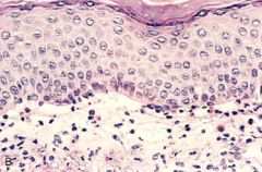
Verruca Vulgaris
-papillomatous epidermal hyperplasia -hyperkeratosis -parakeratosis -hypergranulosis -KOILOCYTES |
What is this?
|
|
|
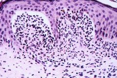
Verruca vulgaris staining for HPV
|
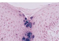
What is seen here?
|
|
|
Flat wart on the face or dorsal surface of hands, smaller than verruca vulgaris; caused by HPV 3
|
Verruca Plana
|
|
|
Warts on the soles & palms caused by HPV 2
|
Verruca Plantaris & Palmaris
|
|
|
Venereal wart caused by HPV 6, 8, 11, 16, 18
|
Condylomata Acuminatum
|
|
|
Condylomata Acuminatum
HPV 6, 8, 11, 16, 18 |
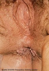
What is seen here? What is the cause?
|
|
|
What is Condylomata Acuminatum associated with?
|
Dysplasia & In-situ Squamous Cell Carcinoma
|
|
|
What is the cause of Molluscum Contagiosum?
|
Poxvirus
|
|
|
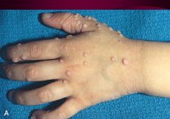
Firm, pruritic, pink to skin-colored umbilicated papules
|
Molluscum Contagiosum
|
|
|
Molluscum Contagiosum
|
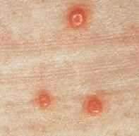
What is seen here?
|
|
|
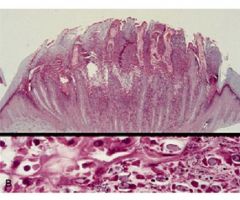
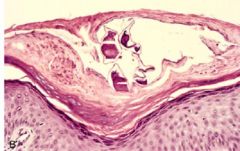
Molluscum Contagiousum
-large, eosinophilic cytoplasmic inclusion bodies (molluscum bodies) in the cells of the Stratum Granulosum & Corneum |
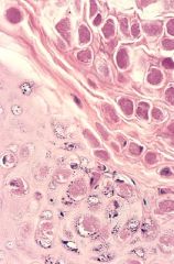
What is this?
|
|
|
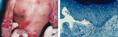
Impetigo:
1. contagious or not? 2. what are most caused by? |
1. highly contagious, usually in children, face, & hands
2. S. aureus & Strep pyogenes (happy kendall?) |
|
|
What is the pathology of Impetigo?
|
Erythematous macule, multiple small pustules, pustules break -> shallow erosions -> covered with drying serum = Honey-colored crust)
|
|
|
What is the characteristic microscopic feature of Impetigo?
|
accumulation of neutrophils beneath the stratum corneum (subcorneal pustule)
|
|
|
What is blister formation in Impetigo related to?
|
a toxin that specifically cleaves the Desmoglein I
-usually due to S. aureus |
|
|
Impetigo
S. pyogenes |
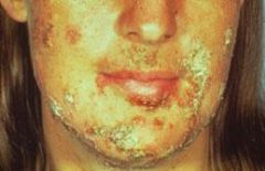
What is seen here? What is the most common cause?
|
|
|
What skin layer are Superficial Fungal Infections confined to? What are they caused by?
|
Stratum Corneum
Dermatophytes |
|
|
Superficial Fungal infections
-mild eczematous dermatitis |
What is shown here?
|
|
|
Superficial Fungal Infections
|
What is seen here?
|
|
|
Superficial Fungal Infection
|
What is seen here?
|
|
|
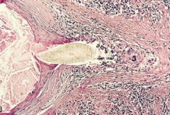
Human mite that causes pruritic erythematous streaks
|
Scabies
|
|
|
Scabies
-epidermal tunnels filled with basophilic granular debris and arthropod eggs |
What is seen here?
|