Reading...
![]()
Play button
![]()
Play button
![]()
Use LEFT and RIGHT arrow keys to navigate between flashcards;
Use UP and DOWN arrow keys to flip the card;
H to show hint;
A reads text to speech;
53 Cards in this Set
- Front
- Back
|
List the anatomic & physiologic features that are uniques to the CNS?
|
1. Rigid skull & spinal column = volume of which is fixed after early childhood
2. Autoregulation of Blood flow = cerebral blood flow is regulated to a large extent independently from systemic blood circulation 3. No lymphatic system 4. CSF system 5. Limited immune surveillance = immunologically secluded from the rest of the body 6. Unique response to injury 7. Vulnerability of lack of perfusion 8. Lack of regeneration |
|
|
Define Vasogenic Edema
|
accumulation of fluid between the neurons & glia & most prominently in the Virchow-Robin spaces around the blood vessels
Develops as a consequence of BBB dysfunction = fluid escapes from the vascular space into the interstitial space of the parenchyma across the cytoplasm of Endothelial cells |
|
|
Define Cytotoxic Edema
|
Fluid accumulates inside cells
Most frequently caused by ischemia &/or hypoxia = lead to hydropic swelling of neurons & glial cells |
|
|
Define Interstitial Edema
|
Result of increased bulk of CSF thru the Ependymal lining = dysfunction of the brain-CSF barrier
Typically a complication of Hydrocephalus |
|
|
What intracranial pressure is associated with edema?
|
> 200 mm water
|
|
|
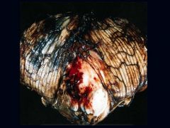
What pathologies are seen in Cerebral Edema?
|
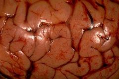
1. Soft brain
2. Flat gyri 3. Narrow slit-like sulci 4. compressed Ventricles 5. possible Herniation |
|
|
What is a Subfalcine Herniation? What is an alternate name? What artery may be compressed?
|
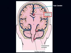
Results from a unilateral hemispheric mass lesion that expands the volume of one hemisphere, dislocates the midline structures & forces the ipsilateral Cingulate gyrus to be compressed underneat the FALX CEREBRI
Alternate = Cingulate Herniation Anterior Cerebral Artery |
|
|
What is a Transtentorial Herniation? What is an alternate name? What can be compressed & how is it manifested?
|
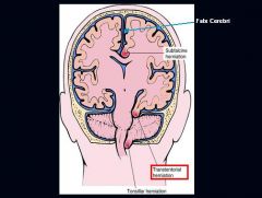
Caused by expansion of one or both Supratentorial tissue compartments. Uncus gyri hippocampi is displaced & herniated underneath the free edge of the Tentorium
Alternate = Uncinate Hernia 3rd Cranial nerve = dilation of the Ipsilateral Pupil + abnormal eye movement on the same side |
|
|
What is a Tonsilar Herniation? What may it cause?
|
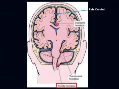
Life-threatening condition b/c the herniated cerebellar tonsils that are forced into the Foramen Magnum compress vital Respiratory & Cardiac centers within the Medulla Oblongata
|
|
|
Duret Hemorrhage
-common with a Trantentorial Herniation -midbrain with blood |
What is seen here?
|
|
|
Midbrain herniation + Oculomotor nerve palsy (eye down & out) + Mydriasis...what herniation?
|

Transtentorial Herniation
|
|
|
Increase in the CSF volume that causes enlargement of the Ventricles
|
Hydrocephalus
|
|
|
What makes the CSF?
|
Choroid Plexus
|
|
|
How is Hydrocephalus manifested in Newborns before the closure of Sutures?
|
Ventricles dilate & enlarge the head circumference
|
|
|
What is "Hydrocephalus Ex Vacuo?
|
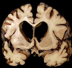
Dilated appearance of the ventricles when the brain mass is decreased
Ex. Alzheimer's disease |
|
|
Hydrocephalus
|

What are these pictures showing?
|
|
|
Describe the flow of CSF
|
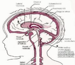
1. Choroid Plexus
2. Lateral Ventricles 3. Interventricular Foramen (Foramen of Monroe) 4. Third Ventricle 5. Cerebral Aqueduct (Aqueduct of Sylvius) 6. Fourth Ventricle 7. Foramen of Magendie & Luschka 8. Subarachnoid Space over brain & Spinal Cord 9. Reabsorption into Venous Sinus blood via Arachnoid Granulations |
|
|
What things cause Focal Lesions?
|

Tumor
Infarct Abscess |
|
|
What things typically cause Multifocal lesions? (3)
|
1. Metastases
2. Small infarcts 3. Abscesses |
|
|
System Degeneration
-typically slowly progressive -Ex: Motor Neuron Disease (ALS) |
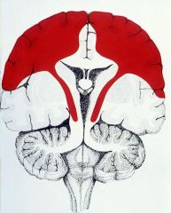
What generally is this showing?
|
|
|
Diffuse Disorder
-Neuronal = Lysosomal storage disease -White Matter = Leukodystrophy |

What generally is this showing? What could be possible causes?
|
|
|
List the types of Glia found in the CNS
|
1. Astrocytes
2. Oligodendrocytes 3. Ependymal cells 4. Microglia |
|
|
Material consisting of granular Endoplasmic Reticulum & Ribosomes & occuring in nerve cell bodies & dendrites
|
Nissl Substance
|
|
|
any of the long, thin, microscopic fibrils that run through the body of a neuron and extend into the axon and dendrites
|
Neurofibrils
|
|
|
cytoplasmic Nissl Substance within neuron
|
What does this picture exemplify?
|
|
|
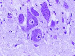
Cerebellar cortex showing large Purkinje cells and smaller dark granule cell neurons (at lower right).
|
What cells are seen in this picture?
|
|
|
What are 2 unique principles of Neurons?
|
1. Selective vulnerability = very sensitive to Ischemia
2. Post-mitotic cells = no regeneration |
|
|
What pathology is seen during Acute Injury of neurons? What most commonly causes it?
|
Pynknotic nuclei & an acidophilic cytoplasm = Red neuron
Ischemia, Anoxia, Hypoglycemia |
|
|
What things occur in the process of "Axonal Reaction"?
|

1. Perikaryon swells
2. Chromatolysis = degranulation of Nissl Substance (RER) -> loss of basophilia |
|
|
What is Wallerian Degeneration?
|
degeneration of nerve fibers Distal to the injury
|
|
|
What is Transsynaptic Degeneration?
|
Atrophy of nerve cells due to loss of input
|
|
|
Neuromelanin
Lipofuscin = 'wear & tear' pigment |

What is the white arrow pointing at? What is seen within the cytoplasm of some cells?
|
|
|
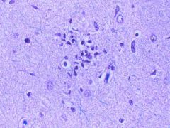
Neurofibrillary Tangle in Hippocampal CA1 neuron
|
What is seen here?
|
|
|
Lewy bodies = Parkinson Disease
|

What are the arrows pointing at?
|
|
|
Which Glia are derived from the Neuroectoderm?
Which Glia is derived from the Mesoderm? |
Astrocytes
Oligodendrocytes Ependymal cells Microglia |
|
|
Glial that are in close contact with neurons & have small oval nuclei with star-like processes
|
Astrocytes
|
|
|
Glia that have cytoplasmic extensions that attach to blood vessels, forming part of the BBB
|
Astrocytes
|
|
|
Glia that contain "Glial Fibrillary Acidic Protein" (GFAP)
|
Astrocytes
|
|
|
What is another term for Gliosis?
|
Astrocytosis
|
|
|
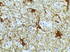
Reactive Gliosis (Astrocytosis)
-pink-staining cells |

What is seen here?
|
|
|
Reactive Astrocytosis
|
What is seen here?
|
|
|
Gemistocytes
-a form of Astrocytosis |

What is seen here?
|
|
|
Metabolic Astrocyte
-clear nuclei in Cerebral Cortex -stimulated to proliferate when there is some sort of metabolic disease occurring |

What is the arrow pointing at?
|
|
|
Rosenthal Fiber
-usually seen in chronic disease -often seen around old sites of injury |
What is the arrow pointing at?
|
|
|
Corpora Amylacea = inclusions within Astrocytes
-seen in aging -gliosis |

What are seen here?
|
|
|
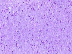
What are the properties of Oligodendrocytes?
|
1. few processes
2. small dark round nuclei 3. make, maintain myelin -1 Oligo: wraps up to 50 axons 4. Injury: myelin loss or abnormal myelin 5. Tumors: Oligodendrogliomas |
|
|
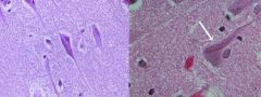
Oligodendrocyte in normal White Matter
|

What is the arrow pointing at?
|
|
|
Columnar ciliated cells lining the Ventricular System
|
Ependymal cells
|
|
|
Normal Ependymal cells with cilia
|
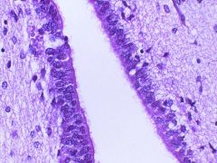
What is seen here?
|
|
|
Patchy Ependymal loss - when they are injured, they don't come back
|

What important property of Ependymal cells is exemplified here?
|
|
|
Choroid Plexus
Secrete CSF |
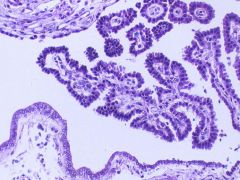
What cells are seen here? What is their functinon?
|
|
|
Phagocytic cells of mesenchymal origin of the CNS
|
Microglia
|
|
|
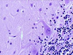
Microglial Nodule
-elongated rods, club-shaped |
What is seen here?
|