![]()
![]()
![]()
Use LEFT and RIGHT arrow keys to navigate between flashcards;
Use UP and DOWN arrow keys to flip the card;
H to show hint;
A reads text to speech;
121 Cards in this Set
- Front
- Back
|
Cardiovascular System General Function |
• Primary purpose: – Deliver oxygen to tissues • Secondary functions: – Removal of CO2, lactate, etc. – Transport of nutrients – Communication system via hormone transport – Acid-Base balance – Body fluid regulation – Thermoregulation |
|
|
Organization of the Circulatory System |
- closed loop (all vessels connected) - blood travels through loop driven by pressure created as the heart contracts - high pressure/low pressure - composed of: blood vessels (transport) heart (pump) blood (transport medium) |
|
|
Arteries |
- these leave the heart |
|
|
Arterioles |
- arteries divide into these |
|
|
Capillaries |
- arterioles divide into these - are the smallest and most numerous type of blood vessel |
|
|
Capillary Membranes |
- where all exchanges between the blood and cells of the body occur |
|
|
Venules |
- where blood is collected after leaving the capillaries |
|
|
Veins |
- venules merge into these - returns blood back to heart |
|
|
Heart |
- 4 chambers - considered "2 pumps in 1" - R + L divided by interventricular septum |
|
|
Blood Flow Through Heart |
- right atrium - right ventricle - lung (via pulmonary arteries) - left atrium (via pulmonary veins) - left ventricle - body (via aorta) |
|
|
Atrioventricular Valve
|
- separates atrium and ventricles - right AV valve: tricuspid valve - left AV valve: mitral or bicuspid valve |
|
|
Right AV Valve |
- tricuspid valve
|
|
|
Left AV Valve |
- mitral or biscupid valve |
|
|
Semilunar Valves |
- separates ventricles and vessels - pulmonary semilunar valve - aortic semilunar valve |
|
|
Pulmonary Circuit |
- low pressure - right ventricle - capillaries of the lung - oxygen loaded onto hemogoblin; CO2 released - capillaries of lung - left atrium |
|
|
Systemic Circuit |
- high pressure - left ventricle - capillaries of tissues - oxygen released from hemogoblin; CO2 taken up - capillaries of tissues - right atrium |
|
|
Epicardium |
- outer layer
|
|
|
Endocardium |
- inner layer
|
|
|
Myocardium |
- muscular middle layer - contracts to force blood out of the heart - blood supplied via coronary arteries - cardiac muscle fibers |
|
|
Differences in Myocardium & Skeletal Muscle Fibers |
- cardiac fibers interconnected via intercalated discs - electrical impulse = one fiber to next - acts as a functional syncytium; no motor units - homogeneous (one fiber type) - resembles slow twitch (fatigue resistant, oxidative, many mitochondria) |
|
|
Similarities of Myocardium and Skeletal Muscle Fibers |
- striated (thick & thin filaments) - contract via sliding filament theory - calcium triggers contractions - length-tension relationship exists - stretched cardiac fibers contract with more force |
|
|
Cardiac Cycle |
- repeated contraction and relaxation of myocardium - contraction phase; systole - relaxation phase; diastole - systole and diastole refer to contraction/relaxation of ventricles - atria undergo systole/diastole as well - 2 step pumping action - atria contracts together first - ventricles contracts together second - ventricles contract 0.1s later; ejects 2/3 of blood from ventricles - at rest: 75 cycles per minute |
|
|
Systole |
- contraction phase of the heart |
|
|
Diastole |
- relaxation phase of the heart |
|
|
Phase of Cardiac Cycle |
- diastole (filling of heart) blood enters atria; flows into ventricles (70%) AV valves are open - atrial systole occurs final push of blood into ventricles (30%) - ventricular systole AV valves close ventricular ejection phase; blood through SL valves |
|
|
Lub Sound |
- 1st heart sound - closing of AV valves - occurs at end of diastole |
|
|
Dub Sound |
- 2nd heart sound - closing of semilunar valves - occurs at the end of systole
|
|
|
What is Blood Pressure?
|
- pressure of blood against arterial walls - expressed as systolic/diastolic pressure |
|
|
Factors that Determine Blood Pressure |
- volume of blood - resistance to blood flow - blood viscosity - blood vessel diameter |
|
|
Sphygmomanometer |
- used to measure blood pressure |
|
|
Systolic Pressure |
- top number - pressure generated during ventricular contraction (systole)
|
|
|
Diastolic Pressure |
- bottom number - pressure in the arteries during cardiac relaxation (diastole) |
|
|
Normal Blood Pressure |
- 120/80 mmHg |
|
|
High Blood Pressure |
- greater than 140/90 mmHg |
|
|
Intrinsically Stimulated |
- self excitable (automaticity) |
|
|
Extrinsically Stimulated |
- stimulated by nerves |
|
|
Syncytium |
- means to contract as a unit |
|
|
SA Node |
- pacemaker |
|
|
Intrinsic Rhythm of SA Node |
- 100 bpm - at rest, slowed by extrinsic nerves |
|
|
Atrial Contraction |
- firing of SA node causing depolarization to spread throughout atria |
|
|
Intrinsic Conduction System |
- impulse carried into ventricles by way of AV node (atrioventricular node) delayed 0.1s - AV node located on floor of right atrium - right and left bundle branches run from AV node down interventricular septum - purkinje fibers branch off of conducting branches to carry impulse into myocardium |
|
|
Extrinsic Conduction System |
- innervated by ANS - sympathetic cardioacceleratory center (NE) - parasympathetic cardioinhibitory center (ACh) |
|
|
Sympathetic Cardioacceleratory Center |
- increases heart rate; norephinephrine |
|
|
Parasympathetic Cardioinhibitory Center |
- lowers heart rate; acetylcholine |
|
|
Bradycardia |
- slow heart rate; < 60 bpm |
|
|
Tachycardia |
- rapid heart rate; > 100 bpm |
|
|
Electrocardiogram (ECG) |
- graphic recording of the electrical activity of the heart - used in the diagnosis of heart disease - deflections called waves p wave qrs complex t wave |
|
|
P Wave |
- depolarization of atrium
|
|
|
QRS Complex |
- depolarization of ventricles |
|
|
T Wave |
- repolarization of ventricles |
|
|
Plasma |
- liquid portion - ions, proteins, hormones |
|
|
Cells |
- red blood cells (hemogoblin to carry oxygen) - white blood cells (immune response) - platelets (blood clotting)
|
|
|
Volume of Blood |
- 5 liters (1.5 gallons) |
|
|
Characteristics of Blood |
- scarlet/dark red - pH: 7.35 - 7.45 (vein - arteries) - temperature: 38 degrees celsius - average volume: 5 L - normal hematocrit: 42% - 45%
|
|
|
pH of Veins
|
- 7.35 |
|
|
pH of Arteries |
- 7.45
|
|
|
Temperature of Blood |
- 38 degrees celsius |
|
|
Hematocrit |
- volume percentage of red blood cells in the blood
|
|
|
Normal Hematocrit Volume |
- 42% to 45% |
|
|
What Increases Cardiac Output |
- the amount of blood pumped per minute by the heart |
|
|
Redistribution of Blood Flow |
- from inactive organs to active skeletal muscle |
|
|
Cardiac Output Equation |
- cardiac output = stroke volume x heart rate |
|
|
Stroke Volume |
- amount of blood pumped per beat - SV |
|
|
Heart Rate |
- beats per minute - HR |
|
|
Average Resting Cardiac Output |
- 5 L x m -1 |
|
|
Endurance Trained Cardiac Output During Exercise |
- 35 L x m -1 |
|
|
Untrained Cardiac Output During Exercise |
- 20 L x m -1 |
|
|
Increasing Cardiac Output |
- achieved by increasing SV - achieved by increasing HR - during exercise both SV and HR increase |
|
|
Regulation of Heart Rate |
- sympathetic and parasympathetic impulses on SA node |
|
|
Sympathetic Fibers |
- accelerator nerve - release norepinephrine |
|
|
Parasympathetic Fibers |
- vagus nerve - releases acetylcholine |
|
|
Parasympathetic Tone (Vagal Tone) |
- resting conditions - intrinsic SA node rate is 100 min -1 - resting HR 75 BPM - HR increased by decreasing parasympathetic tone |
|
|
Increases HR from 75 to 100 BPM |
- withdrawal of parasympathetic (vagal) tone |
|
|
Increases HR above 100 BPM |
- due to stimulation of accelerator nerve (sympathetic) |
|
|
Three Variables that Effect Stroke Volume |
- end diastolic volume (EDV) - mean arterial blood pressure (MABP) - strength of ventricular contractions |
|
|
End Diastolic Volume |
- volume of blood ventricles at end of diastole |
|
|
Blood in Ventricles |
- causes stretching of ventricular myocardium
|
|
|
Frank Starling Law of the Heart |
- stretched fibers contract with more strength equaling greater stroke volume |
|
|
How is EDV Increased During Exercise |
- due to increased venous return to the heart |
|
|
Why Does Venous Return Increase During Exercise? |
- venoconstriction - muscle pump - respiratory pump |
|
|
Venoconstriction |
- veins constrict; squeeze blood toward heart |
|
|
Muscle Pump |
- muscle contract and squeeze veins, blood toward heart |
|
|
Respiratory Pump |
- breathing equals alternating pressure changes between abdominal plus thoracic cavities; milks blood toward heart |
|
|
Minimizes the Increase of MABP During Exercise |
- vasodilation of arterioles |
|
|
This Will Increase Stroke Volume |
- increased strength of contraction |
|
|
Increased Contractility During Exercise Due To |
- increased epinephrine plus norepinephrine realease causing increased Calcium release plus greater cross bridge cycling rate - increased accelerator nerve activity resulting in increased force due to temporal summation |
|
|
Preload (EDV) |
- increased preload = increased SV |
|
|
Afterload (MABP) |
- decrease afterload = increase stroke volume |
|
|
Contractility (force of contraction) |
- increased contractility = increased stroke volume |
|
|
Functional Zones of the Respiratory System |
- conducting zone - respiratory zone |
|
|
Conducting Zone |
- nose pharynx, trachea, bronchi, bronchial tree - essentially all passageways (dead air space) - conducts air to/from respiratory zone - air moved through nose at low flow rates - > 20-30 Lmin-1 mouth becomes primary - filters, humidifies, and warms air |
|
|
Respiratory Zone |
- respiratory bronchioles, alveolar ducts, and alveolie - region where gas exchange occurs - respiratory bronchioles + alveolar ducts - alveoli (convoluted microscopic air sacs; 300 million per lung) - blood gas barrier (2 cell layers thick; rapid diffusion of gases) |
|
|
Boyle's Law |
- relationship between pressure and volume of a gas - increased volume = lower pressure - decreased volume = higher pressure |
|
|
Inspiration |
- contraction of diaphragm + external intercostals - results in increased thoracic cavity size (decreasing pressure, air moved into lungs) - during exercise (labored breathing) - accessory muscles further elevate ribs |
|
|
Muscles Involved in Inspiration |
- scalenes - sternocleiodomastoids - pectoralis minor |
|
|
Expiration |
- relaxation of diaphragm + external intercostals - return to original position (increasing pressure) - no muscular effort at rest - during exercise (forced expiration) - accessory muscles help force air out - increase intra-abdominal pressure - forcefully depress rib cage |
|
|
Muscles Involved in Expiration |
- abdominal muscles - internal intercostals |
|
|
Pulmonary Ventilation (V) |
- amount of air moved in or out of the lungs per minute - product of tidal volume and breathing frequency (Vt + f) |
|
|
Dead-space Ventilation (Vd) |
- unused ventilation - does not participate in gas exchange - anatomical dead space: conducting zone |
|
|
Alveolar Ventilation (Va) |
- volume of inspired gas that reaches the respiratory zone |
|
|
Tidal Volume |
- volume inspired or expired during unforced respiration - 500 mL |
|
|
Inspiratory Reserve Volume |
- volume inspired at end of tidal inspiration - 3100 mL |
|
|
Expiratory Reserve Volume |
- volume expired at end of tidal expiration - 1200 mL |
|
|
Residual Volume |
- air remaining in lungs after maximal expiration - 1200 mL |
|
|
Vital Capacity |
- maximum volume that can be exhaled after a maximum inhalation - 4800 mL |
|
|
Total Lung Capacity |
- total volume in lung after maximum inhalation - 6000 mL |
|
|
FEV1.0 |
- amount of vital capacity that can be expired in 1 second |
|
|
Gas Exchange |
- diffusion between alveolar air spaces and blood across alveolar membranes - o2 enters blood - co2 enters lungs |
|
|
Gas Transport in Blood |
- oxygen and carbon dioxide diffuse from one area to another based on pressure gradient |
|
|
Dalton's Law of Partial Pressures |
- the total pressure of a gas mixture is the sum of the pressures of each independent gas |
|
|
Partial Pressure |
- Pbarometric x gas fraction |
|
|
Alveoli |
- PO2 = 100 - PCO2 = 40 |
|
|
Systemic Arteries |
- PO2 = 100 - PC02 = 40 |
|
|
Systemic Veins |
- PO2 = 40 - PCO2 = 46 |
|
|
O2 Diffuses Where |
- into the blood at the lungs - out of the blood at the tissues |
|
|
Two Mechanisms of Transport of O2 |
- plasma - hemoglobin |
|
|
O2 Transported by Plasma |
- 1% - 0.3 mL dL -1 blood |
|
|
O2 Transported by Hemogoblin |
- 99% - 1.34 mL O2 per gram of Hb - 15 g Hb dL -1 blood - 20.1 mL dL -1 blood |
|
|
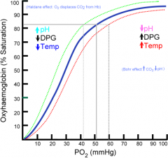
Oxyhemoglobin |
- hemogoblin saturated with O2 |
|
|
Dissociation of O2 from Hb |
- occurs with decrease in PO2 levels in plasma |
|
|
Oxyhemoglobin Dissociaition Curve |
|