![]()
![]()
![]()
Use LEFT and RIGHT arrow keys to navigate between flashcards;
Use UP and DOWN arrow keys to flip the card;
H to show hint;
A reads text to speech;
74 Cards in this Set
- Front
- Back
|
Actinic Keratosis Description |
Pre-malignant lesion typically seen on sun-exposed areas. If untreated can progress to squamous cell carcinoma |
|
|
Actinic Keratosis (pre-cancerous) |
|
|
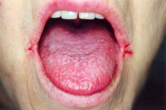
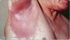
Angular cheilosis description |
inflammation of corners of lips - seen in malnutrition, anemia (Fe-def anemia if also pica!) |
|
|
Angular Cheilosis (malnutrition, anemia) |
|
|
Basal Cell Carcinoma description |
low-grade |
|
|
Basal Cell Carcinoma |
|
|
Gottron's Papules/Sign Description |
Red, scaly papules over MCPs, PIPs and DIPs. |
|

|
Gottron's Papules (Dermatomyositis) |
|
|
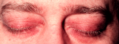
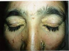
Heliotrope rash Description |
red/violet discoloration of upper eyelids |
|
|
Heliotropic Rash (Dermatomyositis) |
|
|
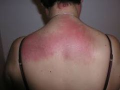
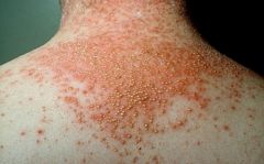
Shawl Sign Description |
widespread, flat, reddened area over upper back, shoulders, and back of neck |
|
|
Shawl Sign (dermatomyositis) |
|
|
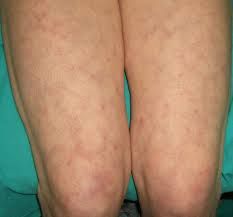
V sign Description |
similar to shawl sign, except reddened skin isinV-neck pattern of chest |
|

|
V Sign (Dermatomyositis) |
|
|
Dermatomyositis findings |
Gottron's Papules, Heliotropic rash, Shawl/V Sign |
|
|
Hyperextensibility description |
ability to stretch skin >4 cm at forearm or neck before feeling resistance - Ehlers-Danlos |
|
|
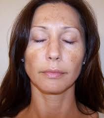
Hyperpigmentation description |
*address patient's background/race/ethnicity/sun exposure* |
|

|
Hyperpigmentation (tricky, I know, but these are the pictures I got when I googled Hyperpigmentation in Addison's Disease) |
|

|
Hyperpigmentation (hematochromatosis) |
|
|
Hyperpigmentation (Also subtle, but this is what hyperpigmentation in hyperthyroidism seems to look like) |
|
|
Livedo reticularis description |
red-blue lacy skin discoloration with central pallor, assoc w/ small vessel changes, small vessel vasculitis, anti-phospholipid antibody syndrome ( in lecture he also mentions Raynaud's syndrome, cholesterol embolism, DIC) |
|
|
Livedo Reticularis (vasculitis) |
|
|
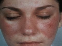
Malar Rash Associated w/ |
SLE |
|
|
Malar Rash - SLE |
|
|
Melanoma Description |
Tumor of melanocytes. Dark. Can be nodular/lentiginous. Can occur on skin that isn't exposed to sun. Even ocular site. |
|

|
Melanoma |
|
|
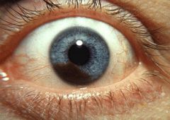
Ocular Melanoma |
|
|
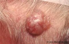
Neurofibroma Description |
Non-tender, soft, fleshy, sessile, or pedunculated skin tumor. Assoc w/ Von Recklinghausen's Disease |
|
|
Von Recklinghausen's Disease Findings |
Neurofibromas |
|
|
Von Recklinghausen's Genetics |
Autosomal Dominant. Also spontaneous mutations are pretty common too! |
|
|
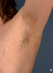
Axillary Freckles, AKA Crowe's Sign, (Von Recklinhausen's disease) |
|
|
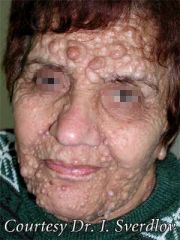
Neurofibromas (Von Recklinhausen's) |
|
|
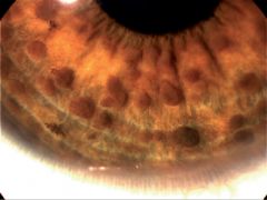
Lisch Nodules (100% has Von Recklinhausen's) |
|
|
Psoriasis (typical description) |
Usually red plaques with silvery scales on extensor surfaces of arms and legs, trunk and scalp. |
|
|
Psoriasis (different presentations) |
Guttate- drop-like |
|
|
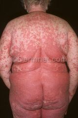
Erythrodermic psoriasis |
|
|
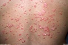
Guttate (drop-like) psoriasis |
|
|
Inverse psoriasis |
|

|
Psoriasis (again, an untraditional spot, but you know how Morehead is) |
|
|
Pustular Psoriasis |
|
|
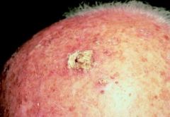
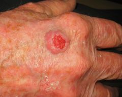
Squamous Cell Carcinoma Description |
Begins as nodule, grows into fungating lesion, sometimes ulcerates. Usually on sun-exposed skin, assoc w/ chronic scarring and inflammation. |
|
|
Squamous Cell Carcinoma |
|
|
Thin Skin w/ visible small cells = |
Ehler Danlos (type 4), assoc w/ arterial rupture (think aortic dissection!!!) |
|

|
Thin Skin (Ehlers-Danlos) |
|
|
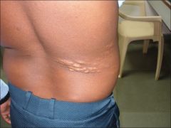
Shagreen Patch |
flesh-colored, orange-peel like CT plaque usually on lower back. |
|
|
Shagreen Patch (Tuberous Sclerosis) |
|
|
Ash-leaf spots (ellipticalmacules) |
hypopigmented spots. Tuberous Sclerosis |
|
|
Ash Leaf Spots (Tuberous Sclerosis) |
|
|
Tuberous Sclerosis Findings |
Angiofibromas |
|
|
Angiofibroma description |
Looks like acne, but firm, not pustular. Sign of Tuberous Sclerosis |
|

|
Angiofibromas (Tuberous Sclerosis) |
|
|
Why important to identify a Tuberous sclerosis patient? |
These patients could have epilepsy, a nueropsychiatric disorder, huge increased risk of cancer, angiomyolipomas, and risk of developing cystic lung disease. |
|
|
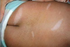
Vitiligo description |
Autoimmune reaction against melanocytes. Diffuse or focal. Think autoimmune, hypothyroidism, Hashimoto's thyroiditis |
|

|
Vitiligo (Autoimmune, hypothyroid, Hashimotos) |
|
|
Xanthomas general description |
Nodular, papular, tumerous lesions (may be confused with rash/infection). Hyperlipidemia, esp familial variants. |
|
|
Xanthoma |
|
|
Variations of Xanthomas |
Xanthelasma - around eyes |
|
|
Eruptive Xanthoma |
|

|
Palmar Xanthoma (he got us on this in a practice question a couple sessions ago... xanthomas can be on the palms!) |
|

|
Tendon Xanthoma (hyperlipidemia definitely familial) |
|
|
Blastomycosis Skin Presentation |
Skin is 2nd most common organ involved |
|
|
Blastomycosis (looks like squamous cell!) |
|
|
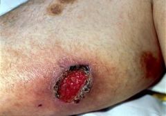
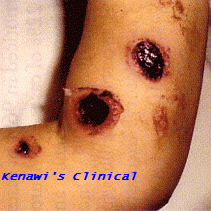
Ecthyma Gangrenosum Presentation |
Hemorrhagic vesicles/pustules, cause microinfarcts, evolve into necrotic ulcers. Manifests from infectious vasculitis due to Pseudomonas aeruginosa (gram neg). |
|
|
Ecthyma Gangrenosum (Pseudomonas infection) |
|
|
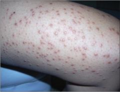
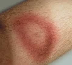
Erythema Chronicum Migrans Presentation |
Painless, Target rash. Lyme Disease |
|
|
Erythma Chronicum Migrans (Lyme) |
|
|
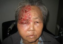
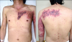
Herpes Zoster Presentation |
burning dysesthesias, burning dermatomal rash that doesn't cross midline. Crusts. Neuropathic pain may persist. Recurrent VZV. |
|
|
Herpes Zoster |
|
|
Herpes Zoster |
|
|
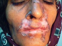
Lupus Vulgaris Presentation (AKA?) |
AKA Tuberculous chancre |
|
|
Lupus Vulgaris (TB) |
|
|
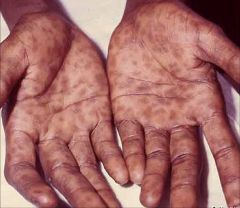
Secondary Syphilis Presentation |
Papular, raised, ulcerated lesions on palms and soles. |
|
|
Secondary Syphillis |
|

|
Secondary Syphillis |