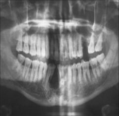
![]()
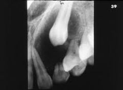
![]()
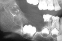
![]()
Use LEFT and RIGHT arrow keys to navigate between flashcards;
Use UP and DOWN arrow keys to flip the card;
H to show hint;
A reads text to speech;
199 Cards in this Set
- Front
- Back
|
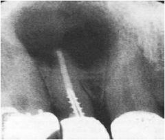
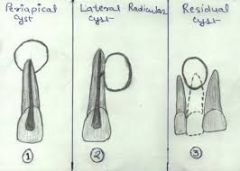
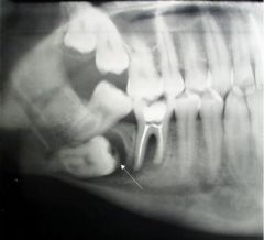
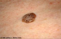
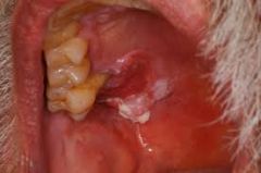
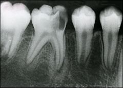
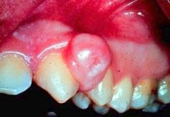
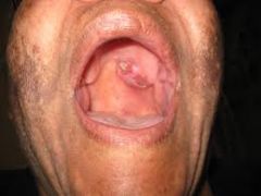
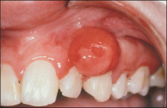
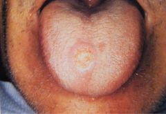
Most common odontogenic cyst Most common cyst of oral cavity at apex of NON VITAL tooth |
Periapical (Radicular) Cyst |
|
|
Types of Odontogenic cysts |
inflammatory developmental neoplastic |
|
|
Types of inflammatory odontogenic cysts |
periapical residual periapical buccal bifurcation |
|
|
Types of developmental odontogenic cysts |
dentigerous/eruption cyst primordial lateral periodontal gingival |
|
|
types of neoplastic odontogenic cysts |
odontogenic keratocyst (keratocytstic odontogenic tumor calcifying odontogenic tumor |
|
|
periapical cyst = inflammatory |
|
|
How do you treat a periapical cyst? |
root canal therapy extraction for non restorable teeth |
|
|
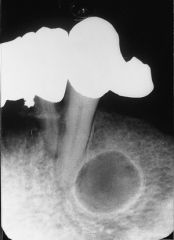
periapical granuloma |
radiographically can't tell diff from cyst histologically = chronically inflamed granulation tissue of non vital tooth |
|

|
Inflammatory |
|
|
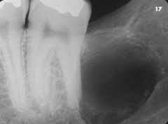
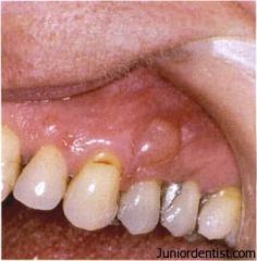
buccal bifurcation cyst |
cyst in furcation of most commonly: Mandibular 1st M INFLAMMATORY response |
|
|
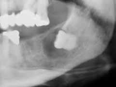
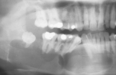
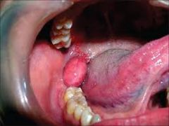
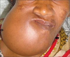
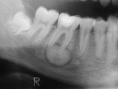
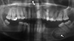
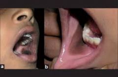
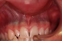
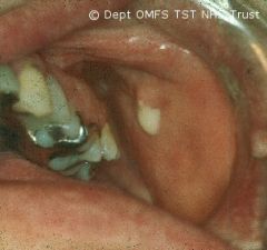
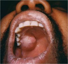
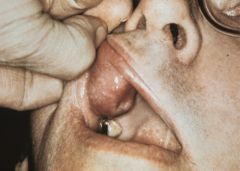
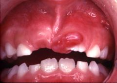
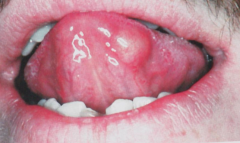
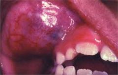
Dentigerous cyst MC DEVELOPMENTAL encloses crown of unerrupted tooth MC 3M and C |
|
|
primordial cyst enamel organ becomes cyst most cases are OKC in place of tooth Developmental |
|
|
Lateral from rest of dental lamina between 2 roots of vital teeth Developmental around transition point mand from ant to post |
|
|
gingival developmental from rests of dental lamina (Serres) mand C and PM |
|
|
Calcifying Odontogenic Cyst |
GORLIN CYST NEOPLASM |
|
|
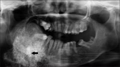
Odontogenic Keratocyst (OKC) |
|
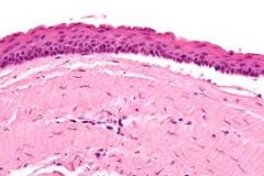
Odontogenic Keratocyst |
histopath feat= most important parakeratotic surface epith cells (Wavy)- horizontal Plaisaded basal cells(fence )- on the bottom borders lighter pink, vertical standing NEOPLASTIC |
|
|
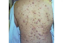
Gorlin Syndrome |
multiple basil cell carcinomas of the skin multiple OKC |
|
|
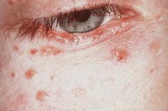
multiple bcc associated with Gorlin syndrome |
|
|
basil cell carcinoma ( usually multiple and associated with gorlin syndrome) |
|
|
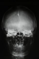
intracranial calcification associated with Gorlin syndrome |
|
|
What are the 2 odontogenic cysts that may act as a tumor? |
OKC can become keratinizing cystic odontogenic turmor (KCOT) calcifying odont cyst (COC) can become Calcifying cystic odonto tumor (CCOT) |
|
|
Tumors of odonto epith without odonto ectomesenchyme |
ameloblastoma calcifying epith odonto tumor squamous odonto tumor |
|
|
Tumors of odonto epith with ectomesenchyme, with or without dental hard tissue formation |
adenomatoid odonto tumor ameloblastic fibroma ameloblastic fibro-odontoma odontoma |
|
|
Tumors of odonto ectomesenchyme with or without epith |
odonto myxoma cementoblastoma |
|
|
Ameloblastoma features |
multilocular radiolucency pericoronal radiolucency extra- osseous (peripheral) |
|
multilocular radiolucency |
can be ameloblastoma (tumor of epith) |
|
pericoronal radiolucency |
can be ameloblastoma (tumor of epith) |
|
extra osseous peripheral |
can be ameloblastoma (tumor of epith) |
|
|
Histo features of Ameloblastoma |
Resembles the enamel organ |
|
|
Calcifying epithelial odontogenic tumor |
|
|
Calcifying epithelial odonto tumor can also look same as ameloblastoma in age location and treatment |
|
|
Calcifying epithelial odontogenic tumor |
histo: cells look like stratum intermedium of enamel organ has uni and multilocular radiolucency Tumor of epithelium |
|
|
Squamous Odontogenic Tumor |
from rest of Malassez in PDL space local radiolucency loss of alveolar bone and lamina dura tumor of epithelium |
|
|
squamous odonto tumor loss of alveolar bone and lamina dura |
|
|
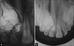
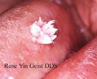
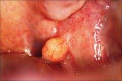
adenomatoid odontogenic tumor tumor of epith and ectomesenchyme involve crown of unerupt tooth younger pt's ( 10-19) Females Maxilla canine snowflake calcification |
|
|
The malignant counter part of ameloblastic fibroma ? |
ameloblastic fibrosarcoma |
|
|
ameloblastic fibroma tumor of epith and ectomesenchyme in young pt's |
|
|
ameloblastic fibro-odontoma tumor of epith and ectomesenchyme ameloblastic fibroma of forming tooth tissue |
|
|
What is the most common type of odontogenic tumor ? |
Odontoma : considered developmental anomaly tumor of epith and ectomesenchyme pt age avg: 14 yrs maxilla |
|
|
compound odontoma (odontogenic tumor) usually in anterior maxilla tooth-like structures |
|
|
complex odontoma posterior jaws irregular calcified mass, radiolucent rim |
|
|
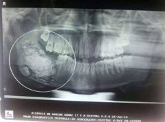
odontogenic myxoma tumor of ectomesenchyme soap bubble local aggressive |
|
|
cementoblastoma tumor of ectomesenchyme benign radio opaque mass around apex of tooth |
|
|
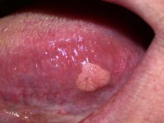
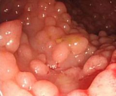
What is oral squamous papilloma caused by ? |
HPV (6 and 11) |
|
|
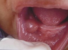
oral squamous papilloma usually solitary |
|
|
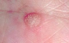
verruca vulgaris aka common wart |
|
|
verruca vulgaris orally often indistinguishable from oral squamous papilloma white |
|
|
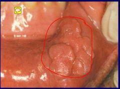
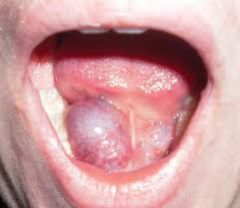
condyloma acuminatum venereal warts - pic in oral cavity multiples, cauliflower like caused by HPV 6 and 11 |
|
|
seborrheic keratosis exclusively skin lesion stuck on look associated with sun exposure |
|
|
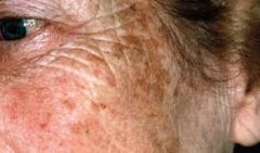
actinic lentigo no intraoral counterpart from sun exposure topical retinoic acid reduces color may be lip lesions |
|

|
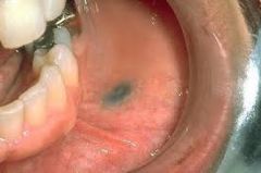
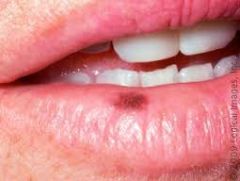
oral melantotic macule <7mm usually on lower lip can occur on soft palate |
|
|
acquired melanocytic nevus mole intraorally- palate and gingiva may never be pigmented |
|
|
actinic keratosis old people sandpaper feel from sun exposure Topical 5-fluorouracil |
|
|
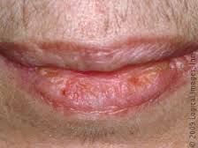
actinic cheilitis scaly look may have ulceration vermillionectomy can become cancerous |
|
|
Tobacco pouch keratosis grey/ grey-white in area of placement textured |
|
|
nicotine stomatitis grey-white mucosa, multiple papules (red spots in it ) and cracked look |
|
|
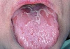
Leukoplakia Definition |
a white patch or plaque which can't be characterized clinically or path as any other disease |
|
|
what is the most common precancerous oral lesion? |
leukoplakie= considered premalignant |
|

|
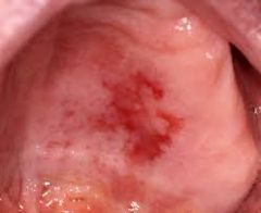
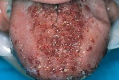
Leukoplakia homogenous and speckled white worry = tongue, floor of mouth, soft palate tobacco and alcohol contribute |
|
|
For which epithelial lesion is a biopsy mandatory? |
Leukoplakia |
|
|
erythroplakia red may be adjacent to leukoplakia most show sever dysplasia tobacco and alcohol etiology |
|
|
Oral squamous cell carcinoma |
sites: tongue, floor of mouth, soft palate minimal pain |
|
|
Oral squamous cell carcinoma |
|
|
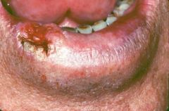
squamous cell carcinoma of lip from chronic sun exposure slow growing |
|
|
Squamous cell carcinoma |
spread by lymphs firm nodes move or fixed spreads to lungs, liver, bones use TMN staging |
|
|
TNM staging |
used with squamous cell carcinoma stage at diagnosis = most important prognostic indicator T=tumor size, N= local node involvement, M= distant metastasis |
|
|
Field cancerization |
people with a carcinoma in one place are at increased risk of developing a second mucosal tumor |
|
|
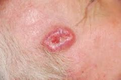
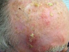
basil cell carcinoma |
low grade malignancy from chronic sun exposure in middle 1/3 of face nodulouclerative = MC form |
|
|
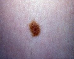
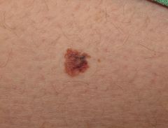
Melanoma |
acute solar damage = really bad sun burn ABCD's asymmetry, irreg borders, color variation, diameter >6mm (eraser), evolution |
|
|
melanoma |
|
|
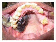
oral melanoma |
rare maxillary gingiva, palate |
|
|
oral melanoma |
|
|
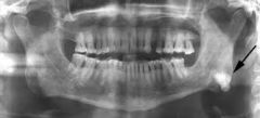
Osteoma benign tumor of compact bone MC site: in posterior mandible |
|
|
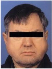
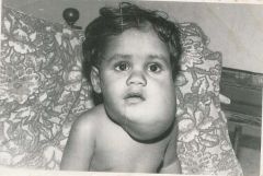
What is the most serious feature of Gardner Syndrome? |
Intestinal Polyps serious because has ability of malignant transformation |
|
|
intestinal polyps associated with Gardner syndrome |
|
|
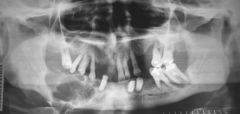
Gardner syndrome: intestinal polyps with malignant potential multiple osteomas multiple odontomas supernumerary teeth |
|
|
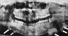
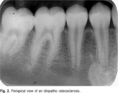
Idiopathic Osteosclerosis |
increased bone density not attributed to any specific cause Mandible PreM and 1st M area adjacent teeth=vital NOT condensing osteitis |
|
|
idiopathic osteosclerosis |
|
|
Idiopathic osteosclerosis vs condensing osteitis |
CO= non-vital tooth widened pdl sclerosis of bone around root from chronic inflam IO=increase bone density adjacent teeth are vital radioopaque mass |
|
|
condensing osteitis |
|
|
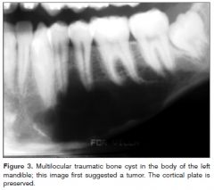
traumatic bone cyst |
|
|
Traumatic bone cyst |
asymptomatic empty cavity assoc with trauma pseudocyst |
|
|
Aneurysmal bone cyst pseudocyst Blood soaked sponge=surgical finding uni/multilocular radiolucency from rupture of local vascular network in a pre existing bone lesion |
|
|
Central giant cell granuloma: non- neoplastic can be either central or peripheral Females Mandible Anterior uni/ multi locular radiolucency assoc with jaw expansion and divergence of tooth roots |
|
|
peripheral giant cell granuloma |
|
|
Post- surgical hyperostosis |
tumor-like reactive growth of bone at surgical site in periosteum after perio surgery ex: gingival graft |
|
|
3 types of benign Fibro-osseous Lesions of the jaws |
Fibrous dysplasia- unknown central ossifying / cementifying fibroma-neoplasm cemento-osseous dysplasia - reactive lesion |
|
|
Fibro-osseous lesions of the jaw |
diseases characterized by the replacement of bone with abnormal fibrous CT interspersed with varying amounts of calcification |
|
|
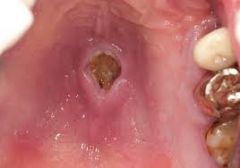
Fibrous Dysplasia: monostotic =affect only 1 bone- MC type maxilla benign and chronic radiograph- ground glass appearance |
|
|
polyostotic fibrous dysplasia |
|
|
Histology of fibrous dysplasia |
chinese letters appearance |
|
|
central ossifying fibroma: true benign neoplasm female mandible well defined unilocular radiolucency with varying radiopacity |
|
|
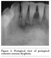
Periapical cemento-osseous dysplasia: Reactive lesion= not neoplasm Female Black Anterior Mandible Involved teeth= vital |
|
|
focal cemento-osseous dysplasia: Female White posterior mandible |
|
|
florid cemento-osseous dysplasia: Multiple quads Black women Osteomyelitis=complication in edentuolous |
|
|
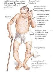
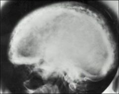
Paget's disease: enlargement and deformity of affected bone spacing of teeth headache dizziness deafness |
|
|
paget's disease |
|
|
cotton wool appearance associated with paget's disease hypercementosis in radiographs as well |
|
|
Histologically, what does paget disease of bone look like? |
mosaic pattern bc prominent reversal lines |
|
|
What are there significantly high levels of in lab serum in paget's disease? |
alkaline phosphatase |
|
|
Osteoblastoma |
benign tumor cells show osteoblastic differentiation bone forming mixed radiolucency |
|
|
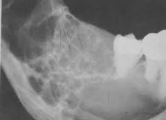
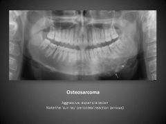
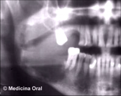
Osteosarcoma Malignant tumor bone forming *sunray or sunburst * widening PDL |
|
|
Where is the most frequent intraoral site for metastatic tumors of the jaw? |
Mandible these tumors have begun because of cancers that began in other areas ex:breast, prostate, kidney |
|
|
Metastatic tumor of jaw radioopaque |
|
|
Frictional keratosis chronic mechanical irritation calloused mucosa linea alba= specific type |
|
|
Endentulous alveolar ridge keratosis |
|
|
frictional keratosis of tongue |
|

|
chronic cheek biting frictional keratosis irregular thick can also occur on tongue |
|
|
Snuff dippers keratosis |
|
|
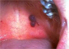
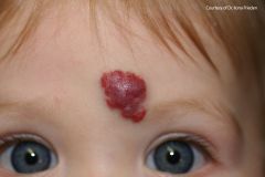
amalgam tatoo |
|

|
graphite tatoo from pencil in kids usually |
|
|
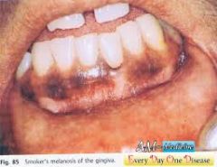
Smoker's melanosis in facial anterior usually femal type of hypermelanosis |
|
|
Lichen Planus type of post inflam hypermalanosis (hyperpigmentation) from inc in melanin synthesis from prostaglandins and other inflam products |
|
|
Oral melantotic macule post- inflam hypermelanosis MC = lower lip |
|
|
Melanoacanthosis in African descent pt after trauma ***need biopsy differ to from melanoma increased in melanocytes |
|
|
Where do drug induced pigmentations usually occur? |
hard palate and gingiva |
|
|
Minocycline pigmentation drug induced |
|
|
Lead line /poisoning drug induced pig |
|
|
Traumatic ulcer MC cause ulcer in OC = trauma physical thermal or chemical |
|
|
Nicotine stomatitis in heavy smokers thermal and chemical injury |
|
|
Cocaine osteonecrosis cocaine abuse manifest: palatal perforations perforated nasal septum cocaine keratosis cocaine dehiscence |
|
|
What drug can cause a chemical burn? |
aspirin = necrotic epithelium |
|
|
Geographic tongue looks like bald spots elevated borders |
|
|
Hematoma submucosal hemorrhage red dark elevated mass trauma related anticoag disorder = predispose |
|
|
Keloid abnorm prolif of scar tissue |
|
|
What is a mucosal alteration that can cause recession of the gingiva ? |
lip barbell |
|
|
What are 2 complications in the oral cavity from chemo? |
mucositis hemorrhage |
|
|
What are 3 complications that arise in the oral cavity in radiation ? |
mucositis xerostomia osteoradionecrosis |
|
|
Chemo related mucositis moveable tissue = affected most |
|
|
Chronic hyperplastic pulpitis growth of granulation tissue from pulp molars of children |
|
|
Apical abscess |
form of liquefactive necrosis inflammatory response to bacterial infect or necrosis of pulp |
|
|
Dental Abscess accum of neutrophils and exudate and necrotic cells =PUS |
|
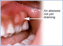
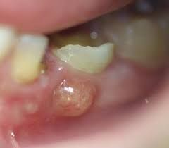
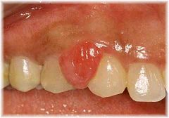
Parulis |
mass of granulation tissue where sinus tract reaches mucosa |
|
|
Untreated dental abscess |
cellulitis (space infection) cavernous sinus thrombosis (infection from maxilla) ludwig angina (infection of mandible ) |
|
|
Periapical granuloma |
mass of inflammed granulation tissue at apex of non vital tooth from pulpal necrosis radiolucent |
|
|
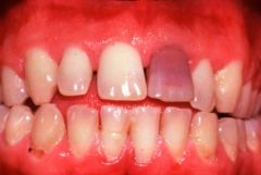
Internal Resorption trauma induced pink crown |
|
|
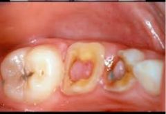
What causes pulpal stones ? |
Chronic inflammation of the pulp 2 types : dystrophic calcification denticles (contain dentinal tubules surrounded by odontoblasts) |
|
|
Abfraction wedge shape notch at cervical region from flex of tooth from pressure |
|
|
What can cause external resorption? |
chronic inflam from trauma infection reimplantation of avulsed tooth orthodontic treatment tumors ex: ameloblastoma |
|
|
Osteoradionecrosis from radiation moth-eaten appear mandible |
|
|
what does the drug bisphosphonate do? |
inhibits activity of osteoclasts= slow bone turnover |
|
|
Clinical indications of bisphosphonate - related osteonecrosis of the jaws? |
osteoporosis cancers that have metastasized to bone multiple myeloma paget's disease of bone ghost outline of sockets 1 yr after extraction |
|

|
Sialadenitis MC= mumps = viral form S. aureus =MC bacterial cause from result of dec saliva flow or duct obstruct = spread bacteria |
|
|
Acute sialadenitis of parotid gland |
|
|
Mucocele mucous spill into soft tissue from rupture of minor salivary gland duct trauma MC= Lower lip |
|
|
Ranula |
mucocele in floor of mouth from trauma |
|
|
Superficial Mucocele vs Mucocele |
Superficial = minor saliva ducts at surface from mucosal inflam clear and bubble like regular= cause by rupture of duct |
|
|
Ranula |
|
|
Salivary duct cyst |
mucous retention phenomenon True cyst looks like mucocele clinically |
|
|
Cyst of blandin nuhn mucocele form in blandin nuhn glands |
|
|
Salivary stones |
partial chronic duct obstruction causes MC= submandibular gland duct |
|
|
Sialadenosis non malig non inflam salivary gland enlarge from peripheral neuropahty of autonomic nerve supply |
|
|
Adenomatoid hyperplasia Inc in # gland acini in minor salivary glands firm mass mimick minor salivary gland tumor MC=hard palate |
|
|
Necrotizing sialometaplasia inflammatory from ischemia-infarction-necrosis and ulcer-slough of necrotic tissue- healing Posterior palate heals spontaneously predisposing: trauma, ill fit denture, adjacent tumor |
|
|
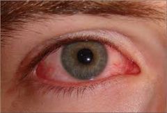
Sjogren Syndrome |
autoimmune disease, affects salivary and lacrimal glands Primary= xerostomia +dry eye secondary = primary +chronic inflammatory CT disease (rheumatoid arthritis) aka dry , irritation |
|
|
Keratoconjunctivitis associated with sjogren syndrome |
|
|
What are 4 signs of Sjogren Syndrome? |
keratoconjunctivitis (dry eye) Swelling of the salivary glands Raynaud's phenomenon Oral: atrophy of the dorsal of the tongue also cervical caries and candidiasis |
|
|
What is the MC site for salivary gland tumors? |
Parotid Gland |
|
|
Where is the MC site for malignant salivary gland tumors ? |
Sublingual gland |
|
|
Where is the most common anatomical position for minor salivary gland tumors? |
Palate |
|
|
What are the 5 types of benign salivary gland tumors? |
Pleomorphic adenoma Warthin tumor canalicular adenoma basal cell adenoma salivary duct papilloma |
|
|
Pleomorphic Adenoma MC salivary neoplasm benign salivary gland tumor firm slow grow Females Recurrant |
|
|
Warthin's tumor MALE most likely to occur bilaterally 2nd MC benign parotid tumor |
|
|
Canalicular Adenoma benign tumor of the salivary gland exclusive minor salivary gland MC= upper lip Female |
|
|
Mucocele vs canalicular adenoma |
Mucocele= from rupture of salivary duct cyst canalicular adenoma= benign tumor of minor salivary gland |
|
|
What are the 5 types of malignant salivary gland tumors? |
Mucoepidermoid carcinoma polymorphoud low-grade adenocarcinoma adenoid cystic carcinoma acinic cell carcinoma carcinoma ex- pleomorphic adenoma |
|
|
What are 3 things that a malignant salivary gland tumor is associated with ? |
paresthesia tumor fixation ulceration |
|
|
Mucoepidermoid Carcinoma |
MC malignant salivary tumor |
|
|
Mucoepidermoid carcinoma parotid= MC site asymptomatic swelling can have cyst formation |
|
|
Polymorphous low grade adenocarcinoma 2nd MC malignant salivary tumor almost exclusive to minor glands Palate **PAIN bone destruction |
|
|
Acinic Cell Carcinoma cells have acinar differentiation parotid |
|
|
Carcinoma Ex- pleiomorphic adenoma malignant salivary gland tumor form of pleiomorphic adenoma formed in an existing one or in a patient that has already had one removed shows sudden rapid growth |
|
|
What are the 3 P's for bump on gums? |
Soft skin lesions= Pyogenic granuloma Peripheral Ossifying fibroma Peripheral Giant cell granuloma |
|
|
Pyogenic granuloma Not a true granuloma non tumor from trauma Pregnancy / hormones bleeds easy Gingiva |
|
|
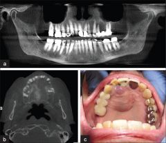
Peripheral Ossifying Fibroma reactive soft skin lesion exclusive on gingiva from PDL Maxilla ulcerated |
|
|
Peripheral giant cell granuloma blue/purple exclusive to gingiva/ alveolar ridge Radiograph= SAUCERIZATION (cupping resorption) |
|
|
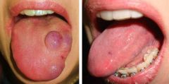
Fibroma usually along bite line buccal mucosa smooth pink raised nodule MC tumor of oral cavity Prob not true neoplasm from trauma/ irritation |
|
|
Traumatic Neuroma smooth sub mucosal nodule in mental foramen area prolif of neural tissue not true neoplasm after disrupt schwann cells |
|
|
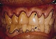
What are common drugs that relate to gingival hyperplasia ? |
phenytoin ca channel blockers cyclosporine |
|
|
Denture stomatitis form of candidiasis |
|

|
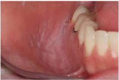
Epulis Fissturatum from ill fit denture single/multi folds fibrous tissue in vestibule fibroepithelial polyp = pedunculated lesion of palate from max denture |
|
|
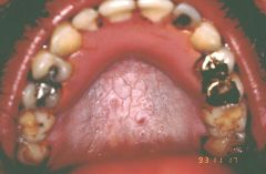
Inflammatory Papillary Hyperplasia hard palate asymptomatic pebbly surface erythematous from: ill fit denture, poor OH, wearing denture all the time and superimposed candidiasis |
|
|
Fibroma benign tumor of fat usually in buccal mucosa normal or yellow color if cut off floats in water |
|
|
Schwannoma benign tumor origin = schwan cell if oral =tongue usually can be in bone = unilocular radiolucency |
|
|
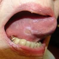
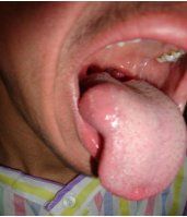
Granular cell tumor solitary dorsal of tongue asymp tumor of schwann cell origin |
|
|
Neurofibroma typical solitary tongue or buccal mucosa can occur in bone multiple=neurofibromatosis assoc- lead to malignant transform |
|
|
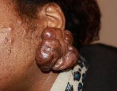
Neurofibromatosis Multiple neurofibromas pleixform neurofibroma cafe au lait pigment Crowe's sign -armpit freckles lish nodules - pigment spot on iris of eyes 5% develop neurofibrosarcoma mental foramen/ alveolar nerve enlarge |
|
|
What are the 3 types of Lymphangioma? |
Capillary-small Cavernous- med Cystic (cystic hygroma)- large in neck and armpit |
|
|
Lymphangioma usually in posterior triangle Oral lesions: TONGUE frog eggs deep tumors ill defined masses |
|
|
Lymphangioma tongue of pt frog egg |
|
|
What is the most common tumor of infancy? |
Hemangioma= benign prolif of blood vessiles |
|
|
Hemangioma MC tumor infancy raised red nodular masses of skin regresses on own |
|
|
Vascular malformation ***present at birth and persists all life port wine stains on skin |
|
|
Hemangioma vs vascular malformation |
H= not present at birth, arise in infancy and goes away VM= present at birth, does not go away |
|
|
What are the 3 types of soft tissue sarcomas? |
Fibrosarcoma Kaposi's sarcoma Rhabdomyosarcoma |
|
|
Fibrosarcoma |
Malignant tumor soft tissue sarcoma tumor of extremities grows slow and is painless Nose and sinus involve most |
|
|
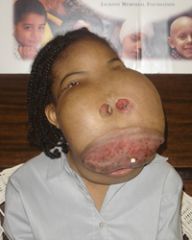
Karposi's sarcoma vascular malignant tumor common in AIDs 4 types: Classic-old ppl, lower extremities, slow Endemic-African, benign nodule aggressive Iatrogenic -organ transplants , aggressive AID's related-homo males , oral lesion common |
|
|
Rhabdomyosarcoma Malignant tumor skeletal muscle 3 types:embryonal, alveolar, pleomorphic(undiff, anaplastic) MC malignant tumor in kids Males orbit, nasal cavity, and nasopharynx metastatis: embryonal>alveolar>pleomorphic |