![]()
![]()
![]()
Use LEFT and RIGHT arrow keys to navigate between flashcards;
Use UP and DOWN arrow keys to flip the card;
H to show hint;
A reads text to speech;
52 Cards in this Set
- Front
- Back
|
1. Which is not a branch of the spinal nerve? Anterior (ventral) ramus Posterior (ventral) ramus Anterior root Posterior root Meningeal branch White communicating branch |
Richtige Antwort: Anterior root Posterior root Outside the vertebral column, the nerve divides into branches. The posterior ramus contains nerves that serve the posterior portions of the trunk carrying visceral motor, somatic motor, and somatic sensory information to and from the skin and muscles of the back (epaxial muscles). The anterior ramus contains nerves that serve the remaining anterior parts of the trunk and the upper and lower limbs (hypaxial muscles) carrying visceral motor, somatic motor, and sensory information to and from the ventrolateral body surface, structures in the body wall, and the limbs. The meningeal branches (recurrent meningeal or sinuvertebral nerves) branch from the spinal nerve and re-enter the intervertebral foramen to serve the ligaments, dura, blood vessels, intervertebral discs, facet joints, and periosteum of the vertebrae. The rami communicantes contain autonomic nerves that serve visceral functions carrying visceral motor and sensory information to and from the visceral organs. Some anterior rami merge with adjacent posterior rami to form a nerve plexus, a network of interconnecting nerves. Nerves emerging from a plexus contain fibers from various spinal nerves, which are now carried together to some target location. Major plexuses include the cervical, brachial, lumbar, and sacral plexuses. |
|
|
2. Which ist not a branch oft he cervical plexus?Lesser occipital nerve Greater auricular nerve Transverse cervical nerve Supraclavicular nerve Suprascapular nerve Phrenic nerve |
Richtige Antwort:Suprascapular nerve The cervical plexus has two types of branches: cutaneous and muscular.Cutaneous (4 branches):Great auricular nerve - innervates skin near concha auricle (outer ear) and external acoustic meatus (ear canal) (C2&C3) Transverse cervical nerve - innervates anterior region of neck (C2&C3) Lesser occipital - innervate the skin and the scalp posterosuperior to the auricle (C2&C3)Supraclavicular nerves - innervate region of supraspinatus, shoulder, and upper thoracic region (C3,C4) [1] Muscular: Ansa cervicalis (loop formed from C1-C3), etc. (geniohyoid (C1 only), thyrohyoid (C1 only), sternothyroid, sternohyoid, omohyoid) Phrenic (C3-C5 (primarily C4))-innervates diaphragm and the pericardium Segmental branches (C1-C4)- innervates anterior and middle scalenes |
|
|
whats the cervical plexus? |
The cervical plexus is a plexus of the ventral rami of the first four cervical spinal nerves which are located from C1 to C4 cervical segment in the neck. They are located laterally to the transverse processes between prevertebral muscles from the medial side and vertebral (m. scalenus, m. levator scapulae, m. splenius cervicis) from lateral side. There is anastomosis with accessory nerve, hypoglossal nerve and sympathetic trunk.It is located in the neck, deep to sternocleidomastoid. Nerves formed from the cervical plexus innervate the back of the head, as well as some neck muscles. The branches of the cervical plexus emerge from the posterior triangle at the nerve point, a point which lies midway on the posterior border of the sternocleidomastoid |
|
|
3. Which can not be seen as a transverse section oft the spinal cord? Posterior funiculus Lateral funiculus Anterior funiculus Anterior horn Dorsal horn Lateral horn Medial horn White commissure |
Richtige Antwort:Medial horn |
|
|
4. A cluster of perikarya of neurons in the central nervous system can be called: Ganglion Neuron Nervus Nucleus Fasciculus |
Richtige Antwort: Ganglion Nucleus |
|
|
was ist ein faszikulus? |
Als Fasciculus (Plural Fasciculi), auch Faszikel (dt. ‚kleines Bündel‘, ‚Päckchen‘; engl. fascicle) genannt, bezeichnet man in der Anatomie und Histologie Substrukturen, die aus mehreren Nerven- oder Muskelfasern bestehen. Die Substruktur stellt ein Bündel aus mehreren Einzelfasern dar. Bei Nervenfasern spricht man auch von einem Nervenfaszikel und bei Muskelfasern von einem Muskelfaszikel.Bei Nervenfasern sind die einzelnen Faszikel vom sogenannten Perineurium, einem kollagenreichem Bindegewebe umgeben. Bei Muskelfasern erfüllt das Perimysium diese Funktion |
|
|
5. A cluster of axons of neurons in the central nervous system can be called: Ganglion Neuron Nervus Nucleus Fasciculus |
Richtige Antwort:Nervus Fasciculus |
|
|
6. A histological section oft he spinal cord shows the typical „butterfly“ formation. This formation ist mainly composed of: Fasciculi only Nuclei only Ganglia only Fasciculi & Nuclei Nerves only |
Richtige Antwort:Nuclei only |
|
|
was ist ein ganglion? |
Ein Ganglion (Plural Ganglien) ist eine Anhäufung von Nervenzellkörpern im peripheren Nervensystem. Ganglien werden auch als Nervenknoten bezeichnet, da sie bei der Präparation als knotige Verdickungen auffallen. |
|
|
7. Which oft he following tracts in the spinal cord is descending? Tractus spinothalamicus anterior Fasciculus cuneatus Tractus spinocerebellaris anterior Fasciculus gracilis Tractus corticospinalis anterior |
Richtige Antwort:Tractus corticospinalis anterior |
|
|
Was ist der Tractus Spinothalamicus? |
Der Tractus spinothalamicus anterior ist eine aufsteigende Faserbahn der Vorderseitenstrangbahn (Funiculus anterolateralis). Über sie werden grobe Mechanosensorik, sowie viszerosensible Stimuli nach zentral vermittelt. Sexuelle Stimuli werden ebenso vermittelt |
|
|
was ist fasciculus cuneatus? |
Der Fasciculus cuneatus (lat., dt. das keilförmige Bündel) ist eine Nervenbahn innerhalb des Rückenmarks. Er leitet epikritische und propriozeptive Informationen von der oberen Extremität und den oberen Rumpfabschnitten zum Gehirn. Der Fasciculus cuneatus gehört mit dem Fasciculus gracilis, dem er sich seitlich anlegt, zu den Hinterstrangbahnen des Rückenmarks.Die Nervenfasern des Fasciculus cuneatus verlaufen gleichseitig (ipsilateral) ohne vorherige Umschaltung zum Nucleus cuneatus in der Medulla oblongata, wo sie auf das zweite Neuron umgeschaltet werden. Nach dieser Umschaltung setzen sie sich als Lemniscus medialis fort, in dem die Kreuzung auf die Gegenseite (kontralateral) erfolgt. |
|
|
was ist der tractus spinocerebellaris? |
Als Tractus spinocerebellaris werden Nervenbahnen bezeichnet, die propriozeptive Informationen aus dem Rückenmark in das Kleinhirn führen. Man unterscheidet zwischen dem Tractus spinocerebellaris posterior (Flechsig-Bündel), der eher dorsal verläuft, und dem Tractus spinocerebellaris anterior, der weiter ventral zu finden ist. Das erste Neuron beider Bahnen liegt im Spinalganglion. ie Fasern des Tractus spinocerebellaris posterior werden im Nucleus dorsalis der Lamina V und VI des Rückenmarks auf das zweite Neuron verschaltet, während die Verschaltung des Tractus spinocerebellaris anterior in den Lamina V-VII erfolgt Die Faserzüge des Tractus spinocerebellaris posterior kreuzen während ihres Verlaufes im Rückenmark nicht, im Gegensatz zu den Fasern des Tractus spinocerebellaris anterior, die zurkontralateralen Seite und wieder zurück kreuzen, so dass das Kleinhirn nur Impulse aus dem ipsilateralen Rückenmark erhält. |
|
|
was ist der fasciculus gracilis? |
Der Fasciculus gracilis (lat., dt. das grazile Bündel) ist eine Nervenbahn innerhalb des Rückenmarks. Er leitet epikritische und propriozeptive Informationen, vor allem von der unteren Extremität zum Gehirn. Der Fasciculus gracilis gehört mit dem Fasciculus cuneatus zu den Hinterstrangbahnen des Rückenmarks.Die Nervenfasern des Fasciculus gracilis verlaufen gleichseitig (ipsilateral) ohne vorherige Umschaltung zum Nucleus gracilis in der Medulla oblongata, wo sie auf das zweite Neuron umgeschaltet werden. Nach dieser Umschaltung setzen sie sich als Lemniscus medialis fort, in dem die Kreuzung auf die Gegenseite (kontralateral) erfolgt. |
|
|
Was ist der Tractus corticospinalis (pyramidenbahn)? |
Als Pyramidenbahn wird die Efferenz des Motokortex (Gyrus praecentralis) bezeichnet. Sie ist die größte absteigende Bahn und innerviert die Alpha-Motoneurone. Zur Pyramidenbahn zählt man funktionell zwei motorische Faserbahnen: den Tractus corticospinalis und den Tractus corticonuclearis Der Tractus corticonuclearis zählt allerdings nur im erweiterten Sinn zur Pyramidenbahn, da er nicht durch die Pyramide in der Medulla oblongata zieht, sondern Fasern an die motorischen Hirnnervenkerne entsendet. In der Pyramidenkreuzung (Decussatio pyramidum), die sich am Übergang zwischen Medulla oblongata und Rückenmark befindet, kreuzen 70 bis 90 Prozent der Axone als Tractus corticospinalis lateralis auf die kontralaterale Seite, die restlichen Fasern steigen ungekreuzt als Tractus corticospinalis anterior ab. |
|
|
8. The perikaryon of the third neuron of the nociception pathway lies mainly here: Nucleus ventralis posteromedialis Nucleus gracilis Gyrus postcentralis Nucleus cuneatus Nucleos ventralis posterolateralis |
Richtige Antwort:Nucleus ventralis posteromedialisNucleos ventralis posterolateralis |
|
|
was sind die nuclei ventralis posteromedialis und posterolateralis? |
Über den Tractus spinothalamicus werden vor allem Schmerz und Temperatursinn aus dem Rumpf und aus den Extremitäten geleitet. Über den Lemniscus medialis werden vorwiegend der Tastsinn und Empfindungen über die Gelenkstellung aus Rumpf und Extremitäten (Propriozeption) geleitet. Diese beiden Bahnen enden im äußeren Anteil des Nucleus ventralis posterior. Dieser Anteil wird daher auch Nucleus ventralis posterolateralis (VPL) (Syn. Nucleus ventrocaudalis externus) genannt.Über den Lemniscus trigeminalis werden Schmerz, Temperatursinn, Tastsinn und Propriozeption aus dem Gesicht (über den fünften Hirnnerven, den Nervus trigeminus) übermittelt. Diese Bahn endet im mittleren Anteil des Nucleus ventralis posterior. Dieser Anteil wird daher auch Nucleus ventralis posteromedialis (VPM) (Syn. Nucleus ventrocaudalis internus) genannt |
|
|
9. The information of vibration from the left arm is travelling within this structure: Fasciculus gracilis dexter Fasciculus cuneatus dexter Tractus spinothalamicus anterior sinister Fasciculus cuneatus sinister Fasciculus gracilis sinister |
Richtige Antwort:Fasciculus cuneatus sinister |
|
|
10. The chronic atonic palsy of the lower extremity is likely not caused by the deficiency of this structure: Cervical spinal cord Peripheral nerve Alpha-motoneuron Spinal nerve Sympathetic chain |
Richtige Antwort:Cervical spinal cord |
|
|
1. How many pairs of spinal nerves can usually be found in the human body? 2829303132 |
Richtige Antwort:31 |
|
|
2. Which of the following structures is exclusively afferent? Radix anterior Radix posterior Ramus comunicans albus Ramus anterior / ventralis Ramus communicans griseus Ramus dorsalis |
Radix posterior (hintere nervenwurzel) |
|
|
was macht der ramus posterior? |
Ramus posterior (Ramus dorsalis) für die Versorgung der wirbelsäulennahen Haut und Muskulatur (Autochthone Rückenmuskulatur). |
|
|
was macht der ramus anterior? |
Ramus anterior (Ramus ventralis) für die Versorgung der Haut und Muskulatur des wirbelsäulenfernen Rückens, sowie der seitlichen und bauchseitigen Körperabschnitte. |
|
|
was macht der ramus meningeus? |
Ramus meningeus für die Innervation der Rückenmarkshäute |
|
|
3. What is the correct sequence of brachial plexus structures from central to peripheral? Nerve – Cord – Division – Root – Trunk Nerve – Division – Root – Cord – Trunk Division – Trunk – Branch – Cord – Nerve Division – Trun – Branch – Nerve – Cord Root – Trunk – Division – Cord – Nerve Cord – Nerve – Root – Division – Trunk Cord – Root – Division – Trunk – Nerve Division – Root – Nerve – Trunk – Cord Root – Cord – Trunk – Division – Nerve |
Richtige Antwort:Root – Trunk – Division – Cord – Nerve |
|
|
Was ist der Plexus Brachialis? |
Der Plexus brachialis[1] (lat. „Armgeflecht“) ist ein Geflecht aus den ventralen Ästen der Spinalnerven der letzten vier Hals- und des ersten Brustsegments (C5–Th1). Beim Menschen sind auch kleinere Bündel des vierten Halswirbelsegmentes (C4) und des zweiten Brustwirbelsegmentes (Th2) an der Bildung des Plexus brachialis beteiligt. Diese Spinalnerven vereinigen sich nach ihrem Durchtritt durch die Musculi scaleni zu drei Hauptstämmen (Trunci; genauer Truncus superior, Truncus medius und Truncus inferior) und anschließend zu mehreren, untereinander verbundenen Strängen (Fasciculi; Fasciculus lateralis, Fasciculus medialis und Fasciculus posterior). Diese Stränge treten entlang der Arteria subclavia und Arteria axillaris in die Achselgegend. Aus ihnen bilden sich wiederum Nerven, die durch den Faseraustausch im Plexus nun immer Anteile von mehreren (2–3) Spinalnerven besitzen. Diese Nerven innervieren die gesamte obere (bei Tieren vordere) Extremität sowie Teile der Brustwand. Das gleiche Prinzip zeigt das Beingeflecht (Plexus lumbosacralis). |
|
|
4. Which oft he below listed reflexes is monosynaptic? Cremaster reflex Corneal reflex Hot-plate reflex Patellar-tendon reflex Moro reflex |
Patellar-tendon reflex |
|
|
What's a monosynaptic reflex? |
monosynaptic reflex A simple reflex that involves transmission of information from a sensory neuron to the appropriate motor neuron across a single synapse in the spinal cord. The knee-jerk reflex action is an example of a monosynaptic reflex |
|
|
5. Which relation „reflex – spinal segment“ is not correct? Brachioradialis reflex – C5 to C7 Biceps reflex - C5 and C6 Cremaster reflex – L1 and L2 Triceps reflex – Th1 and Th2 Patella reflex – L2 to L4 Plantar reflex – L5 to S2 |
Richtige Antwort:Triceps reflex – Th1 and Th2 Richtig wäre: The triceps reflex, a deep tendon reflex, is a reflex as it elicits involuntary contraction of the triceps brachii muscle. It is initiated by the Cervical (of the neck region) spinal nerve 7 nerve root (the small segment of the nerve that emerges from the spinal cord).[1] The reflex is tested as part of the neurological examination to assess the sensory and motor pathways within the C7 and C8 spinal nerves. |
|
|
6. A patient’s complains include numbness and tickling in a dermatome. What ist he most likely pathology? Lesion of a peripheral nerve Lesion of Spinal nerve root Lesion of a cortical area Lesion of dermal receptors Lesion of an ascending spinal pathway |
Richtige Antwort:Lesion of Spinal nerve root |
|
|
7. What ist he last thing you do when you find someone with an acute shoulder dislocation? Repositioning Calling for help Pain management Basic life support Armfixation |
Richtige Antwort:Repositioning |
|
|
8. Which is not a named nerve emerging from the brachial plexus? Axillary nerve Ulnar nerve Radial nerve Medial cutaneous nerve of the arm Medial cutaneous nerve of the forearm Musculocutaneous nerve Intercostobrachial nerve Thoracodorsal nerve Suprascapular nerve |
Richtige Antwort:Intercostobrachial nerve |
|
|
what are intercostal nerves? |
The intercostal nerves are part of the somatic nervous system, and arise from the anterior roots of the thoracic spinal nerves from T1 to T11. The intercostal nerves are distributed chiefly to the thoracic pleura and abdominal peritoneum and differ from the anterior roots of the other spinal nerves in that each pursues an independent course without plexus formation.The first two nerves supply fibers to the upper limb in addition to their thoracic branches; the next four are limited in their distribution to the walls of the thorax; the lower five supply the walls of the thorax and abdomen. The 7th intercostal nerve terminates at the xyphoid process, at the lower end of the sternum. The 10th intercostal nerve terminates at the navel. The twelfth (subcostal) thoracic is distributed to the abdominal wall and groin. |
|
|
9. Which is not a named nerve emerging from the lumbar plexus? Iliohypogastric nerve Ilioinguinal nerve Genitofemoral nerve Lateral cutaneous nerve of the thigh Femoral nerve Obturator nerve Sciatic nerve |
sciatic nerve (It contains fibres from both the anterior and posterior divisions of the lumbosacral plexus.) |
|
|
10. Which is not a named nerve emerging from the sacral plexus? Sciatic nerve Superior gluteal nerve Inferior gluteal nerve Sacral nerve Pudenal nerve Posterior cutaneous nerve of the thigh Coccygeal nerve |
sacral nerve |
|
|
what do sacral nerves do? |
The sacral nerves have both afferent and efferent fibers, thus they are responsible for part of the sensory perception and the movements of the lower extremities of the human body. From the S2, S3 and S4 arise parasympathetic fibers whose electrical potential supply the descending colon and rectum, urinary bladder and genital organs. These pathways have both afferent and efferent fibers and, this way, they are responsible for conduction of sensory information from these pelvic organs to the central nervous system (CNS) and motor impulses from the CNS to the pelvis that control the movements of these pelvic organs. |
|
|
1. Which is usually not considered part of the brainstem? Diencephalon Mesencephalon Pons Medulla oblongata Cerebellum |
Richtige Antwort:Diencephalon Cerebellum |
|
|
2. Which structure represents the caudal border of the medulla oblongata? Olive Colliculus inferior Decusatio pyramidum FlocculusPyramis Suboccipital nerve |
Richtige Antwort:Decusatio pyramidum |
|
|
was ist das decussatio pyramidalis? |
Als Pyramidenbahnkreuzung bezeichnet man die im Bereich der ventralen Medulla oblongata auf die Gegenseite kreuzenden Anteile der Pyramidenbahn, die als Pyramidenseitenstrangbahn (Tractus corticospinalis lateralis) absteigen, während ungekreuzte Fasern direkt als Pyramidenvorderstrangbahn (Tractus corticospinalis anterior) weiter nach kaudal ziehen. |
|
|
3. Which of the below listed nuclei in the brainstem lies most medial? Ncl. Spinalis (sensorius) nervi trigemini Ncl. (motorius) nervi abducentis Ncl. Principalis (pontinus n. trigemini) Ncl. Cochlearis Ncl. Vestibularis |
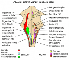
abducentis??? |
|
|
4. Which of the below listed nuclei in the brainstem lies most cranial? Ncl. nervi oculomotorii Ncl. nervi trochlearis Ncl. nervi abducentis Ncl. nervi hypoglossi Ncl. spinalis nervi accessorii |
Richtige Antwort:Ncl. nervi oculomotorii |
|
|
5. Which of these arterie is not involved in the blood supply of the brainstem? Anterior spinal Posterior spinal Anterior inferior cerebellar Posterior inferior cerebellar Basilrvertabral Posterior cerebral Middle cerebral Pontine branches Superior cerebellar |
Richtige Antwort:Middle cerebral |
|
|
6. Ischemia in the brainstem causes symptoms which can best be summarized as: Deadly syndrome Jumping syndrome Alternans syndrome Paresis syndrome Turning syndrome |
Richtige Antwort: Alternans syndrome |
|
|
7. Alternans syndromes are characterized by these symptoms: Ipsilateral nuclear dysfunction – ipsilateral paresis Ipsilateral nuclear dysfuction – contralateral hemiparesis Contralateral nuclear dysfunction – ipsilateral hemiparesis Contralateral nuclear dysfunction – contralateral hemiparesis Contralateral nuclear hemiparesis – contralateral dysfunction |
Richtige Antwort:Ipsilateral nuclear dysfunction – contralateral hemiparesis |
|
|
8. What is the medial lemniscus? motoric tract connecting cortex and olive sensory tract conecting thalamus and cortex motoric tract connecting cortex and cerebellumsensory tract connecting nuclei and thalamusmotoric tract connecting cortex and nuclei |
Richtige Antwort:Sensory tract connecting nuclei and thalamus |
|
|
9. What is the function of the corticobulbar (-nuclear) tract? Modulation of alpha-motoneurons in the spinal cord Modulation of the sensory part of the capsula interna Modulation of motor nuclei in the brainstemModulation of excitatory cells in the cerebellum Modulation of the nuclei in area 4 |
Richtige Antwort:Modulation of motor nuclei in the brainstem |
|
|
10. which of the below listed motoric nucleus is not completely supplied by both (right and left) corticobulbar tracts? ncl. nervi oculomotorii ncl. nervi trochlearis ncl. nervi trigemini ncl. nervi abducentis ncl. nervi facialis ncl. nervi ambiguus ncl. nervi accessorii ncl. nervi hypoglossy |
Richtige Antwort:ncl. nervi facialis |
|
|
1. Which is not a part of the diencephalon? Epithalamus Thalamus Parathalamus Subthalamus Hypothalamus |
Richtige Antwort:Parathalamus |
|
|
2. Which statement about the epithalamus is not true? The epithalamus consists of the hebanula, habenular commissure, stria medullaris, pineal gland and postrior commussre The habenula is involved in pain processing, reproduction, nutrition, sleep-wake cycle, stress sesponse, and learning The pineal gland is located at the inferior tip of the fourth ventricle The pineal gland produces melatonin The habenula receives olfactory afferences The hebanula receives retinal afferences |
Richtige Antwort:The pineal gland is located at the inferior tip of the fourth ventricle |
|
|
4. Which statement about the abducent nerve (VI) is true? The nucleus lies in the mesencephalon, near the tip of the rhomboid fossa The innervated eye muscles include the medial and lateral rectus The nerve exits the brainstem ventral between the pons and the pyramids The nerve enters the orbit through the inferior orbital fissure The nerve carries parasympathetic fibers fort he ciliar muscle |
Richtige Antwort:The nerve exits the brainstem ventral between the pons and the pyramids |
|
|
5. A patient is unable to form a grin with his left mouth, but he can still wrinkle his left forehead muscles. Which neural structure is probably affected? Left facial nerve Right facial nerve Left motor nucleus of the facial nerve Left motorcortex for the facial nerve Right motorcortex for the facial nerve |
Richtige Antwort:Right motorcortex for the facial nerve |
|
|
11. Which statement about the optical reflexes is true? Afferent fibers to the pretectal area switch in the lateral geniculate body from 3rd to 4th order neurons The accessory nucleus of the oculomotor nerve (Edinger-Westphal) recieves 4th order neurons from the ipsi- and from the contralateral pretectal area A lesion of the optic nerve does not affect the consensual optic reflexes The efferent fibers of the accessory nucleus of the oculomotor nerve (Edinger-Westphal) travel with the optic nerve A lesion of the primary visual cortex does not affect the accomodation reflex. |
Richtige Antwort:The accessory nucleus of the oculomotor nerve (Edinger-Westphal) recieves 4th order neurons from the ipsi- and from the contralateral pretectal area |