![]()
![]()
![]()
Use LEFT and RIGHT arrow keys to navigate between flashcards;
Use UP and DOWN arrow keys to flip the card;
H to show hint;
A reads text to speech;
33 Cards in this Set
- Front
- Back
|
ID these ox tarsal bones
|

key
|
|
|
ID these horse tarsal bones
|

key
|
|
|
What bones make up the equine tarsus?
|
Calcaneus
Talus Central tarsal bone Fused first and second tarsal bones Third tarsal bone Fourth tarsal bone |
|
|
What bones make up the ruminant tarsus?
|
Calcaneus
Talus Fused central and fourth tarsal bones First tarsal bone Fused second and third tarsal bones |
|
|
What are the extensor retinacula of the pelvic limb of the large animal? What do they hold in place?
|
There are three.
Proximal (crural) extensor retinaculum Long digital extensor, peroneus tertius, and cranial tibial muscle tendons Middle extensor retinaculum Long digital extensor tendon May or may not be present in the ruminant Distal (metatarsal) extensor retinaculum Long digital extensor and lateral digital extensor tendons |
|
|
What is the horse peroneus m of the pelvic limb?
In the ruminant? |
Horse: Peroneus tertius m.
Ox: Peroneus tertius m. Peroneus longus m. |
|
|
What are the OIAI of the long digital extensor m. of the horse pelvic limb?
|
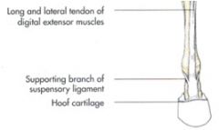
O: extensor fossa of the femur
I: extensor process of the distal phalanx; dorsal surface of the proximal and middle phalanges Action: extend the digit and flex the hock; assist in fixing the stifle joint Innervation: peroneal / fibular n. Extensor branches of the interosseus ligament join the long digital extensor tendon in the digit |
|
|
What are the OIAI of the peroneus tertius m. of the pelvic limb of the horse?
|
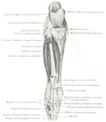
Consists of a strong tendon located between the long digital extensor and cranial tibial mm.
O: extensor fossa of the femur I: Dorsal tendon – proximal extremity of metatarsal III and the 3rd tarsal bone I: Lateral tendon – calcaneus and the 4th tarsal bone Action: flex the hock when the stifle joint is flexed Innervation: peroneal / fibular n. The proximal part of peroneus tertius is fused with the long digital extensor tendon while the distal part of peroneus tertius is fused with the cranial tibial tendon |
|
|
ID 1 and 2
|

Peroneus tertius m. (1)
Dorsal tendon Lateral tendon Middle extensor retinaculum arises from this tendon Cranial tibial m. (2) Dorsal tendon Medial tendon (cunean tendon) Inserts on fused tarsal bones 1 & 2 Cunean bursa |
|
|
WTF is wrong with this horse?!
|
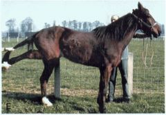
She has a ruptured peroneus tertius m.
A ruptured peroneus tertius allows flexion of the stifle and extension of the hock at the same time May occur if a horse slips and lands on the dorsal part of the leg Does not affect the stay apparatus |
|
|
What are the OIAI of the cranial tibial m. of the horse?
|
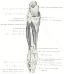
O: lateral condyle and border of the tibia, small area on the lateral surface of the tibial tuberosity, crural fascia
I: Dorsal tendon – dorsal aspect of the proximal end of metatarsal III I: Medial (cunean) tendon – fused 1st and 2nd tarsal bones Action: flex the hock joint Innervation: peroneal / fibular n. |
|
|
Where is the cunean bursa located in the equine crus?
|

Cunean bursa located between the cunean tendon and the medial collateral ligament of the hock
|
|
|
What is the OIAI of the lateral digital extensor m. of the equine pelvic limb?
|
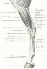
O: lateral collateral ligament of the stifle joint, fibula, lateral border of the tibia, interosseus ligament
I: long digital extensor tendon distal to the tarsus Action: assist the long digital extensor m. Innervation: peroneal / fibular n. |
|
|
What is the OIAI of the short digital extensor m. of the equine pelvic limb?
|
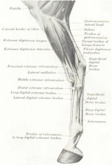
O: lateral tendon of peroneus tertius, middle extensor retinaculum, lateral collateral ligament of the hock
I: tendons of long and lateral digital extensor muscles Action: assist the long digital extensor muscle Innervation: deep peroneal / fibular n. Present in horse and ruminant |
|
|
cancel
|
cancel
|
|
|
What is the OIAI of peroneus tertius m. of the equine pelvic limb?
|

(10)
O: extensor fossa of the femur I: Lateral tendon – dorsomedial aspect of the proximal end of metatarsal III/IV I: Medial tendon – medial aspect of the first tarsal bone, fused second and third tarsal bones, plantar aspect of the proximal end of metatarsal III/IV Action: flex the hock joint Innervation: deep peroneal / fibular n. |
|
|
What is 10, 10', and 10" in the ruminant pelvic limb? What are its OIAI?
|
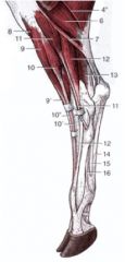
Long digital extensor m. (10, 10’, 10’’)
O: extensor fossa of the femur I: Medial tendon – middle and distal phalanges of digit 3 I: Lateral tendon – extensor processes of the distal phalanges of digits 3 and 4 Action: extend the digits and flex the hock joint Innervation: deep peroneal / fibular n. |
|
|
What is the OIAI of cranial tibial m. of the ruminant pelvic limb?
|
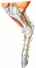
Cranial tibial m. (7)
O: lateral surface of the tibial tuberosity, cranial border of the tibia, proximal portion of the body of the tibia; lateral condyle of the tibia and head of the fibula I: first tarsal bone, fused second and third tarsal bones, proximomedially on metatarsal III/ IV Action: flex the hock joint Innervation: deep peroneal / fibular n. |
|
|
What is the OIAI of peroneus longus m. in the ruminant pelvic limb?
|
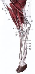
Peroneus longus m. (11, 11’)
O: lateral condyle of the tibia, lateral collateral ligament of the stifle joint, lateral meniscus I: first tarsal bone, proximal end of metatarsal III/IV Action: flex the hock joint and rotate it medially Innervation: deep peroneal / fibular n. |
|
|
What is the OIAI of the lateral digital extensor m. of the ruminant pelvic limb?
|
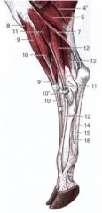
Lateral digital extensor m. (12)
O: lateral collateral ligament of the stifle, lateral condyle of the tibia, vestigial head of the fibula I: middle and distal phalanges of digit 4 Action: extend the fourth digit Innervation: superficial peroneal / fibular n. |
|
|
What are the OIAI of gastrocnemius m. in large animal pelvic limbs?
|

O: Lateral head – lateral supracondylar tuberosity (and lateral epicondyle of the femur in the Ruminant)
O: Medial head – medial supracondylar tuberosity (and medial epicondyle in the Ruminant) I: plantar aspect of the tuber calcanei Action: flex the stifle joint and extend the hock – both actions cannot occur simultaneously in the horse due to the tendinous nature of the peroneus tertius Innervation: tibial n. |
|
|
What are the OIAI of soleus m. of the large animal pelvic limb?
|
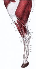
O: head of the fibula
I: tendon of gastrocnemius Action: assist gastrocnemius in extending the hock Innervation: tibial n. |
|
|
What is the OIAI of superficial digital flexor m. of the equine pelvic limb?
|
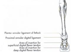
O: supracondylar fossa of the femur
I: tuber calcanei; proximal end of the middle phalanx, distal end of the proximal phalanx Action: flex the digit and extend the hock joint Innervation: tibial n. No proximal check ligament – SDF attaches to tuber calcanei |
|
|
What is the reciprocal apparatus of the equine pelvic limb?
|
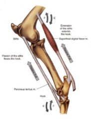
Superficial digital flexor m.
-Mostly collagenous Peroneus tertius m. -Mostly collagenous Extending the stifle extends the hock (if the hock is extended, the hock should be extended) Flexing the stifle flexes the hock (vice versa) |
|
|
What are the OIAI of the superficial digital flexor m. of the ruminant pelvic limb?
|

(23)
O: supracondylar fossa of the femur I: tuber calcanei; plantar surfaces of the middle phalanges of digits 3 and 4 Action: flex the digit and extend the hock joint Innervation: tibial n. |
|
|
Where is the calcaneal bursae in the pelvic limb?
|
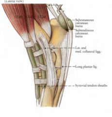
Subcutaneous
Between the skin and the tendon of the superficial digital flexor tendon as it crosses the tuber calcanei Subtendinous Between the superficial digital flexor tendon and the tuber calcanei |
|
|
What five muscles come together to make up the common calcaneal tendon?
|
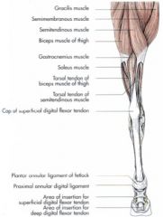
Gastrocnemius
Superficial digital flexor Biceps femoris (Horse) / Gluteobiceps (Ruminant) Semitendinosus Soleus |
|
|
What is the OIAI of popliteus m. of the large animal pelvic limb?
|
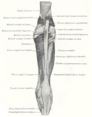
O: popliteal fossa on the lateral condyle of the femur
I: caudomedial border of the tibia Action: flex the stifle and rotate the leg medially Innervation: tibial n. |
|
|
What are the three heads of the equine deep digital flexor m. of the pelvic limb? What are the OIAI of this muscle?
|
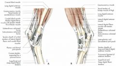
O: Lateral head (lateral digital flexor m.) – lateral condyle and caudal surface of the tibia
O: Medial head (medial digital flexor m.) – lateral condyle of the tibia O: Caudal head (caudal tibial m.) – caudolateral surface of the tibia I: flexor surface of the distal phalanx Action: flex the digit and extend the hock joint Innervation: tibial n. |
|
|
What forms the structure similar to the distal check ligament in the equine pelvic limb? Where does it course to?
|
Accessory ligament of the deep digital flexor tendon
Courses between the fibrous layer of the tarsal joint capsule and the DDF tendon in the proximal metatarsal region Similar to the distal check ligament of the thoracic limb except may not be as well-developed |
|
|
What are the three heads of the deep digital flexor m. of the ruminant pelvic limb? What are the OIAI?
|
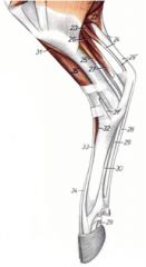
O: Lateral head (lateral digital flexor m.) (27) – lateral condyle and caudal surface of the tibia
O: Medial head (medial digital flexor m.) (25) – lateral condyle of the tibia O: Caudal head (Caudal tibial m.) (26) – lateral condyle of the tibia I: flexor surface of the distal phalanges of digits 3 and 4 Action: flex the digits and extend the hock joint Innervation: tibial n. |
|
|
ID 11-20 of these sheep crural muscles from the lateral view
|
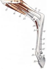
Craniolateral muscles
Cranial tibial m. (17) Peroneus tertius m. (19) Long digital extensor m. (20) Peroneus longus m. (16) Lateral digital extensor m. (18) Caudal muscles Lateral head of deep digital flexor m. (15) Caudal head of deep digital flexor m. (caudal tibial m.) (14) Lateral head of gastrocnemius m. (11) Common calcaneal tendon (12, 13) Superficial digital flexor tendon (13, 13’) |
|

Name 20-31 of the lateral view of the sheep pelvic limb.
|

Craniolateral muscles
Cranial tibial m. (27, 27’) Peroneus tertius m. (28) Long digital extensor m. (29,30) Caudal muscles Medial head of deep digital flexor m. (24) Lateral head of deep digital flexor m. (25) Medial head of gastrocnemius m. (20) Lateral head of gastrocnemius m. (21) Common calcaneal tendon Superficial digital flexor muscle and tendon (23, 23’) Deep digital flexor tendon (26) Interosseous m. (31) |

