![]()
![]()
![]()
Use LEFT and RIGHT arrow keys to navigate between flashcards;
Use UP and DOWN arrow keys to flip the card;
H to show hint;
A reads text to speech;
56 Cards in this Set
- Front
- Back
|
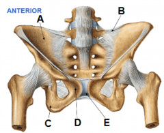
A: ileum B: sacroiliac joint C: ischium D: pubis E: pubic symphysis |
|
|
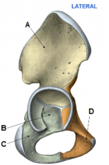
A: ileum B: acetabulum C: ischium D: pubis |
|
|
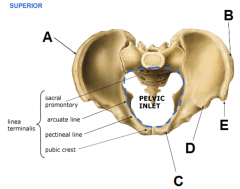
A: linea terminals B: sacral promontory C: arcuate line D: pectineal line E: pubic crest |
|
|
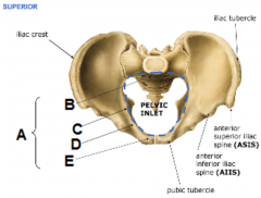
A: iliac crest B: iliac tubercle C: pubic tubercle D: anterior inferior iliac spine (AIIS) E: anterior superior iliac spine (ASIS) |
|
|
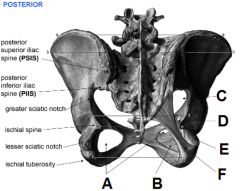
A: Posterior Superior Iliac Spine (PSIS) B: Posterior Inferior Iliac Spine (PIIS) C: greater sciatic notch D: ischial spine E: lesser sciatic notch F: ischial tuberosity |
|
|
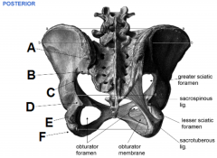
A: obturator foramen B: obturator membrane C: greater sciatic foramen D: sacrospinous ligament E: lesser sciatic foramen F: sacrotuberous ligament |
|
|
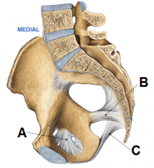
A: obturator membrane B: sacrospinous ligament C: sacrotuberous ligament |
|
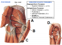
Identify the foramina |
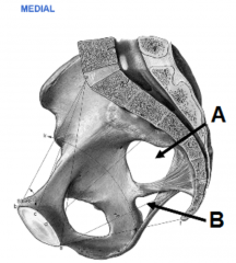
A: greater sciatic foramen B: lesser sciatic foramen |
|
|
Which muscle is found in the greater sciatic foramen, and what foramina are found either side of it? |
piriformis suprapiriform foramen and infraspiriform foramen |
|
|
Name the muscles, nerves and arteries found in the lesser sciatic foraman? |
Muscle: obturator internus Nerves: pudendal nerve Arteries/veins: internal pudendal artery and vein |
|
|
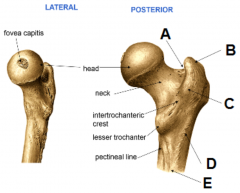
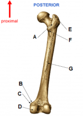
A: fovea capitis B: head C: neck D: intertrochanteric crest E: lesser trochanter F: pectineal line |
|
|
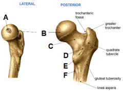
A: trochanteric fossa B: greater trochanter C: quadrate tubercle D: gluteal tuberosity E: linea aspera |
|
|
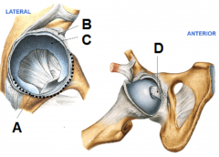
A: transverse acetabular lig. B: acetabular labrum C: horseshoe shaped cartilage D: ligamentum teres |
|
|
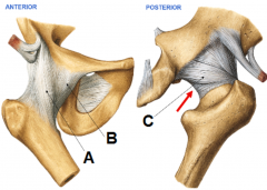
A: iliofemoral lig. B: pubofemoral lig. C: ischiofemoral lig. |
|
|
A: intertrochanteric crest B: adductor tubercle C: medial epichondyle D: medial condyle E: greater trochanter F: lesser trochanter G: linea aspera |
|
|
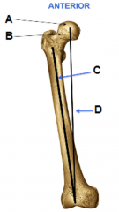
A: head B: neck C: anatomical axis D: mechanical axis |
|
|
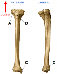
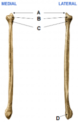
A: Gerdy's (iliotibial) tubercle B: tibial tuberosity C: medial malleolus D: fibular notch |
|
|
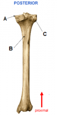
A: tibial plateau B: soleal line C: fibular articular facet |
|
|
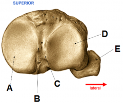
A: apex B: head C: neck D: lateral malleolus |
|
|
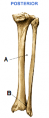
A: interosseous membrane B: distal tibiofibular joint |
|
|
A: medial condyle B: medial intercondylar tubercle C: lateral intercondylar tubercle D: lateral condyle E: fibula |
|
|
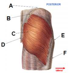
A: iliac crest B: posterior superior iliac spine C: gluteus maximus D: sacrum E: greater trochanter of femur F: iliotibial band/fascia lata |
|
|
A: gluteus medius B: piriformis C: obturator internus D: gluteus minimus |
|
|
What test might you administer to assess whether someone has trapped their superior gluteal nerve? |
Trendelenberg test |
|
|
What can the Trendelenberg test be used to assess? |
Superior Gluteal nerve entrapment or injury
Gluteus Medius m. (and gluteus minimus m.) rupture or weakness |
|
Error: "C" is for arrow above |
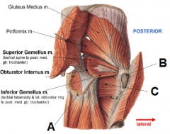
A: gluteus medius B: piriformis C: superior gemellus D: inferior gemellus |
|
|
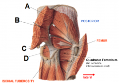
A: ischial tuberosity B: femur C: quadratus femoris |
|
|
Which muscles are involved in external rotation of the hip, and which is most powerful? |
Obturator Internus
ObturatorExternus Quadratus Femoris Piriformis Gluteus Maximus (most powerful) |
|
|
Which muscles are involved in internal rotation of the hip?
|
Gluteus Medius & Minimus
|
|
|
Which muscles are involved in flexion of the hip? |
Psoas major and iliacus |
|
|
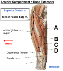
A: tensor fascia lata B: end of gluteal region C: quadriceps tendon D: patella E: patellar tendon |
|
|
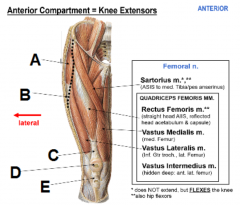
A: sartorius B: rectus femoris C: vastus medialis D: vastus lateralis |
|
|
What is the main extensor of the knee? |
quadriceps femoris |
|
|
Which three factors help prevent lateral dislocation/sublux of the patella? |
obliquus genu lateral patella facet lateral femoral condyle |
|
|
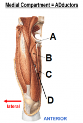
A: pectineus B: adductor longus C: gracilis D: adductor/subsartorial canal |
|
|
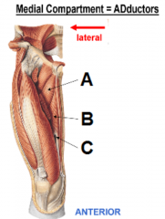
A: adductor brevis B: adductor magnus C: adductor hiatus |
|
|
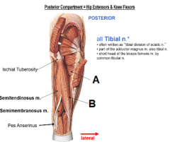
A: ischial tuberosity B: semitendinous muscle C: semimembranous muscle D: pes anserinus |
|
|
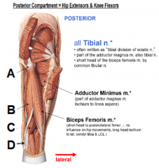
A: abductor minimus B: biceps femoris |
|
|
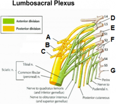
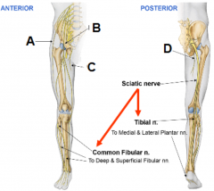
A: anterior B: posterior C: sciatic nerve D: tibial nerve E: common fibular (peroneal) nerve |
|
|
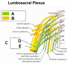
A: superior gluteal nerve B: inferior gluteal nerve C: nerve to piriformis D: lumbosacral trunk E: gray rami communicantes F: pelvic splanchnic nerve G: coccygeal nerve |
|
|
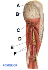
A: superior cleneal nerves B: medial cluneal nerves C: inferior cluneal nerves D: posterior femoral cutaneous nerve E: sciatic nerve F: perforating arteries |
|
|
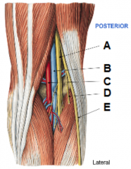
A: tibial nerve B: popliteal vein C: popliteal artery D: lateral sural cutaneous nerve E: common fibular nerve |
|
|
A: lateral femoral cutaneous nerve B: femoral nerve C: obturator nerve D: posterior femoral cutaneous nerve |
|
|
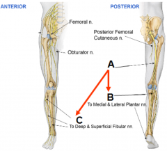
A: sciatic nerve B: tibial nerve C: common fibular nerve |
|
|
Where in the gluteal region should you administer an injection? |
superolateral |
|
|
Where in the spine do the following nerves originate; femoral obturator tibial common fibular (peroneal) |
L2-L4 L2-L4 L4-S3 L4-S2 |
|
|
Where regions of the lower limb do the following nerves provide sensory input; femoral obturator tibial common fibular (peroneal) |
femoral: anterior/medial thigh and medial/posterior leg/foot obturator: medial thigh tibial: posterior leg, lateral foot and plantar foot common fibular (peroneal): lateral leg and dorsal foot |
|
|
Which muscles do the following nerves innervate; femoral obturator tibial common fibular (peroneal) |
femoral: anterior thigh muscles
obturator: adductor muscles of the thigh tibial: all posterior thigh muscles except short head of biceps femoris common fibular (peroneal): short head of biceps femoris, lateral leg muscles, anterior leg and dorsal foot muscles |
|
|
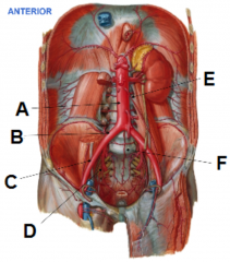
A: abdominal aorta B: common iliac artery C: external iliac artery D: internal iliac artery E: left gonadal artery F: median sacral artery |
|
|
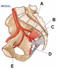
A: superior gluteal artery B: piriformis C: inferior gluteal artery D: internal pudendal artery E: obturator artery |
|
|
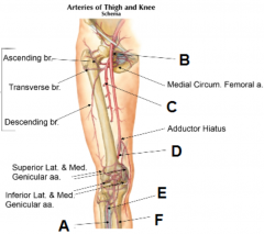
A: fibular artery B: femoral artery C: deep femoral artery D: popliteal artery E: anterior tibial artery F: posterior tibial artery |
|
|
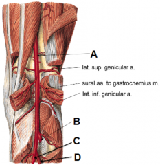
A: popliteal artery B: anterior tibial artery C: posterior tibial artery D: fibula artery |
|
|
What is the first way you would identify a popliteal artery aneurysm? |
Palpation. Would find pulsatile swelling in the popliteal fossa. |
|
|
What might you confuse a popliteal artery aneurysm with? |
Baker's cyst (popliteal cyst) |
|
|
What is a Baker's cyst and what can cause it? |
Collection of synovial fluid which has escaped from knee joint & formed synovial-lined sac in popliteal foss. Caused by knee joint trauma or degenerative changes. |
|
|
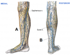
A: great saphenous vein B: small saphenous vein |