Reading...
![]()
Play button
![]()
Play button
![]()
Use LEFT and RIGHT arrow keys to navigate between flashcards;
Use UP and DOWN arrow keys to flip the card;
H to show hint;
A reads text to speech;
139 Cards in this Set
- Front
- Back
|
Is air or bone conduction bette for hearingr? Why?
|
air conduction
bone conduction doesn't give sound the time and speace to be properly processed |
|
|
What basic PE test takes advantage of this for testing hearing?
Mnemonic? |
The Rinne test
You hear a ringing in your ears! |
|
|
Explain it. How and why?
|
It's the one where you put a vibrating tuning for to the mastoid so the vibrations bypass the middle ear and stimulate hair cell directly.
Then when they can't hear it anymore, hold it up to their ear to see if they can hear it. They should be able to. |
|
|
WHat is the dx if they can't hear it though the ear (through air)
|
conductive hearing loss
|
|
|
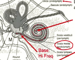
What is the fluid in the cochlea?
|
endolymph
|
|
|
How does sound travel through the cochlea?
|
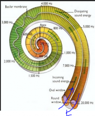
start in oval window, spiral in, spiral out, exit in round window
|
|
|
What IS the organ of corti?
|
It is the thing in the cochlea that houses the actual hair cells
|
|
|
What does the cochlea look like if you cut in up?
|

3 compartments.
scala |
|
|
What is each compartment called?
What one had the hairs of corti? |
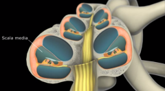
scala... vestibul, media, and tympani
scala media had the organ of corti in it |
|
|
Is the same fluid in all of them?
mnemonic? |

endolymph in media
perilymph in the others. The non- media ones are on the outside so they get perilymph |
|
|
But how do these three compartments stack up with one another?
show it. |
they are all continuous in a spiral by themselves
|
|
|
Which hair cells are good for high vs low freq sounds?
Mnemonic? |
base is better for high frequencies
apex is better at low freq sound first touches the base and we tend to lose high freq hearing first! |
|
|
Is endolymph intra or extracellular flkuuid? what is unique about it's composition?
|
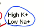
it is EFC, but has oppsit composition for K and Na
|
|
|
Are the perilymph like this too?
Why is it like this? |
No, they are normal
as the hair cells vibrate through the compartments, the differnt ECF compositions cause them to depolarize and repolarize quickly |
|
|
What is presbycusis? What structures do we lose in it?
|
hearing loss as we age when we lose hair cells at the base.
|
|
|
What happens when you vibrate the oval window?
|
endolymph creates fluid waves in the scala media
|
|
|
Mnemonic for scala?
|
we use them to hear out SCALES!
|
|
|
What is the static labryith supposed to do?
|
part of the vestibular system that helps us sense linear acceleration
|
|
|
Linear acceleration in layman's terms
|
linear acceleration is basically a fancy way of saying to maintain balance according to gravity
|
|
|
What structures compose it? (show)
|
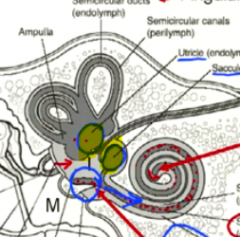
Two sacs called the Utricle and the Saccule
|
|
|
What fluid and functional cells are in these sacs?
|
endolymph and a macula of hair cells
|
|
|
What are the semicircular canals called?
|
the kinetic labrynth
|
|
|
How are they arranged in space and why is this good?
Why are they called kinetic and static labryiths? What is this referring to? |
they are in the 3 planes of space, which is good to detect angular acceleration and decelleration
the labrynth's names are referring to the movement of the head when they record their data. |
|
|
Where are the hair cells in the semicircular canals?
|
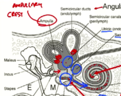
in the opening of them called the ampulla/ampullary crest
|
|
|
What vestibular reflex are these semicircular ducts responsible for?
(think i phones!) |
adjusting the eyes in response to changes in head position.
The iphone automatic video stabilization technology uses these semicucular canals toooooo! |
|
|
What are the hairs of inner hair cells made of? Can they move?
Is Kartagener's syndrome associated with hearing loss for this reason? |
stereocilia coming off epithelial cells which do not move
kartagener's syndrome is associated with hearing loss because the ear cilia have trouble cleaning the hears out, resulting in a CONDUCTIVE hearing loss. the hair cells are not affected. |
|
|
What is the fx of stereocilia everywhere?
|
to increase surface area
|
|
|
What are sterocilia essentially?
|
tall microvilli
|
|
|
Where are the cell bodies of the sensory neurons of hearing and balance? Why?
|
outside the CNS because they are derived from neural crest cells
|
|
|
How many vestibular and cochlear nuclei on each side of the brainstem?
|
v- 4 (conglomerated into 1)
c- 2 (one on each of the ICP) |
|
|
Show where these nuclei are on a transection
|
|
|
|
Show where on the neural pathway the cochlear and vestibular ganglion are. Name them too.
(is this before or after the internal auditory meatus?) |
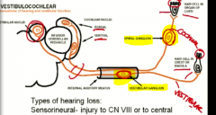
Cohlear- spiral ganglion
Vestibular- vestibular ganglion both before the IAM |
|
|
What are 3 causes of conduction deafness?
|
1. obstruction (ear wax)
2. Otosclerosis (genetic overgrowth of middle ear bones) 3. Otitis media (middle ear infection) |
|
|
Mnemonic for spiral ganglion?
|
the cochlea is a spiral so it makes sense that it terminates at the spiral ganglion as soon as the cochlea ends
|
|
|
When do pretty much all sensory neurons cross in the CNS?
|
As soon as they enter
|
|
|
What is the only exception?
|
dorsal column/medial leminiscus crosses at the medulla
|
|
|
Does hearing just go straight to the cortex? What additional processing does it get and where?
|
No.
It undergoes additional procesing for LOCALIZATION at the SUPERIOR OLIVARY NUCLEUS. |
|
|
What input does each superior olivary nucleus get?
|
input from both ears
|
|
|
What structure is made from auditory fibers crossing to get to the CL Superior olivary nucleus?
|
the trapezoid body
|
|
|
So what sx would you get if you lesioned one superior olivary nucleus?
|
you could still hear out of both ears, but you wouldn't be able to loacalize sound
|
|
|
When along the auditory pathway would you lose hearing in one ear with a lesion?
|
at or before the cochear nucleus
|
|
|
What does the inferior colliculus do?
|
additonal sound procesing and localization
|
|
|
Outline the path of auditory pathway to the cortex from one ear's hair cell to cortex.
|
1. inner hair cell
2. spiral ganglion 3. cochlear nuclei 4. BOTH superior olivary nuclei 5. BOTH lateral leminisci 6. BOTH inferior colliculi 7. BOTH inferior brachium 8. BOTH medial genticulate bodies of the thalamus 7. BOTH superior temporal gyri |
|
|
Would you get ANY hearing loss with a lesion after the cochlear nucleus?
|
yes, but only slight bilateral loss
|
|
|
What is the vestibular-occular relfex? (VOR)
|
reflex eye movement to head movement
|
|
|
What sensory cells are being stimulated and inhibited when you turn your head to the right?
|
you move endolymph to stimulate hair cells in the right vestibular system while inhibiting the left
|
|
|
What is the goal of the vestibular system when you turn your head to the right?
|
to stimulate cranial nerves to make you look to the left (maintain gaze)
|
|
|
State the pathway by which we coordinate gaze when turning our head to the right. (include sides)
|
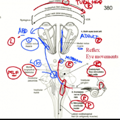
1. Right vestibular system activated and sends this to right CN 8 nuclei
2. CN 8 activates CL CN 6 and inhibits ipsilateral CN 6 3. CN 6 activates LR and also goes via MLF to activate right CN 3 |
|
|
What are the two main fx of the vestibular system?
|
1. eye movement with head turning
2. antigravity muscles to stay up while moving in gravity |
|
|
Which parts of the vestibular system is responsible for each?
What type of acceleration is associated with each? |
1. semicricular canals- angular acceleration - eye movement
2. vestibular sacs- linear acceleration- body movements to stand up |
|
|
what is the general sx of someone with a vestibular lesion?
|
1. dizzy
2. can't stand straight 3. nausea from being so dizzy |
|
|
what sign would you find with someone with a left vestibular nuclei lesion?
|
vestibular nystagmus
|
|
|
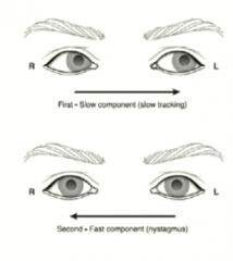
Define nystagmus
|
an irragular movement in one or both eyes inresponse to a pathology with a distinctive fast or corrective phase in opposite direction
Greek for "to nod" |
|
|
What can you compare the slow vs fast phase of nystagmus to?
|
slow- fell asleep in class with head nodding down
fast- quickly jerk awake when you realize your mistake |
|
|
What is going on in the brain that is responsible for the slow vs fast phase?
|
slow- some pathology causing deviation or bad movement
fast- the cortex attempting to correct this movement |
|
|
In a vestibular lesion, what direction is the slow phase eye drift and why?
|
toward the side of the lesion because the OTHER vestibular nucleus is ok and so the balance is shifted as if they were stimulated
|
|
|
What direction is the fast phase relative to the side of the vestibular lesion?
|
since it is deviating toward the lesion in the slow phase, the correction will be in the direction away from the lesion
|
|
|
What test would you use to see if they has vestibular lesion?
|
ask them to move their head from right to left and check for nystagmus
|
|
|
What test for the vestibular system should you use for an unconscious person?
|
the caloric test of putting warm or cold water in their ear to check their nystagmus reaction
|
|
|
What was warm vs cold water simulate?
|
warm water- makes endolymph flow so imitates turning to that side
cold water- makes endolymph not flow to imitates lesion to that side/inhibition from turning to the other side |
|
|
Say and explain the mnemonic for interpreting the test results.
|
COWS (Cold Opposite, Warm Same)
If you pour _____ water in one ear, the direction of the fast phase of evoked nystagmus should be in the ______ direction. COWS NEED A LOT OF CALORIES!!! (TO BE FAST!) |
|
|
Why do you get this fast phase?
|
because the eyes are slowing deviating and the cortex senses this and tries to correct it
|
|
|
What is you pour water in one ear and there is no eye movement?
|
there is a lesion in the vetibular system because it isn't responding
|
|
|
What happens if you put warm water in the left, the eyes turn right and STAY there?
|
there is some sort of cortical damage or cortical connection defect
|
|
Where is the lesion?
|
Left- fast phase is away from the lesioned side
|
|
|
NOW WE TALK ABOUT THE WIRING THAT ALLOW THIS TO HAPPEN
BEGIN "CONJUGATE GAZE LECTURE" |
FANTASTIC MR. FOX!
|
|
|
Describe the pathway we take to create voluntary conjugate gaze to the right.
|
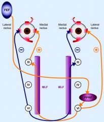
Follow the orange line for what is activated
1. initiate plan in left visual eye field 2. send UMN fibers down to right paramedian pontine reticular formation (PPRF) - push button 3. PPRF sends UMN's to the CN 6 right next to it and to the left CN 3 4. smooth, conjugate gaze to the left |
|
|
What disease threatens the MLF heavily? Why?
|
MLF needs to send signal quickly to congugate gaze to it is heavily myelinated
|
|
|
What condition do you get with a MLF lesion?
|
internuclear opthamoplegia
|
|
|
dissect internuclear opthamoplegia
|
internuclear- between CN 3 and CN 6 nucleus
opthalmoplegia- paralysis of the eyes |
|
|
What sx would a pt experience with internuclear opthamoplegia? Why?
|
blurry vision when looking to the direction of the lesion (if you count the MLF position as where it starts in CN 6)
because your other eye doesn't adduct to keep a stable image |
|
|
Define strabismus
|
means squinting
but refers to when a eye is deviated to one side |
|
|
Why would someone quint if an eye was deviated to one side?
|
to prevent the weird gaze from creating blurry vision
|
|
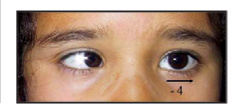
Why would someone have a medial/internal strabismus?
|
If their lateral rectus wasn't working because of a same sided CN 6 lesion.
|
|
|
Aren't there other muscles that can abduct though? Which ones?
|
yes, the obliques can, but not nearly as strongly as the lateral rectus.
|
|
|
What other 2 structures would you affect if you lesioned the abducens nucleus?
|
the same sided PPRF and the facial nerve
|
|
|
So what sx would you get with a right abducens lesion?
|
1. CN 6- inability to make right eye look to right
2. PPRF- inability to make left eye look to the right 3. CN 7- right facial paralysis/hyperacusis Can't look to the right at all! |
|
|
Does it seem if the cortex can make the eye adduct without going through MLF?
|
so far, no
|
|
|
How can you tell an MLF lesion apart from a CN 3 lesion?
|
In CN 3 lesion, you lose many more functions and would see things like that eye's pupil being extremely dilated
|
|
|
What will the convergence test be in a MLF lesion?
|
totally intact.
|
|
|
What would you see if you had a left frontal eye field lesion?
Nejeeb Mnemonic? |
You wouldn't be able to look to the right and there may be a slow drift to the left as a result of the imbalance
When a person irritates you, you look away, but whe they destroy your stuff, you look towards them (deviation from unoppose action of CL FEF) |
|
|
What other associated sx may you see? (think surrounding cortex areas)
|
1. weakness/UMN paralysis of RIGHT LOWER face (only CN that is not bilateral)
2. UMN weakness with RIGHT UPPER LIMB |
|
|
VISUAL SYSTEM
|
YEAHH!
|
|
|
What nerves run through the choiroid of the eye?
|
all the S and PS motor control
and the V1 of CN 5 sensory |
|
|
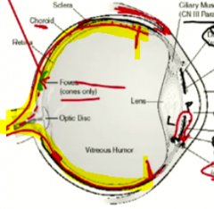
What is the goal of the cornea and lens?
|
to concentrate light rays to the fovea of the retina
|
|
|
Where is the fovea and what is in it?
What does it let us do? |
it is in the macula and is a concentration of cones
lets us see objects in our central vision and colors |
|
|
What sx do people with age related macular degeneration get?
Is there a cure? |

1. loss of central vision, but preservation of peripheral vision
2. loss of color no cure |
|
|
What makes the aqueous humor and what drains it?
|
ciliary body creates it and canal of schlemm drains it
|
|
|
In glaucoma, where do you get intraoccular pressure?
|
it is distributed all along the inside of the eye
|
|
|
When the whole eye is exploding with pressure, which neurons are most affected?
|
the ones at the outside of the choiroid, which are the longer ones.
|
|
|
What sx does this result in?
|
peripheral vision loss
|
|
|
What layer of the retina are the rods and cones in?
|
the most outer of the 3 lsyers
|
|
|
When they receive light, how to the rods and cones sens this message to the brain?
|
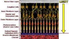
1. send it back up the layers via bipolar cells to the ganglion layer of tertiary sensory neurons
2. teritary neurons send this through the optic tract back to the lateral genticulate culeus and suprchiasmatic nucleus |
|
|
What supplies nutrient to the rods and cones?
What kind of nutrients especially? |
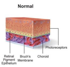
outer layer of retina consisting of epithelium called the pigment epithelium
regular nutrients plus vitamin A! |
|
|
What happens to the photoreceptors if you get punched and a part of your retina dettaches?
|
those cells will no longer get nutrients and will degneerate
|
|
|
How many types of rods vs cones do we have?
|
1 type of rod
3 types of cones for each type of light |
|
|
How active are rods vs cones in bright and dark light? Why does this make sense?
|
Rods are better in the dark because they are very sensitive and also get more inut in dark
COnes work under brigh light when we constrict the pupil to focus light at the fovea |
|
|
So how does everything look in the dark?
|
greyish
|
|
|
What is the 3 neuron rule for all sensory processing?
|
There must be 3 neurons in the processing pathway
1. terminates on peripheral ganglia 2. crosses midline and goes to thalamus 3. goes to cortex |
|
|
So are the rods and cones counted as neurons?
Show it |
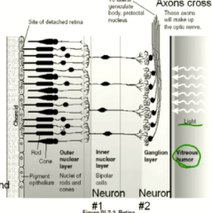
Nope
|
|
|
What is the 1st neuron in the viual pathway system?
|
the bipolar neurons
|
|
|
Define fovea
|
a small fossa
|
|
Why is the eye fovea shaped like this?
|
to provide direct access of light to the cones there
|
|
|
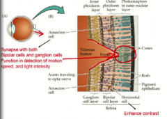
Show a horizontal cell. what do they do? where are they?
|
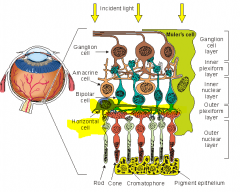
connect the bipolar cells and photoreceptors horizontally
responsible for enhancing contrast |
|
|
Show a amacrine cell. what do they do? where are they?
|
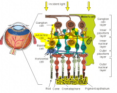
synapse with bipolar and ganglion cells to help detect motion, speed, and light intensity
|
|
|
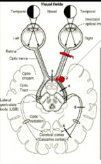
How does the image get projected onto the retina?
|
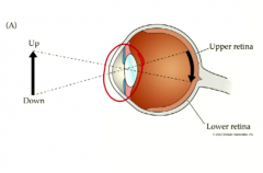
it is inverted both horizontally and vertically
|
|
|
What is central vision? alternate name?
|
the half of your vison closest to your nose.
you NASAL HEMIFIELDs |
|
|
What hemifield compose your peripheral vision?
|
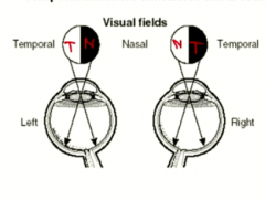
you TEMPORAL HEMIFIELDS (closest to your temporal bone)
|
|
|
if you see something in your temporal hemifield, which side of the retina is it projected on? Show.
|
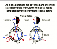
the nasal retina
|
|
|
some secondary visual neurons cross and some don't. guess and show which ones cross.
mnemonic? |
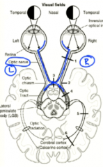
The ones from the central/nasal part of the retina cross at midline.
since they are closer to midline already, they are the ones that cross. |
|
|
Trace an object in the left nasal field all the way tot the calcarine cortex.
|
1. left nasal hemifield
2. left temporal retina 3. left optic tract 4. left lateral genticulate nucleus 5. left calcarine cortex (primary visual cortex) |
|
|
What are the optic radiations?
|
the neurons that carry the 3rd order neurons from the thalamus to the visual cortex
|
|
|
What field of vision info is crossing at the chiasm?
|
the info from both temporal fields
|
|
|
define an-opsia
|
lack of vision
|
|
|
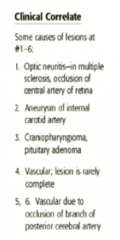
What are two things that can cause a right eye anopsia?
|
something that lesions the right optic tract- MS or an opthalmic infarct
|
|
|
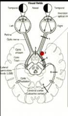
what does a right nasal hemianopsia look like?
|

|
|
|
What type of tumor tends to always compress the optic ciasm? why?
|
pituitary tumors because bone is beneath them so they must go up!
|
|
|
What kind of deficit would you get with an optic chiasm compression?
|
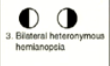
BITEMPORAL HETERONYMOUS HEMIANOPSIA
|
|
|
Describe the route of the pupillary light reflex to unilateral light stimulus.
Does it involve the thalamus? Why? |
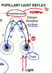
No thalamus because this is a reflex that doesn't need cortical input.
1. light in one eye 2. travels via CN 2 to the pretectal area 3. pretectal area bilaterally innervated nucleus of edinger westphal 4. both pupils constrict |
|
|
What 2 pathologies tend to decrease the pupillary light reflex?
|
1. pineal tumor
2. neurosyphillis |
|
|
When does the CN 2 fibers go to the pretectal area?
|
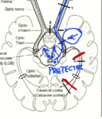
they branch off right before the thalamus
(if they go to the thalamus then they would go to the cortex and not be a reflex) |
|
|
so what kind of CN 2 lesion would create a decrease in pupillary reflex?
Would a single lesion eliminate it? Why? |
anywhere before the thalamus
no because light from both eyes go to both tracts |
|
|
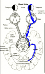
where does the carotid artery pass in relation to the optic tract? show
what is it headed to? |

lateral to the chiasm
headed to the circle of willis |
|
So what would the first sx be here?
|
right nasal hemianopsia
|
|
|
Would the pupillary light reflex be completely gona in an CN 2 lesion? what would it look like?
|
no it would just be more sluggish
|
|
|
What is a craniopharyngioma?
how do you dissect this? |
anterior pituitary tumor bcause thhis is the only crnaial structure arising from the pharynx tissue
|
|
Name some reaons for each of these lesions
|
|
|
|
What are the two lobe destinations for one side of optic radiations?
|
an upper cuneus gyrus and a lower lingual gyrus
|
|
|
Describe the route to the cuneus gyrus vs the lingual gyrus.
What lobe do they pass? How direct? Show |

cuneus- goes through a direct path in the parietal lobe
lingual- goes through path in temporal lobe with a tight hairpin turn called "meyer's loop" |
|
|
What separates the two gyri?
|
the calcarine fissure!
|
|
|
where is central vs peripheral vision processed in the cortex?
|
fovea more posterior
peripheral is more anterior |
|
|
what half of the visual field does each gyrus process?
|
again, the oppsite
cuneate on top sees the bottom visuual field lingual sees the top visual field |
|
|
What would you get with a lesion to the left optic radiation in the temporal lobe?
|
loss of input to right lingual gyrus and thus...
left homonymous superior quadrantanopia |
|
|
What is the rule of L's for the Lateral optic radiation fibers?
|
Loop (meyer's)
Lower quadrants of retina (upper of visual field) Lower gyri Lateral fibers |
|
|
What kind of insult tends to happen to the visual fibers after the thalamus?
|
infarct/vascular insult
|
|
|
what does macular sparing correspond to on the retina?
|
stuff seen in the fovea
|
|
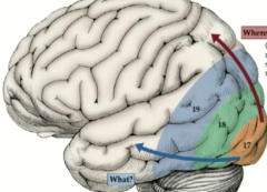
How does the parietal lobe help with vision analysis?
|
WHERE? helps with analysis of motion and spatial relations
(it is involved in a lot of spatial stuff!) |
|

How does the temporal lobe help with vision analysis?
|
WHAT? helps with analysis of form and color
|
|
|
What part of the visual cortex would have more connection to the temporal lobe then?
|
the part receiving the macula/fovea
|