Reading...
![]()
Play button
![]()
Play button
![]()
Use LEFT and RIGHT arrow keys to navigate between flashcards;
Use UP and DOWN arrow keys to flip the card;
H to show hint;
A reads text to speech;
51 Cards in this Set
- Front
- Back
|
Sexual Dimorphism and Evidence of it
|
Definition: morphological differences between m and f
-apparent in the lumbar region -hypothesized to be an evolutionary response to changes in center of mass that occur during pregnancy associated with obligate bipedalism - women have wedging in the lumbar region |
|
|
Palpable Skeletal landmarks and Points of Reference for Back and Spinal Column
|
Cervicothoracic Junction - located at spinous process of C7
Scapular spine - located lateral to spinous process of T3 Inferior Scapular Angle - located lateral to spinous process of T7 12th Rib - lateral of spinous process of T12 Iliac crests - spinous process of L4 |
|
|
Number of Cervical Vertebrae and Cervical Nerves
|
There are 7 cervical vertebrate (C1 and C2 atypical)
There are 8 Cervical Nerves First cervical nerve exits between cranium and C1 Second one exits between C1 and C2 Eighth one exits between C7 and T1 |
|
|
Typical Cervical Vertebrae
|
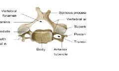
C3-C7
Body: small wider side to side Vertebral Foramen: triangular shaped Transverse Foramen: have foramina Movements: flexion, extension, lateral flexion, and rotation (greatest ROM for vertebral column) |
|
|
Cervical Ribs (supernumerary ribs)
|
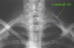
An extra rib or pair of ribs arising from the 7th cervical vertebrae
This can cause Thoracic Outlet Syndrome (not always) |
|
|
Thoracic Outlet Syndome (TOS)
|
Impingement of Brachial Plexus and Large Arteries in the region superior to the clavicle
Anything that impinges neurological vascular structures in the region where the Thoracic Outlet is as it goes into the upper limb. Could be caused by extra ribs, fractured clavicle, extra muscle or scar tissue in the region of scalene muscles, poor posture of the neck and shoulder regions |
|
|
Thoracic (12 vertebrae)
|
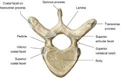
Body: Larger, heart shaped, costal facets
Spinous Process: long and sharp, projecting inferiorly Vertebral foramen- circular in shape Transverse process - facets for the ribs except T1 and T12 Articular Processes : superior facets directed posteriorly and inferior facets directed anteriorly Movements: rotation, lateral flexion is limited, flexion and extension are prevented |
|
|
Lumbar Vertebrae (5 Vertebrae)
|
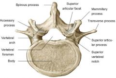
Body: Massive kidney shaped
Spinous Process: short and blunt, rectangular shape, projects posteriorly Vertebral Foramen: thin and tapered Articular Facets: superior directed posteromedially and inferior directed anterolaterally Movements: flexion and extension, some lateral flexion, rotation prevented |
|
|
Sarcum: Properties and such
|
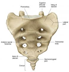
Five fused vertebra, used to provide strength and stability to pelvis. Houses and anchors inferior part of spinal cord.
Has the coccyx(made of 3-4 small fused bones) attached to the inferior portion of it. |
|
|
Articulated Vertebral column: What contributes to stability?
|
Stability of articulated vertebral column depends on the articular discs between the vertebral bodies, surrounding ligaments, and surrounding musculature.
|
|
|
Supraspinous Ligament
|

Ligament that covers the spinous processes longitudinally and superiorly running down the entire length of the vertebral column
Starts at T1 |
|
|
Interspinous Ligament
|
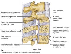
Ligament that spans in between the inferior portion of the spinous process to the superior portion of the spinous process below it.
|
|
|
Ligamentum Flavum
|

Connects the lamina of adjacent vertebrae. Each vertebra has them and there is also one going to the first segment of the sacrum
|
|
|
Ligamentum Nuchae (or nuchal ligament)
|
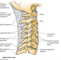
Runs from the External Occipital Protuberance and runs to the spinous process of C7
This is a well developed portion of the Supraspinous LIg |
|
|
Anterior longitudinal ligament
|
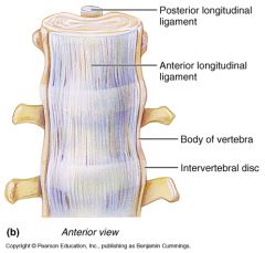
The ligament that spans longitudinally on the anterior aspect of the vertebral column. Spans across the entire column covering the fronts of the vertebra and helps reinforce vertebral discs
Prevents hyperextension |
|
|
Posterior longitudinal Ligament
|

Spans Across the posterior aspect of the bodies of the vertebra and runs along the posterior aspect of the bodies of the vertebra longitudinally throughout the whole vertebral column and helps reinforce vertebral discs
Prevents hyperflexion Smaller than the Anterior Longitudinal Ligament |
|
|
Annulus Fibrosus
|
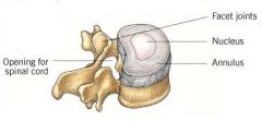
The outer layer of the intervertebral disc. Part of it supports the joints in between the vertebral bodies.
encloses the nucleus pulposus |
|
|
Intertransverse Ligament
|
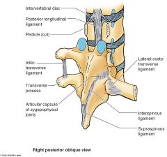
Ligament that is present in between transverse processes
Runs longitudinally |
|
|
Herniation of Disks: Why does it occur and where?
|
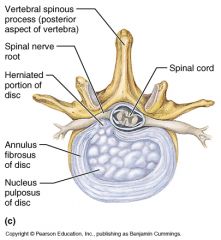
Herniated disks are most common in the lumbar region in the posterolateral direction mostly because the posterior olongitudinal ligament is not broad. Herinated material will take path of least resistance
Also occurs with cervical vertebrae |
|
|
Stenosis and Herniation of Cervical Region: What types are there?
|
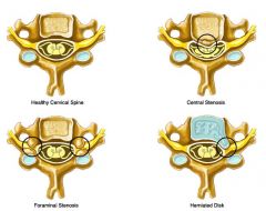
Central stenosis- when the anterior portion of the spinal cord is pressed up against by the posterior aspect of the body of the vertebra
Foraminal Stenosis - when nerves are squished in between intervertebral foramina Herinated disk - contents of the intervertebral disk push out and cause pressure on nerves |
|
|
Thoracic Region Herniation: How does it happen?
|
Not common, usually result of wear and tear/disk degen
Sudden and forceful twisting of midback region could contribute Conditions that predispose: abnormal kyphosis (Scheuerman's disease) |
|
|
Lumbar region disc herniation: How does it happen and what?
|
Common herniation here because it is most weight bearing
Bending, twisting, improper lifting increases load on tendons and discs Sudden injury contributes to this herniation Levels of herniation correlate to areas of pain |
|
|
Soft Tissue of Vertebral Column: What is it and where?- ACCORDING TO VASU
|

This is referred to as Thoracolumbar Fascia
Posterior Lamina -encompasses posterior aspect of erector spinae muscles. Middle lamina- occurs between the intermediate and deep layers of muscle (b/t ESM and QuadratusL Anterior layer is deepest layer lining the anterior aspect of the deepest muscle (use some common sense dummy) |
|
|
Soft Tissue of Vertebral Column:
|

Posterior lamina-where LatD arises from
Anterior lamina-anterior aspect of musculature (posterior side of body wall) Posterior and middle lamina surround ESM Middle lamina and anterior lamina surround QuadL |
|
|
Spinal Cord and Nerves: numbers, pairs, ventral rami, dorsal rami
|

31 pairs of spinal nerves with 8 cervical pairs, 12 thoracic pairs, 5 lumbar pairs, 5 sacral pairs, 1 coccygeal pair
Ventral Rami-larger portion of nerves that innervate lateral and anterior body walls and limbs (hypaxial nerves) Dorsal Rami- innervate the intrinsic back muscles (epaxial nerves) |
|
|
Segmental Innervation, Dermatomes, Myotomes
|

Segmentally-nerves in a region will tend to innervate things in that region.
Dermatome-area of skin innervated by sensory fibers of one single nerve root Myotome-a group of muscles that is primarily innervated by motor fibers of a single nerve root These two overlap-don't always correspond on a one to one basis |
|
|
Denticulate Ligaments
|

Tooth like ligaments that come off of the spinal cord laterally. They anchor the spinal cord to the dura mater
|
|
|
Ventral Horn
|

ALWAYS THINK SAME DAVE SENSORY AFFERENT MOTOR EFFERENT DORSAL AFFERENT VENTRAL EFFERENT
Horn on anterior side of Spinal cord that is responsible for motor control |
|
|
Dorsal Horn
|

Horn on the posterior side of spinal cord responsible for most sensory functions of spinal cord.
SAME DAVE |
|
|
Dura Mater
|

The most outer layer that will surround spinal cord which has the Arachnoid mater anchored to it medially
Encompasses the brain and spinal cord |
|
|
Arachnoid Mater
|

One layer medial to dura mater, translucent and spider web like.
|
|
|
Pia Mater
|
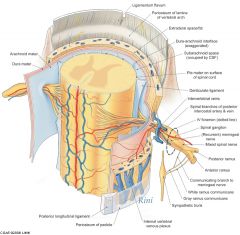
Adheres tightly to the spinal cord tissue itself
|
|
|
Subarachnoid Space (including CSF)
|

Space in between the arachnoid and the pia mater that contains cerebrospinal fluid
|
|
|
Gray matter
|

Corresponds to the neuronal cell bodies in the middle of the spinal cord. with white matter insulating those gray matters.
|
|
|
ANATOMY OF SPINAL CORD
|
brainstem comes out and becomes part of spinal cord.
There is a cervical enlargement, lumbosacral enlargement, ends with a filum terminale |
|
|
Cervical Enlargement
|
Area where the cervical plexus and brachial plexus come from
|
|
|
Lumbar Enlargement/Lumbosacral Enlargement
|
Area where lumbar and sacral plexuses come from
|
|
|
Filum Terminale
|

Terminal end of spinal cord that appears as a thin string which helps anchor the spinal cord.
It is a piece of pia mater that comes off the tip of the conus medullaris which travels down the sacral canal to anchor inferiorly. |
|
|
Conus Medullaris
|
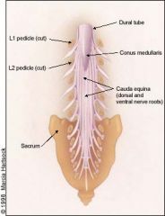
End of spinal Cord Ending at about L2
|
|
|
Cauda Equina
|

bundle of spinal nerves and spinal nerve roots which originate in the Conus Medullaris
|
|
|
Growth of Vertebral Column vs Growth of Spinal Cord?
Why do we have a cauda equina? |
The spinal cord grows at a slower rate than the spinal column. Therefore, these nerves get pulled down and elongate while the conus medullaris retracts/stays where.
|
|
|
Neural Tube Defect/Incomplete Development of Vertebral arch: what happens
|
You get spina bifida-many different types of spina bifida,
May or may not have protrusion of spinal contents May have a meningocele or meningomyelocele |
|
|
Dorsal Ramus/Rami Information and what it does and composition
|

Dorsal Rami is expaxial and smaller. It travels posteriorly
On the posterior side of spinal cord: You have dorsal rootlets forming the dorsal root/ganglion This dorsal root will come together with the ventral root to form the Spinal Nerve at that level of vertebrae REMEBER SAME DAVE |
|
|
Ventral Ramus/Rami Information and what it does and composition
|

hypaxial: larger and goes lateral and anteriorly
Rootlets from the ventral/anterior side come together to form the ventral root ganglion which will combine with the dorsal root ganglion to form the spinal nerve at that level. ALWAYS REMEMBER SAME DAVE> |
|
|
Types of nerves at RAMI/RAMUS
|
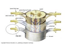
At the Ramus, there are both sensory and motor neurons.
All the nerves from the upper limb will be coming from spinal nerve roots which have a mixture of sensory and motor nerves. |
|
|
Kyphosis and Lordosis: what is it and what can cause it?
|
Kyphosis is convexity posteriorly of the thoracic region while Lordosis is concavity posteriorly of the cervical and lumbar regions.
Lordosis-associated with congenital abnormalities, musculoskeletal problems, degenerative disease. Kyphosis-results from developmental abnormalities, trauma, or degenerative disease. |
|
|
Spondylolysis and Spondylolisthesis: What is it?
|
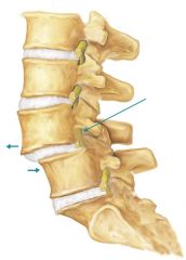
Broken vertebtra caused by trauma or degenerative disease.
It is a common cause of spondylolisthesis: anterior displacement of vertebra or vertebral column in relation to the vertebra below. |
|
|
Pars Interarticularis: what is it?
|
Pars is located in between superior and articular facets: in between the lamina and pedicles of vertebrae so to speak.
Fractures here are associated with spondylolsis KNOWN AS SCOTTIE DOG WITH DOG COLLAR |
|
|
Compression Fracture: what is it and where does it happen?
|
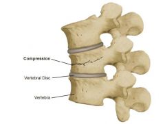
It is a collapse of bone of the vertebral body. This can be caused by trauma or degenerative disease
|
|
|
Sacral Abnormalities: What happens?
|
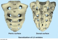
Fusion or partial fusion of L5 to the sacrum.
This would be a congenital defect. There can also be a condition where S1 does not fuse with the rest of the sacral bodies. |
|
|
Osteopenia and Osteoporosis: what are they?
|
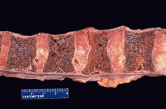
Osteopenia-process of thinning or decrease in bone mass
Osteoporosis - condition of having diminished bone densitry making bones prone to fracture These are both degenerative diseases. |