Reading...
![]()
Play button
![]()
Play button
![]()
Use LEFT and RIGHT arrow keys to navigate between flashcards;
Use UP and DOWN arrow keys to flip the card;
H to show hint;
A reads text to speech;
101 Cards in this Set
- Front
- Back
|
What level is the jugular notch at?
|
T2
|
|
|
What level is the sternal angle at?
|
T4/T5 (2 below T2 sternal junction)
|
|
|
What is the subcostal angle?
|
the angle between the midline and the costal margin. (different on the left and right)
|
|
|
What is Larrey's point?
|
the point between the costal margin and the xiphoid process?
|
|
|
Why is larrey's point important?
|
it's the landmark you would use to withdraw pericardial fluid.
|
|
|
Where is traube's semilunar space?
|
on the left and sitting right above rib 6.
|
|
|
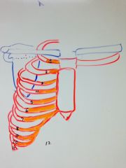
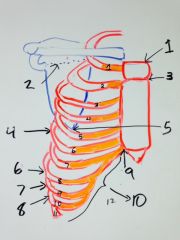
Draw the anterior view of the ribs. get the angles and the attachments right and include the scapula for reference.
|
|
|
|
1. jugular notch at rib 1/T3
2. spine of the scapula at Rib 1/T3 3. Sternal angle at rib 2/T4 4. Rib 6 which is parallel with the oblique fissure 5. Inferior angle of the scapula which is at rib 5/T7 6. Rib 8, which is the widest rib 7. Rib 9, which is the most oblique rib 8. rib 10, which is the most inferior at the midaxial line 9. Larrey's point between rib 7 and the xiphoid process 10. the costal margin made up of the costal cartilage of ribs 7-10 |
|
|
what borders the interscalene traingle? What is in the triangle?
|
Borders: The anterior and middle scalene and the 1st rib
In it: the subclavian artery and the brachial plexua |
|
|
What is in front of the trangle? How do you rmember this?
|
The subclavian vein because veins are always more superficial to arteries.
|
|
|
What would happen if you had compression of the triangle? What is this called?
|
This is thoracic outlet syndrome and involves paleness of the arm and tingling from the artery and plexus being affected.
|
|
|
What nerve is affected by horner's syndrome?
|
T1, the last nerve of the brachial plexus
|
|
|
What happens in a pancoast tumor?
|
all the structures in the triangle are compressed and you have thoracic outlet syndrome.
|
|
|
Is horners syndrome a part of thoracic outlet syndrome?
|
yes! T1 is part of the plexus.
|
|
|
What is the movement of the upper ribs and what is it caused by?
|
pump handle going anterior caused by the anterior scalenes
|
|
|
What is the movement of the lower ribs and what is it caused by?
|
bucket handle going lateral caused by the diaphragm pushing abdominal contents out.
|
|
|
What happens if you have paralysis of the intercostal muscles during inspiration?
|
the thoracic cage will depress because they are passively dragged down by the diaphragm
|
|
|
When is this normal? Why?
|
In babies because they have undeveloped intercostal muscles (no shell notching on radiograph)
|
|
|
WHat rib is lowest on the axillary line?
|
rib 10
|
|
|
Which rib is directly over the posterior oblique fissure?
|
rib 6
|
|
|
Describe the collateral vasculature in the intercostal regions.
|
each of the vessels/nerves for one rib makes a collateral going in the opposite direction for the superior margin of the rib below. (VAN NAV)
|
|
|
What nerves would you anesthetize to operate on rib 7?
|
collateral of rub 6 and nerve of rib 7.
|
|
|
How are the anterior intercistal arteries compared to the veins?
|
they run the same routes
|
|
|
what does the interna thoracic artery branch into and where?
|
It braches at rub 7 into the musculophrenic and the epigastric artery.
|
|
|
Where do the anterior inteercostal arteries for ribs 7-9 come from?
|
the musculophrenic artery
|
|
|
Where do the anterior inteercostal arteries for ribs 10-11 come from?
|
from the posterior intercistals
|
|
|
Where do the posterior intercostal arteries come from?
|
1-2 from the costocervial trunk off the subclavian
3-12 off the aorta |
|
|
Where do the 1st ribs veins drain into?
|
the bracheocephalic vein?
|
|
|
Where do the intercostal veins of ribs 2-4 drain into? Where does that vein drain into?
|
the superior intercostal vein which drains into
R- bracheocephlic vein L- azygous vein |
|
|
which rib levels are drained into the accessory hemiazygous and the hemiazygous?
|
5-8- accessory
9-12- hemiazygous |
|
|
Where do the intercostal nerves come from?
|
the ventral rami from the thoracic vertebrae
|
|
|
which are the exceptions?
|
intercostal nerves 1-2 which can cause referred pain
|
|
|
Which intercostal arteries are longer?
|
The right because the aorta is slightly on the left
|
|
|
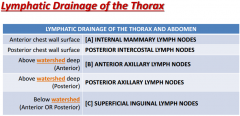
Where does the lymph of the breast drain into?
|
your axillary lymph nodes
|
|
|
What is the watershed line and why is it significant?
|
Everything above it drains predictably into the anterior vs posterior axillary nodes depedning on which side they are on
|
|
|
What drains below the watershed line?
|
superficial inguinal nodes
|
|
|
What drains the anterior chest not including breasts?
|
the centrally located interal mammary nodes
|
|
|
Where is the pulmonary vein always located on the hilum, both left and right?
|
most inferior and anterior
|
|
|
Does the pleural cavity and the lungs line up?
|
No, the pleural cavity is 2 ribs below the lung?
|
|
|
What level is the pleural line from front midclavicular to back scapular line?
|
12-10-8
|
|
|
why does lung cancer grow for so long without pain?
|
there is no painreceptors on the visceral pleura so you won't feel it until it gets to the parietal.
|
|
|
What is the normal total angle of the carina?
|
65 degrees
|
|
|
What is the left and right angles respectively?
|
left- 40 degrees
right- 25 degrees |
|
|
What could cause the angle to widen?
|
cardiac atrial hypertrophy
upper lobe atelectasis carinal lymphadenopathy |
|
|
which bronchi is larger?
|
thje right bronchi is larger, making it doubly as likely to get foreign bodies stuck into it.
|
|
|
What is the hyparterial bronchus? Why is it named this?
|
the left main bronchus because it sits right behind the aorta
|
|
|
What is the eparterial bronchus? Why is it named this?
|
the superior branch of the right bronchus. It is the only bronchus that is above a pulmonary artery.
|
|
|
The following questions are about pneumonia of the different lobes. What structure will RUL PNA opbscure?
|
superior right heart border
|
|
|
What structure will RML PNA opbscure?
|
right atrial border
|
|
|
What structure will any LL PNA opbscure?
|
the hemidiaphragm
|
|
|
What compartment of the heart is most affected by a crushed sternum?
|
the right ventricle because it sits right below it.
|
|
|
What happens when you damage the right ventricle?
|
bleeding into the pericardial sac and cardiac tamponade
|
|
|
what sx come about from cardiac tamponade?
|
Beck's triad
|
|
|
What is beck's triad?
|
1. hypotension from not being able to fill up the heart
2. distant heart sounds from the heart being submerged in blood 3. JVD from blood backing from the right side |
|
|
Where are the aortic and pulmonary valves heard?
|
2nd right and 2nd left intercostal space respectively
|
|
|
Where is the tricuspid valve sound heart?
|
the right 5th intercostal space by the sternum
|
|
|
Where is the bicuspid valve sound heart?
|
the left midclavicular line right below the nipple
|
|
|
Where are the pectinate muscles?
|
on the anterior wall of the atria only
|
|
|
What is the sinus venarum?
|
the smooth posterior wall of the atria
|
|
|
What is the thesbian and eustachian valve respectively?
|
The thin menbrane bump on the coronary sinus (thesbian) and the inferior vena cava (eustachian) respectively.
|
|
|
Where is the SA node?
|
in the crista terminalis
|
|
|
Where is the sulcus terminaline?
|
running inferiorly from the crista terminalis in the atria.
|
|
|
What are the superficial atrial fibers?
|
neural fibers that wrap like an infinity sign threough both atria so that the SA node signal can be spread quickly
|
|
|
How must you cut the heart so you can see both the ventricles?
|
You cut it obliquely to the left.
|
|
|
What is the moderator band?
|
the muscular band in the right ventricle that anchors the right papillary muscles to the septum.
|
|
|
Describe why pruning and plumping happens in the pulmonary arteries?
|
There is constriction from some cause of pulmonary HTN causing the vessels to taper off (pruning) build up of pressure before this causes proximal dilation (plumping) of central branches
|
|
|
what is ischemia compared to infarction?
|
infarction is complete blokage and ischemia can just be partial.
|
|
|
occlusion of LAD leads to dying of which parts of the hear?
|
anterior part of the left ventricle and the interventricular septum
|
|
|
occlusion of Left circumflex leads to dying of which parts of the hear?
|
lateral part of the left ventricle
|
|
|
occlusion of right coronary artery leads to dying of which parts of the hear?
|
posterior part of the left ventricle and the interventricular septum (and also the right ventricle)
|
|
|
What is the most common site of heart attacks?
|
the left ventricle.
|
|
|
What are the fibers of the ventricular myocardium for?
|
they twist around to facilitate a wringing motion for ventricular contraction.
|
|
|
What determines if you are right or left heart dominant?
|
which coronary artery your posterior descending artery (PDA) comes off of.
|
|
|
What is the only innervation in the pacemaker cells?
|
the vagus nerve in the SA node
|
|
|
What is the blood supply to all the pacemaker cells?
|
the right coronary artery supplying the SA, AV, and bundle of His
|
|
|
Why does the septum close up after birth?
|
blood flow starts to go from left to right and snaps the septum primum shut against the septum secondum.
|
|
|
Describe the setup of the breast in terms of lobes.
|
it is kind of like the kidney where you have 15-20 lobes with lobule segments all going towards the nipple like the pyrimids
|
|
|
Describe the structures along the drainage of exogenous secretions of the breast?
|
you have individual acini glands that drain into lactiferous ducts which go to the nipple, but balloon into a sinus right before reaching it.
|
|
|
what is the axillary tail of the breast?
|
the fibrous portion of the left outer quadrant that extends into the armpit.
|
|
|
What quadrant of the breast is cancer most likelt to arise in and why?
|
the left outer quadrant (near the armpit) because it has the most amount of fibroglandular tissue that is always remodeling itself and is prone to cancer for this reason.
|
|
|
When will you have maximal fibroglandular tissue growth? WhY?
|
right before ovulation because that is when nourishing estrogen and progesterone spike
|
|
|
What is the difference in breat tissue between people who have been pregnant vs have been pregnant?
|
the breast of a never pregnant woman (nulleparis) has more glandular tissue because after pregnancy, glandular tissue regresses to a point that is far less than before pregnancy
|
|
|
Which vertebral bodies don't have transverse costal facets?
|
T11 and 12
|
|
|
What other articulations do ribs 11 and 12 not have?
|
and articulation with the vertebral body in the level above. they are only ataches to the vertebral body of their own level by one attachment point.
|
|
|
Where does rib 1 articulate in the spine?
|
inferior costal facet of C7
superior costal facet of T1 Transverse facet of T1 |
|
|
Draw this pattern.
|

|
|
|
Where does the axillary nodes drain on the left vs right?
|
left- thoracic duct
right - lymphatic duct |
|
|
Where is the level 1 mammary nodes located and called?
|
these are the pectoral nodes which are lateral to the coracoid insertion of pec minor.
|
|
|
Where is the level 2 mammary nodes located and called?
|
central nodes- right behind pec minor
rotter's nodes- between pec minor and pec majoe both at the coracoid insertion |
|
|
Where is the level 3 mammary nodes located and called?
|
The apical nodes which are medial to pec minor
|
|
|
How can I use omm to remember that rib 9 is the most oblique/slanted rib?
|
The rule of 3's. From 10-12, they start decreasing in angle severity.
|
|

|
|
|
|
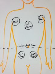
Draw where the groups of major lymph nodes are on the anterior body.
|
|
|
|
Draw the hilum of the left lung
|
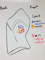
|
|
|
Draw the hilum of the right lung
|
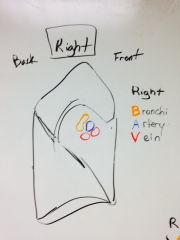
|
|
|
Will the bronchi run posterior or anterior to the vessels? How do you know this?
|
posterior because the heart sits anteriorly.
|
|
|
Where do the pulmonary arteries run in relation to the bronchi in the right vs left?
|
Right - in front
Left- above |
|
|
How do you remember this?
|
2 ways:
1. Right in front 2. Remember LAB V and know that the artery is above for the left. |
|
|
How do you remember how the lobes of the lungs sit on the back?
|
Imagine two sets of angel wings on top of each other.
|
|
|
Draw the anterior and posterior views of the left and right lung lobes
|
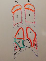
|
|
|
Draw the main structures of the heart that are seen in a cxr (include the arteries).
|
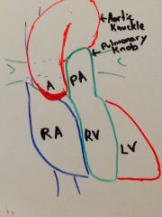
|