![]()
![]()
![]()
Use LEFT and RIGHT arrow keys to navigate between flashcards;
Use UP and DOWN arrow keys to flip the card;
H to show hint;
A reads text to speech;
25 Cards in this Set
- Front
- Back
Why do organisms need special exchange surfaces? |
- Oxygen: Aerobic respiration - Glucose: Source of Energy - Proteins: Growth and Repair - Fats: Make membranes and store energy - Water - Minerals: Water Potential, enzyme action and metabolism |
|

What makes an efficient exchange surface? |
- Large surface area to provide more space - Thin barrier to reduce the diffusion distance - Maintain a steep diffusion gradient |
|
What are some examples of specialised exchange surfaces? |
- Small intestine where nutrients are absorbed - Liver: levels of sugars in the blood adjusted - Root hairs of plants: water&minerals absorbed - Hyphae of plants where nutrients are absorbed |
|
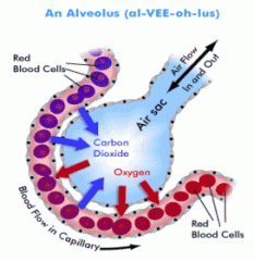
List three ways in which the lungs are adapted for efficient exchange |
- Large surface area (thousands of individual alveoli, total area=70m squared) - A barrier permeable to oxygen and carbon dioxide (thin cystoplasm) - Thin barrier to reduce diffusion distance |
|
What is surfactant? |
The lungs must produce a substance called a surfactant to reduce the cohesive forces between the water molecules. Without the surfactant the alveolus would collapse due to the cohesive forces of the water lining the air sac |
|
The process of inhaling (inspiration) |
- Diaghram contracts to become flatter, digestive organs pushed down - External intercostal muscles contract, raise ribs - Volume of chest cavity increases - Pressure in chest cavity drops below Atmospheric Pressure - Air Moves into lungs |
|
The process of exhaling (expiration) |
- Diaghram relaxes and is pushed up by displaced organs underneath - External intercostal muscles relax, ribs fall - Volume of chest cavity decreases - Pressure in lungs go above Atmospheric Pressure - Air moves out of lungs |
|
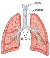
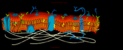
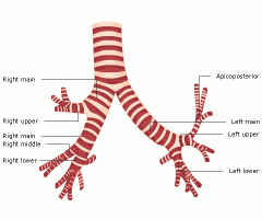
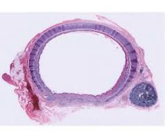
Structure of Trachea and Bronchi |
- Relatively thick walls, several layers - Cartilage as incomplete rings, less regular in bronhi - Inside cartilage is glandular tissue, connective tissue, elastic fibres, smooth fiber and vessels - Inerlining is ciliated epithelium
|
|
Structure of the Bronchioles |
- Much narrower than the bronchi - Larger bronchioles may have some cartilage but small ones have none - The wall is mostly made up of smooth muscle and elastic fibres - The smalles bronchioles have alveoli |
|
What is the role of Cartilage? |
- Structural role - Supports trachea and bronchi stay open - This prevents collapsing when pressure is low - Doesn't form complete rings for flexibility - This allows you to move your neck - Also allows oesophagus to expand when swallowing
|
|
What is the role of smooth muscle? |
- They can contract - This constricts the airway - The lumen of the airway gets narrower - This can restrict the flow of air to and from the alveoli - Not a voluntary act - Constricted bronchioles is a cause of asthma
|
|
What is the role of elastic fibers? |
- When smooth muscle contracts, the diameter of the lumen reduces - The smooth muscle cannot reverse this - Airway constriction deforms elastic fibers - As smooth muscle relaxes elastic fibers recoil - This helps dilate (widen) the airway |
|
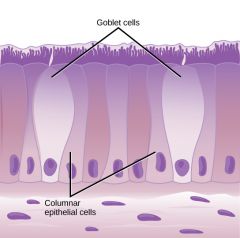
What is the role of the Goblet cells and Glandular tissue? |
- They lie under epithelium and secrete mucus - The mucus traps tiny particles in the air - These particles may contain pollen or bacteria - Trapping the bacteria means it can be removed and will reduce the risk of infection |
|
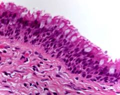
What is the role of ciliated epithelium? |
- This consists of ciliated cells - They have tiny hair-like structures projecting from their membrane - These are the cilia - They move in a synchronised pattern to waft mucus up the airway up the back of the throat - Once there the mucus is swallowed and the acidity in the stomach will kill any bacteria |
|
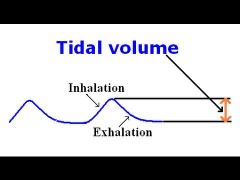
What is tidal volume? |
The volume of air moved in and out of the lungs with each breath when you are at rest. Approximately 0.5dm cubed |
|
|
What is Vital capacity? |
The largest volume of air that can be moved into and out of the lungs in one breath. Approximately 5dm cubed but can vary between genders |
|
|
What is Residual volume? |
The volume of air that always remains in the lungs even after the biggest possible exhalation. Approximately 1.5dm cubed |
|
|
What is Dead space? |
The air in the bronchioles, bronchi and trachea. There is no gas exchange between this air and the blood |
|
|
What is the Inspiratory reserve volume? |
How much more air can be breathed in over and above the normal tidal volume when you take in a big breath. You call on this reserve this when exercising |
|
|
What is the Expiratory reserve volume? |
How much more can be breathed out over and above the amount that is breathed in a tidal volume breath |
|
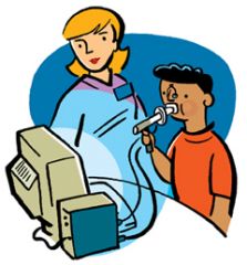
How does a Spirometer works? |
- A person breathes from a disposable mouthpiece attached to a tube connected to a chamber of medical grade oxygen - Breathing in takes oxygen from the chamber which then sinks down - Breathing out pushes air into the chamber which then floats up - Movements recorded using datalogger |
|
How is the intake of dangerous amounts of carbon dioxide prevented when using a spirometer? |
Soda lime is used to absorb the carbon dioxide that is exhaled |
|
Blood |
Blood is held in the heart and blood vessels |
|
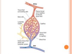
Tissue Fluid |
Tissue fluid bathes the cells of individual tissues |
|

Lymph |
Lymph is held within the lymphatic system |