Reading...
![]()
Play button
![]()
Play button
![]()
Use LEFT and RIGHT arrow keys to navigate between flashcards;
Use UP and DOWN arrow keys to flip the card;
H to show hint;
A reads text to speech;
98 Cards in this Set
- Front
- Back
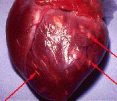
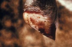
What disease can cause these lesions in calves?
|
Foot and mouth disease, apthovirus, tiger heart lesions; may also appear as gray ib color and round foci
|
|
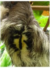
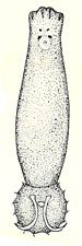
a. name the genus of this animal
b. what sex is this animal |
a. Bradypus spp. three-toed sloth
b. male d/t coloration, females don't have as much color |
|
Name the foot conformation
|
Anisodactylic
|
|
Name the foot conformation
|
Zygodactylie
|
|
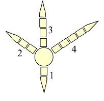
Name the foot conformation
|
Tridactyle
|
|

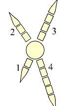
Name the foot conformation
|
Didactyle
|
|
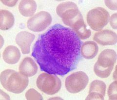
1. What type of cell is this?
2. In what two species can it be found? |
1. Kurloff cell
2. Guinea pigs and capybaras |
|
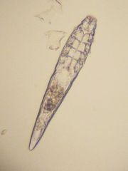
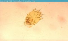
Name the etiologic agent
|
Sarcoptes scabei
|
|
What disease is most likely in this animal.
|
Seal Pox
|
|
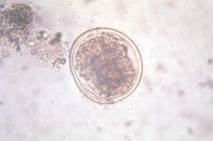
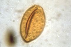
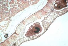
Name the organism.
|
Anisakis egg
|
|
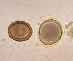
Name the organism in the left and the one in the right.
|
Baylisasrcaris procyonis on Left
Toxocara cani on right |
|
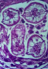
Name the organism seen in this slide.
|
Baylisascaris procyonis
(cross sections with "wing" like features) |
|

Name the organism
|
B. procyonis larvated egg
|
|
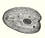
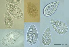
Name the organism
|
Chillodonella fish parasite
|
|

Provide the name, toxin type, and clinical signs.
|
Name: crown vetch
toxin:nitroglycoside signs:slow growth or paralysis; detoxified by ruminants |
|
Name the organism
|
Dactylogyrus
|
|
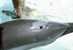
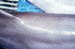
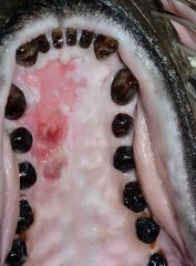
What is the most likely etiology for this lesion in a dolphin?
|
Candida
|
|
Name the most likely etiology
|
Candida
|
|
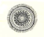
Name in what disease is this organism commonly seen in conjunction with in fish.
|
This is Epystilis which is found commonly with Aeromonas hydrophila cases.
|
|
Name the disease
|
Erysipelothrix rhusiopathiae
|
|
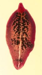
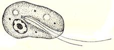
Name the organism by Genus and species
|
Fasciola hepatica
|
|
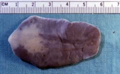
Name the genus and species
|
Fasciola magna
|
|
What caused these lesions
|
Erysipelothrix rhusiopathiae
|
|
Name the organism
|
Gyrodactylus
|
|
Name the organism
|
Ichthyobodo
|
|
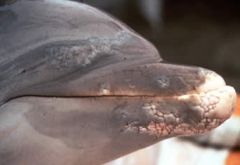
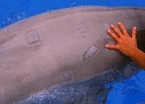
Name the likely etiology and the species it affects
|
Lacazia loboi
Dolphins and humans |
|
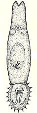
What is the organisms and where does it usually infect
|
Nasitrema, nasal sinus fluke of cetaceans
|
|

What is the structure labeled by the black line.
|
Otolith in fish
|
|

Name the organism that affects phocids
|
Otostrongylus
|
|
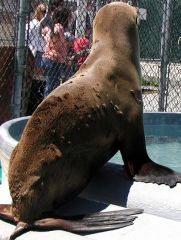
What is the most likely etiology and species affected
|
San miguel sea lion calicivirus in a California sea lion
|
|
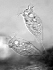
Name the organism
|
Tetrahymena
|
|

Name the organism
|
Trichinella larvae
|
|
Name the organism
|
Trichodina
|
|

Name the organism
|
Zalophotrema hepaticum, liver fluke from california sea lion
|
|
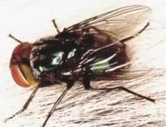
Name the genus and species
|
Cochliomyia hominivorax, New World Screw worm
|
|
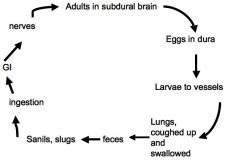
Name the organism with this cycle
|
Parelaphostrongylus tenius - meningeal worm of WTD
|
|
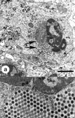
What is the disease showing inclusions in the WBC's of a chelonian
|
Iridovirus
|
|
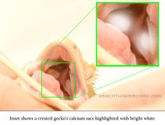
What structure?(s) is identified?
|
Calcium sacs
|
|
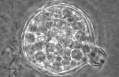
Name the genus and species of the disease.
|
Batracochytrium dendrobatidis
|
|
H & E stain of anuran skin. What is the diagnosis?
|
Batracochytrium dendrobatidis
|
|
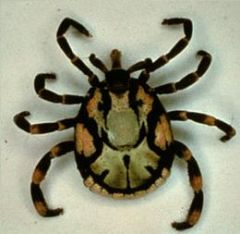
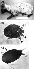
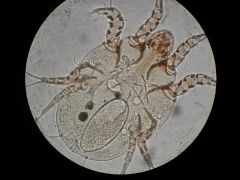
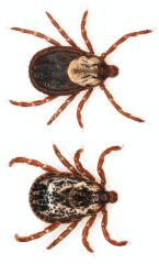
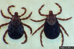
You find this tick in an animal imported to your facility. What is the genus and species of the tick and the disease of concern.
|
Amblyomma hebraeum one of the vectors for Cowdria ruminatum together w/ Amblyomma variegatum
|
|
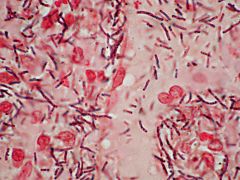
This slide was made from a blood sample obtained from the nose of a white tailed deer found dead in west Texas. What is the organism?
|
Bacillus anthracis
|
|
Give one differential for this presentation.
|
Blue tongue virus
Foot and mouth disease Vesicular stomatitis Malignant catarrhal fever |
|

This duckling presented in the summer time with flaccid paralysis. What is your top differential?
|
Clostridium botulinum
|
|
Give the top differential for a mink with flaccid paralysis.
|
Clostridium botulinum
|
|
What is the top differential for this caribou with a hygroma?
|
Brucella abortus
|
|
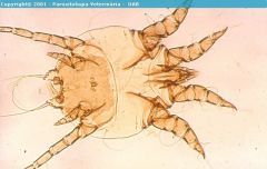
Specify the class this organism belongs to.
|
Chorioptes mite
|
|
What is the organism (Species not needed)
|
Demodex mite
|
|
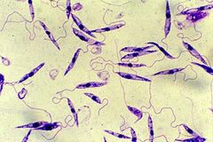
What is the organism (Genus)
|
Leishmania
|
|
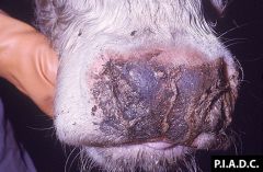
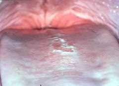
What is the top differential for this presentation in cattle?
|
Malignant catarrhal fever
|
|
|
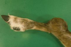
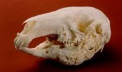
Name the genus and species.
|
Odocoielus hemionus
Mule deer |
|
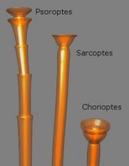
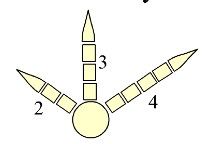
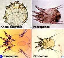
Pedicles of the different mites
|
Mites
|
|
Name the class/type of organism.
|
Psoroptes mite
|
|
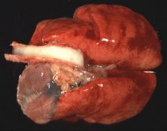
Lungs from Oryctolagus cuniculus. What is the top differential.
|
Rabbit hemorrhagic disease-calicivirus
|
|
Name the type of organism.
|
Sarcoptes mite
|
|
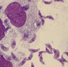
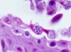
Aspirate of tissue cyst from a small rodent. What is the diagnosis?
|
Toxoplasma gondii
|
|
Mites
|
mites
|
|
Give the genus and species.
|
Dermacentor variabilis
American dog tick or wood tick |
|
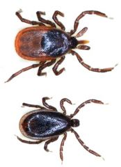
Give the genus and species.
|
Ixodes scapularis
Blacklegged or deer tick |
|
Give the genus and species
|
Rhipicephalus sanguineus
Brown dog tick |
|
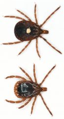
Give the genus and species.
|
Amblyomma americanum
Lone star tick |
|
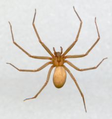
Give the genus and species or common name. Venomous or not?
|
Loxosceles reclusa
Brown recluse spider venomous |
|

What is this condition?
|
Corneal lipidosis.
|
|

Slide from box turtle. What is the diagnosis?
|
Iridovirus
inclusions in WBC's |
|
What is the diagnosis?
|
Ranavirus
|
|

What is the diagnosis and proposed etiology.
|
Spindly leg syndrome.
Hypovitaminosis B but has not been proven. Other etiologies such as genetics, incubation, etc.. are also proposed. Likely multifactorial. |
|
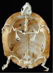
Name A, B, and C in the skeleton of a turtle.
|
A. scapula
B. acromion C. coracoid |
|
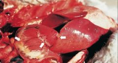
Necropsy of a coot that died suddenly after capture. There was a mass mortality in the area. The bird was in good body condition. What is the top differential.
|
Pasteurella multocida - Avian cholera
Heart petechiation and liver lesions are hallmarks together with the sudden death of a bird in good body condition. Manual Wild Dz Ch7 |
|
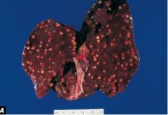
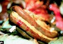
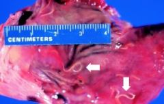
This is the liver from a duck. It palpates firm. What is your top differential.
|
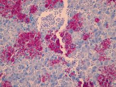
slide from the same liver
Mycobacterium avium |
|
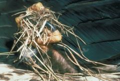
This bald eagle was found dead. Necropsy revealed a proventriculus full of large amount of ingesta. What is the most likely diagnosis?
|
organophosphosrus or carbamate toxicity
-common for birds to die acutely while struggling and hence clinched substrate on their feet and full GI Man Wild Dz Ch 39 |
|
name the type of parasite. Intestine of duck.
|
Acantocephalan
|
|
Duck intestine. What is the most likely diagnosis?
|
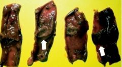
Duck plague-duck herpes virus 1
This image shows the lumen of the intestine and the appearance of the tissue beneath the bands. |
|
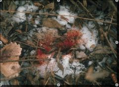
During a survey of a wildlife management area various patches like this are found after ducks fled the area.
What should be a differential diagnosis of concern? |
Duck herpes virus 1 - duck plague
It is common to see fresh blood left over in a patch where the animal was. You may also see bloody feathers on the ventrum. Man Wild Dz |
|

House finch. What is the most likely diagnosis?
|
Mycoplasma gallisepticum
|
|
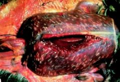
Goose liver. What causes these characteristic lesions?
|
Sracocystis
|
|
Heart of swan. What is the organism/diagnosis?
|
Heart worms - Sarconema eurycera
|
|

Give the genus and species of the organism.
|
Syngamus trachea -gape worm or tracheal worm
|
|
Name the organism.
|
Trichomonas
T. gallinae in most birds |
|
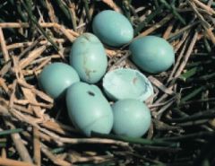
What type of chemical compound is most likely to cause this problem.
|
organochlorines - chlorinated hydrocarbons such as DDT, DDE cause thin egg shells that collapse or brake easily during incubation
|
|
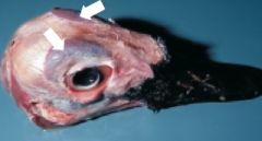
What organ/structure is pointed by the arrows in this head of a waterfowl.
|
Salt glands
|
|
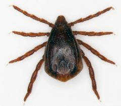
In what species is this parasite more important?
|
Alces alces - moose. This is Dermacentor albipictus which causes severe winter anemia in moose. looks similar to Rhpicephalus sanguinesu but has more light colored clear legs
|
|
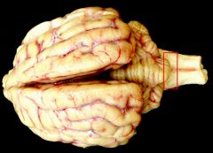
Name this section of the brain and what disease is diagnosed from testing of this site?
|
This is the obex (dorsal to medulla oblongata) and must be collected for diagnosis of chronic wasting disease/ TSE.
|
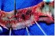
|
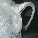
Adult, female Asian elephant. What does this image indicate?
|
Impending parturition. Bulge under the tail created by the limbs of the calf move over the hip girdle into the vestibule.
http://www.elephanttag.org/ |
|
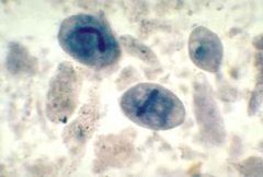
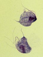
Fecal sample form a young gorilla recently introduced into a group. What is the diagnosis?
|
Balantidium coli
these are trophozoites which are more likely to be found in watery feces vs cyst found in more formed feces |
|
Gill biopsy. What is the diagnosis?
|
Ichthyophirius multifiliis
note the large macronucleus |
|

Egg in the urine of a wolf with hematuria.
What is the diagnosis? What is the typical location of the adult? What are the IH's? |
Dioctophyma renale = giant kidney worm
Live sin the right kidney. Annelids, fish, amphibians, inverts are IH. |
|
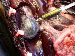
This is a necropsy of Zalophus californianus that presented with abdominal discomfort and hind limb paresis. Necropsy also revealed a full urinary bladder. What is the most likely diagnosis?
|
Leptospira interrogans pomona
Leptopsirosis |
|
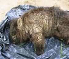
Name the etiology for this presentation in a Vombatus ursinus:
|
Wombat with Sarcoptes scabie wombati
|
|

Name the disease and the genus and species of the animal affected.
|
Devil facial tumor
Sarcophilus harrisii |
|
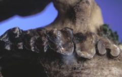
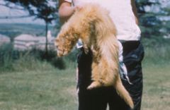
Name the Genus and species of the animal. Is it young, middle age or elderly?
|
Koala - Phascolarctos cinereus
Elderly animal due to teeth degradation which is their ultimate demise. |
|
Name two bacteria commonly implicated in this presentation in marsupials and ruminants.
|
Fusobacterium necrophorum
Bacteroides nodosus |
|

7. On necropsy, the following image is noted involving the serosa of the intestinal tract of a snowy egret. What parasite is likely involved? List intermediate, parentenic, definitive hosts.
|
Eustrongylides tubifex, E. ignotus, and E. excisus. oligochaete worms-1st IM, small fish—2nd IM, larger fish/reptiles/amphibians—parantenic, birds—definitive
|
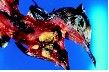
|
Give the scientific names of the two likely parasites that could represent those shown below, and what types of birds each species would be most likely to occur in:
|
Cyathostoma bronchialis—Anseriformes and Syngamus trachea—land birds
|
|
8. A client was out hunting with her Cooper’s hawk, which caught a mourning dove. After taking the mourning dove from her hawk, she noted the lesions below. She said she has fed several other doves with similar lesions to her hawk before, but has become concerned, as he seems to need greater amounts of food to keep him at hunting weight than usual. What is likely going on in this dove (4 rule outs), how would you definitively diagnose, is it a concern for her hawk, how would you treat it if it is?
|
a. Trichomonas gallinae, poxvirus, candidiasis, capillariasis, vitamin A def
b. direct smear of lesions, gram’s stain, and histopathology c. yes if trichomoniasis (maybe Capillariasis?) d. carnidazole, metronidazole |
|
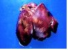
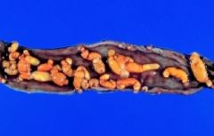
9. A client who fancies himself a gamebird aficionado contacts you because he has lost several wild turkeys (some of which had passed sulfur yellow feces). His bobwhite quail and Hungarian partridges are fluffed up and not eating as well. His recently added ring-necked pheasants are fine. On necropsy you find the following gross lesions involving the liver and cecal plug. What is your tentative diagnosis? What should he do to treat the sick birds and also manage the flock long term?
|

Histomoniasis is capable of causing catastrophic losses in the wild turkey. Histomonas meleagridis, utilizes cecal worm, Heterakis gallinarum (nematode), as a vector for entry into the bird hosts. The disease commonly called blackhead. Earthworms feed on fecal-contaminated soil that contains cecal worm eggs infected with histomonads. The cecal worm larvae and histomonads are stored in the body of the earthworm and are transmitted. However, earthworms are not required for the life cycle; cecal worm larvae that contain histomonads may be ingested. Turkey, grouse, chicken, and partridge develop severe disease and suffer high mortality rates. Disease is less severe in Hungarian partridge and bobwhite quail. In contrast, pheasant and some other species often do not exhibit signs of disease, but they instead become carriers that maintain the disease cycle. Wild turkeys affected with this disease often are listless, have an unthrifty appearance of ruffled feathers, and stand with drooped wings. The birds may appear depressed, and their feces are often sulfur-yellow in color. This fecal coloration generally occurs early in the disease and, combined with other field signs, it is highly suggestive of histomoniasis. The primary gross lesions seen upon necropsy of infected birds are numerous large, pale grey, discrete circular crater-like areas of necrosis or tissue death within the liver and thickened caecal walls that often also become ulcerated and hemorrhagic. The lumen of the ceca may also be obstructed by aggregations of yellowish necrotic debris referred to as cecal cores. Elimination of pheasant carriers would be important and removal of top soil. Because chickens are often carriers of H. gallinarum (cecal worms), and often shed histomonads, spreading uncomposted chicken manure onto fields can distribute cecal worm eggs to wild and susceptible species. So do not do that!
|
|
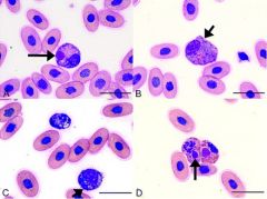
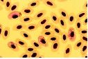
Name the hemoparasite, list a vector of that parasite, and describe populations with document morbidity and mortality associated with it.
|
a. Stained blood smear from an apapane infected with Plasmodium relictum. Some red blood cells contain multinucleated, asexually-reproducing stages of the parasite called schizonts. These are diagnostic for Plasmodium infections and contain one or more centrally-located pigment granules. Hawaiian birds.
|
|
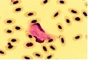
Name the hemoparasite, list a vector of that parasite, and describe populations with document morbidity and mortality associated with it.
|
b. Stained blood smear from a turkey infected with Leucocytozoon smithi. This parasite causes enlargement and distortion of the infected blood cell. The red blood cell nucleus is divided in two halves that lay on either side of the parasite. Membrane of infected cell stretched into 2 hornlike points. Ducks & geese.
|
|
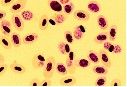
Name the hemoparasite, list a vector of that parasite, and describe populations with document morbidity and mortality associated with it.
|
c. Stained blood smear from a turkey infected with Haemoproteus meleagridis. Gametocytes (G) contain a single pink-staining nucleus and contain black or golden brown pigment granules. Generally considered nonpathogenic.
|