![]()
![]()
![]()
Use LEFT and RIGHT arrow keys to navigate between flashcards;
Use UP and DOWN arrow keys to flip the card;
H to show hint;
A reads text to speech;
135 Cards in this Set
- Front
- Back
|
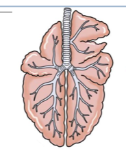
Anatomically and physiologically, the respiratory tract is divided into 3 independent, but continuous systems... |
1. Conducting system 2. Transitional system 3. Exchange system |
|
|
Describe where the conducting system is and what cells are in it |
Nasal cavity, sinuses, larynx, trachea, and bronchi Lined by ciliated epithelium and goblet cells |
|
|
Describe where the transitional system is and what cells are in it |
Bronchioles Lined by clara cells, non-ciliated secretory cells, and only a few ciliated cells. Healthy bronchioles do not have goblet cells. Clara cells produce surfactant which helps to keep the airways open. They also act as stem cells for the respiratory tract |
|
|
Describe where the exchange system is and what cells are in it |
Alveoli Lined by Type l (membranous) and Type ll (granular) pneumocytes |
|
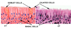
Explain what each of these cells does |
Goblet cells make mucus Ciliate cells distribute the mucus |
|
|
4 roles of Clara cells |
Detoxification Surfactant Innate immunity (collectins) Act as stem cells |
|
|
3 portals of entry for pathogens that target the respiratory system |
Aerogenous Hematogenous Direct extension |
|
|
3 types of macrophages that perform phagocytosis |
Pulmonary alveolar macrophages Pulmonary intravascular macrophages Non-specific (endogenous) |
|
|
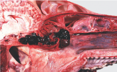
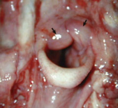
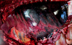
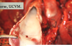
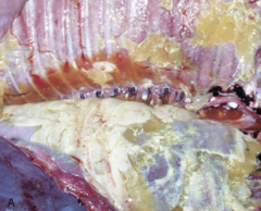
ethmoidal hematoma - causes epistaxis in horses A pendunculated, tumor like lesion in older horses. Etiology unknown. Chronic unilateral epistaxis |
|
|
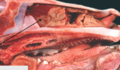
Fibrino-necrotic rhinitis. Calf with IBR (Bovine herpesvirus 1) |
|
|
Granulomatous rhinitis due to Rhinosporidium seeberi. Also infects people (gross mouthed man) |
|
|
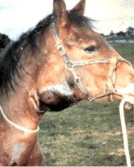
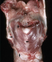
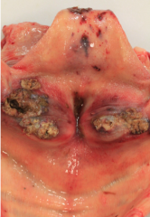
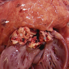
Equine strangles due to Streptococcus equi - causes suppurative rhinitis and lymphadenitis of the retropharyngeal and submandibular lymph nodes - mostly in foals and young horses |
|
|
Name 3 of the possible 5 other strangles sequelae |
Bastard strangles - disseminated abscesses Roaring (laryngeal hemiplegia) Horner's Syndrome - compresses sympathetic nerves Purpura hemorrhagica - antigen/antibody complexes Fistulation and cellulitis |
|
disease? synergistic with? gross pathology? |
Infectious Bovine Rhinotracheitis (IBR) - synergistic with Mannheimia haemolytica in causing pneumonia - not limited to resp system - Fibrino-necrotic inflammation of resp epithelia |
|
|
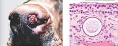
Oestrus ovis myiasis - a nasal bot in sheep with worldwide distribution |
|
pigs may grossly have a bent nose |
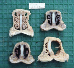
Porcine atrophic rhinitis - combined infection of Bordetella and Pasteurella - histologic lesion is osteoclastic hyperplasia and osteopenia |
|
|
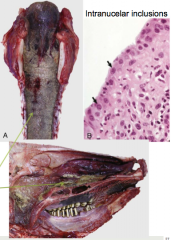
Inclusion body rhinitis of pigs - Porcine cytomegalovirus infection (herpes) - Affects pigs 3-5 weeks old causing fatal systemic infection - nasal submucosal glands will have large basophilic intranuclear inclusions |
|
|
3 causes of upper resp problems in cats |
1. Feline Viral Rhinotracheitis (FVR), FHV-1 2. Feline Calicivirus (FCR) 3. Chlamydiosis (Chlamydia felis) |
|
|
nasal carcinoma of dog |
|
|
Enzootic Nasal Carcinoma - affects sheep - oncogenic retrovirus induced! |
|
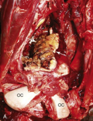
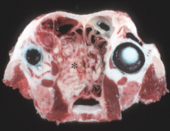
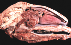
What species causes this? What neighbours should we be worried about? Can lead to what 3 other problems? |
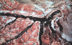
Guttural Pouch Mycosis - caused by Aspergillus - neighbours are internal carotid, CN 7, 9, 10, 11, 12, and atlanto-occipital joint - Can lead to dysphagia, Horner's syndrome, or Laryngeal hemiplegia |
|
|
guttural pouch mycosis. Stain shows Aspergillus hyphae |
|
|
Laryngeal hemiplegia - atrophy of laryngeal muscles in horses (and some dogs) - left cricoarytenoideus dorsalis and lateralis muscles affected - due to primary (idiopathic) neuropathy or secondary neuropathy (Wallerian degeneration) |
|
|
Calf diphtheria - Secondary infection by Fusobacterium necrophorum following trauma or viral infection (IBR, VS) |
|
|
Necrotic laryngitis in a bison due to Mycoplasma bovis and F. necrophorum infection |
|
|
Laryngeal edema in a bovine - causes dypnea because of "rima glottidis" (occluded larynx) - caused by acute inflammation, edema disease (pigs), purpura hemorrhagica (horses), AIP (cattle), anaphylaxis, or irritation - Idiopathic version causes tracheal edema and hemorrhage. Called "honker" syndrome in feedlot cattle |
|
|
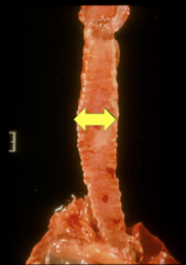
Tracheal collapse - mainly in toy and mini dogs. Sometimes seen in horses, cattle, goats - congenital or acquired - dorso-ventral flattening of trachea |
|
|
What bacteria causes Canine Infectious Tracheobronchitis (Kennel cough) What are the lesions? |
Bordetella bronchiseptica - rhinitis, conjunctivitis, tonsilitis, lymphadenitis, tracheitis, bronchitis |
|
|
Oslerus osleri in canids - nematode - causes foreign body reaction (granulomatous inflammation) |
|
|
Atelectasis vs emphysema |
Atelectasis = collapsed air spaces Emphysema = over-inflated airspaces |
|
|
Atelectasis |
|
|
Emphysema is always secondary to what? |
Airway obstruction! |
|
|
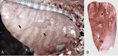
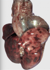
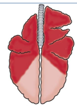
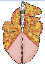
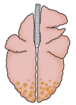
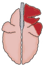
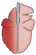
What is EIPH? |
Exercise induced pulmonary hemorrhage - follows strenuous exercise in horses - marked pressures in the lungs causes hemorrhage in the dorso-caudal portion of caudal lobes |
|
|
EIPH |
|
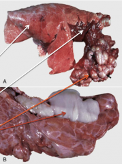
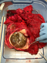
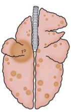
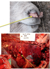
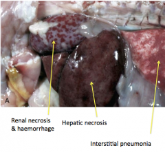
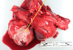
what would cause this in cattle? |
generally due to grain overload. Abscesses build in the liver which shower the vena cava. Causes epistaxis and pulmonary hemorrhage. (PVCT at FHMS) |
|
pathogenesis of pulmonary edema? |
1. increased hydrostatic pressure or decreased osmotic pressure (cardiogenic edema) 2. increased vascular permeability causes injury to the blood-air barrier 3. obstruction to lymphatic drainage (neoplasia) |
|
|
severe interstitial pulmonary edema |
|
|
What is ARDS? |
Acute Respiratory Distress Syndrome - aka "shock lung" - etiology: sepsis, toxemia, aspiration of gastric contents, trauma, pancreatitis, or severe burns - cytokine mediated reaction causes acute alveolitis lesion. This creates pneumocyte necrosis and endothelial damage/leakage. The fibrin, surfactant, and debris in the alveoli involved form hyaline membranes. |
|
|
Explain 5 sources of pulmonary emboli |
- Parasites in blood vessels (Dirofilaria immitis or Angiostrongylus vasorum) - Tumours (metastatic sarcomas) - Septic (liver abscesses or right sided endocarditis) - Fat (bone fractures) - Deep vein thrombosis (downer cows, anesthesia) |
|
|
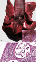
Bronchiectasis |
dilation by exudate and inflammatory destruction of bronchi walls - where a bronchiole gets so dilated with exudate that it completely blocks off - due to chronic injury |
|
|
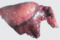
Bronchiectasis - calf with bronchopneumonia (see upstairs/downstairs) |
|
|
How do alveoli respond to injury? |
Type l pneumocytes are highly susceptible to damage. Type ll cells with proliferate and recover the basement membrane with cuboidal cells. They are more resistant to pathogens than type l. Called type ll pneumocyte hyperplasia or epithelialization. Chronic injury will show proliferation of fibroblasts and myofibroblasts |
|
|
Fibrinous bronchopneumonia - caused by Mannheimia haemolytica |
|
|
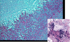
interstitial pneumonia in a pig |
|
|
embolic pneumonia |
|
|
Granulomatous pneumonia |
|
|
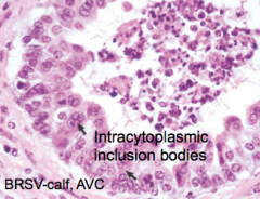
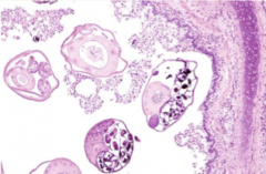
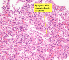
Bovine Respiratory Syncytial Virus (BRSV) - forms eosinophilic intracytoplasmic inclusion bodies in airways. Forms syncytial cells |
|
|
Chronic suppurative bronchopneumonia (upstairs/downstairs) - Primarily caused by Mycoplasma, Chlamydophila, or Ureaplasmas - Followed by opportunistic bacteria: Pasteurella, Arcanobacterium, Histophilus, Mannheimia |
|
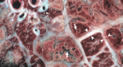
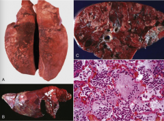
What pathogen caused this? What cells will you see histologically?
|
Marbling appearance typical of Mannheimia haemolytica (pneumonic mannheimiosis). Will see "oat cells" histologically, which are elongated degenerating neutrophils |
|
|
Mycoplasma bovis |
|
|
Mycoplasma bovis |
|
|
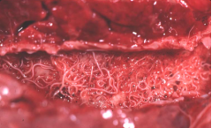
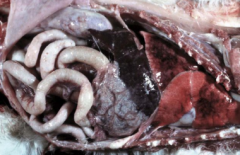
Dictyocaulus viviparus |
|
|
AIP is characterized by the presence of what 5 things |
Edema Interstitial emphysema Hyaline membranes Type ll pneumocyte hyperplasia Interstitial fibrosis with cellular infiltrates |
|
|
5 common syndromes of AIP |
1. Fog fever 2. Extrinsic Allergic alveolitis (hypersensitivity) 3. Reinfection syndrome (Dictyocaulus or BRSV) 4. Milk allergy (type l hypersensitivity) 5. ingestion of moldy sweet potatoes |
|
|
Explain the pathogenesis of Fog Fever |
Cattle grazing lush pasture ingest L-tryptophan. This is metabolized to 3-methylindole in the rumen and absorbed into the blood. It goes to the lungs and is metabolized by Clara cells into a highly pneumotoxic compound that causes extensive necrosis of bronchiolar epithelial cells and type l pneumocytes |
|

|
Bovine tuberculosis caused by Mycobacterium bovis |
|
|
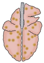
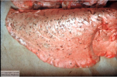
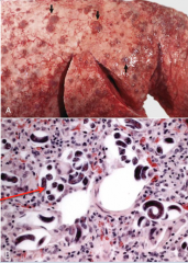
Ascaris suum. Will also cause milk spots on liver. Interstitial parasitic pneumonia |
|
|
Hydatid cysts |
|
|
Rhodococcus equi - Intracellular gram positive bacteria that causes problems in the intestines and bronchopneumonia |
|
|
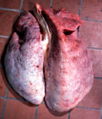
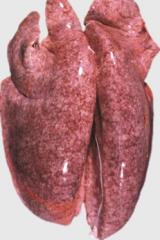
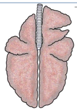
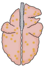
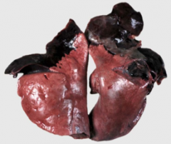
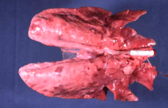
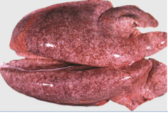
normal lung |
|
|
suppurative bronchopneumonia (enzootic pneumonia). Bacterial |
|
|
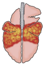
fibrinous bronchopneumonia (shipping fever). Bacterial |
|
|
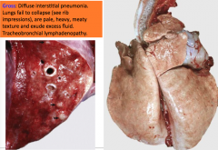
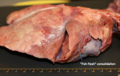
Interstitial (diffuse) pneumonia. Viral influenza |
|
|
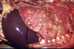
Embolic pneumonia (bacterial endocarditis) |
|
|
Granulomatous pneumonia (tuberculosis). Bacterial |
|
|
Tumour metastases from a nonpulmonary primary site (mammary carcinoma) |
|
|
Primary lung tumor with secondary metastases |
|
|
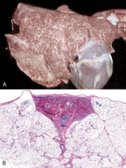
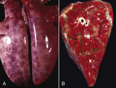
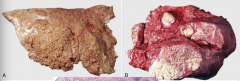
Locally extensive dorsal-diaphragmatic pneumonia (porcine fibrinous pleuropneumonia). Bacterial |
|
|
Veriminous (parasitic) pneumonia. (lung worms) |
|
|
Aspiration pneumonia (improper stomach tubing) |
|
|
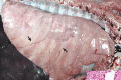
Hypostatic congestion (prolonged recumbency) |
|
|
Bibersteinia trehalosi |
|
|
Maedi/Ovine Progressive Pneumonia (OPP) - caused by retrovirus - may also cause encephalitis - lifelong persistent disease |
|
|
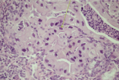
Whats the histological distinction between OPP and CAE? |
With CAE the alveoli are filled with proteinaceous eosinophilic material like surfactant. Type ll proliferation is a feature of CAE but not OPP |
|
This is in a sheep. What was the bacteria? what is the disease? What other bacteria are etiologic agents? |
Chronic enzootic pneumonia due to Pasteurella multocida.
- Multifactorial disease that is rarely fatal. Affects animals under a year old. Combination of poor husbandry, stress, and many bacterial agents at work. - Etiologic agents include Mannheimia, PI-3, Pasteurella, adenovirus, reovirus, RSV, Chlamydophila, and Mycoplasma ovipneumoniae. |
|
|
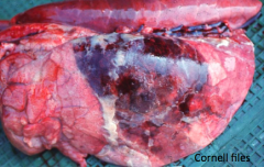
Acute fibrinous bronchopneumonia |
|
|
4 clinical presentations of Mannheimosis (Mannheimia haemolytica) in sheep |
1. ovine pneumonic mannheimiosis (similar to shipping fever in cattle) 2. septicemia in young lambs 3. ovine enzootic pneumonia 4. gangrenous mastitis |
|
|
What agent causes Septicemic Pasteurellosis in animals under 2 months old? What about in lambs over 5 months old? |
Under 2 months = Mannheimia haemolytica (biotype A) Over 5 months = Bibersteinia (Pasteurella) trehalosi |
|
|
What are the lesions seen with Septicemic Pasteurellosis? |
Basically think of lesions that arise from septicemia with severe pulmonary involvement: - necrotizing pharyngitis and tonsilitis - septicemia with disseminated intravascular thrombosis and bacteremia - severe pulmonary congestion with edema and hemorrhage |
|

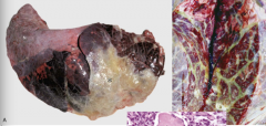
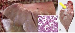
What did this big horn sheep have? |
Bibersteinia trehalosi (Justin Bieber loves big horn sheep) |
|
|
Bibersteinia trehalosi |
|
|
Which two bacteria cause tuberculosis in ovids? |
Mycobacterium bovis Mycobacterium avium NOT MYCOPLASMA. Mycoplasma does not cause TB (MycobacTerium = TB). If it doesn't have a t in it, it didn't cause TB. |
|
|
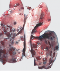
Parasitic-caused multifocal sub-pleural granulomatous pneumonia due to nodular lungworm (Muellerius capillaris) |
|
|
Similar to Dictyocaulus filaria causing lungworm in cattle, which species causes it in sheep? |
Dictyocaulus filaria |
|
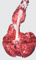
what is it? Describe the lesions in sheep vs goats |
Muellerius capillaris (sheep lung) - Will see dorso caudal distribution of nodules in sheep - In goats the distribution is diffuse interstitial pneumonia (looks like CAE) |
|
|
What type of virus causes PRRS (porcine reproductive and respiratory syndrome) |
arterivirus (which causes immunosuppression) |
|
|
What is the causative agent of PMWS (post-weaning multisystemic wasting syndrome) |
circovirus (PCV-2) |
|
|
interstitial pneumonia in pig - highly suggestive of a viral cause |
|
|
Pig. Enzootic pneumonia due to Mycoplasma hypopneumoniae |
|
|
Porcine enzootic pneumonia: Mycoplasma hypopneumoniae |
|
|
Fibrinous pleuritis and peritonitis caused by Glasser's disease (Haemophilus parasuis) - Haemophilus is similar to Histophilus in cattle in that it likes to affect many systems |
|
|
Porcine contagious pleuropneumonia agent: Actinobacillus pleuropneumoniae |
|
|
Describe the disease Porcine Contagious Pleuropneumonia |
Highly contagious often fatal fibrinous bronchopneumonia of pigs 2-5 months old. Dorsal area of the caudal lung lobes are often affected. Lesions are severe with hemorrhage, necrosis, and thrombosis. - Except for the distribution, it looks similar to lesions from Mannheimia in cattle |
|
|
Porcine contagious pleuropneumonia |
|
What is this? What pathogen caused it? What problem can it cause in the lungs? |
Vegetative valvular endocarditis. Streptococcus suis. Embolic pneumonia can occur if pieces of it break off |
|
|
Ascaris suum - creates edema and sub-pleural hemorrhage Remember to look for milk spots on the liver |
|
|
Metastrongylus - will create nodules in the caudal lung lobe and adults will be in the bronchi - will see lobular atelectasis and catarrhal bronchitis |
|
|
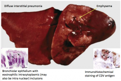
Canine distemper virus |
|
name 2 diseases which you could see this on histology |
CDV BRSV |
|
Cause? Where does it lie dormant? |
CHV-1 Virus is dormant in the trigeminal ganglion |
|
|
2 types of Mycotic pneumonia |
1. Opportunists (ex. Aspergillus fumigatus) 2. Systemic (deep) mycoses (ex. Blastomyces, Histoplasma, Coccidioides, Cryptococcus) |
|
|
Blastomycosis - pyogranulomatous pneumonia (multifocal to coalescing) - seen in Atlantic Canada and Great Lakes region |
|
|
3 protozoal parasites that can cause pneumonia in dogs |
- Toxoplasma gondii (cat is definitive host) - Sarcocystis canis - Pneumocystis carnii |
|
|
Name 3 of the possible 5 Helminths that can cause pneumonia in dogs |
- Filaroides hirthi - Crenosoma vulpis - Eucoleus aerophila - Paragonimus kellicotti - A. vasorum - D. immitis |
|
What organ failure causes this? What's it called? How will the lungs present? |
Uremic pneumonopathy - due to chronic renal failure - pulmonary edema and calcified vascular smooth muscle and basement membranes in alveoli. The lungs will fail to collapse, they'll feel gritty or brittle, and are pale, heavy, and edematous. |
|
Type and cause of this cats pneumonia |
Fibrinopurulent bronchopneumonia due to P. multocida |
|
|
What is the most common fungal cause of mycotic pneumonia in cats? |
Cryptococcus neoformans or gatti |
|
|
Name 2 of the 4 parasites that can cause pneumonia in cats |
- Toxoplasma gondii - Dirofilaria immitis - Aelurostrongylus abstrusus - Paragonimus kellicotti |
|
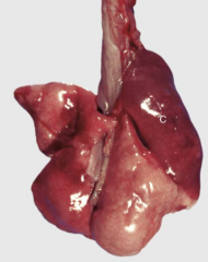
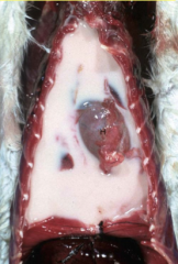
What caused this rabbit to have rhinitis/sniffles? |
Pasteurellosis (P. multocida) |
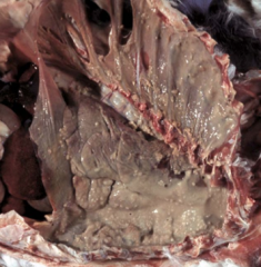
|
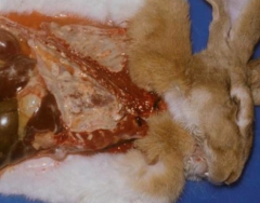
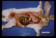
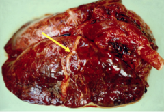
What can you see here that is a common presentation with Pasteurellosis? |
Fibrinosuppurative pleuropneumonia |
|
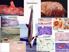
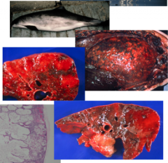
Harbour seal lung |
Phocine distemper virus (PDV). A morbillivirus pneumonia |
|
|
Cetacean morbillivirus (CeMV) |
|
|
Cryptococcal pneumonia due to C. neoformans gatti |
|
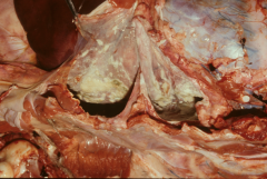
What did this emu have? |
Granulomatous airsacculitis due to Aspergillus |
|
|
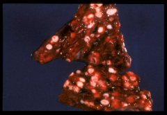
emu again. Granulomatous pneumonia due to Aspergillus |
|
|
Are primary pulmonary neoplasias common in domestic animals? |
no. The majority are malignant and appear as solitary masses. Carcinomas are more common |
|
|
What is a common way for lungs to get neoplasia? |
Via metastases (ex. osteosarcomas, melanoma, mammary adenocarcinomas) |
|
|
Marie's disease!!! (ties in well with MSK) |
|
How can you easily diagnose this disease in sheep? |
Ovine pulmonary adenocarcinoma (Jaagsiekte) - easy diagnosis is the wheelbarrow test. Lift up the sheep by its hind legs. If fluid comes out the nose then it's probably Jaagsiekte. |
|
|
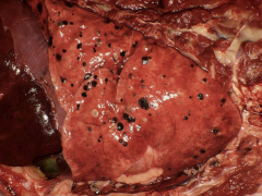
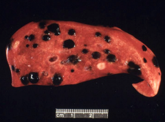
Hemangiosarcoma metastases |
|
|
Thyroid carcinoma |
|
|
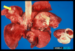
Metastatic mammary carcinoma with pulmonary and renal involvement. Note the umbilicated nodule |
|
|
Melanoma that metastasized to the lungs from the oral cavity |
|
|
oral melanoma again |
|
|
2 causes of pleural calcification |
uremia vitamin D toxicity |
|
|
2 types of pneumothorax |
Spontaneous (no apparent trauma or disease) Secondary (trauma, emphysema, parasite cysts) |
|
|
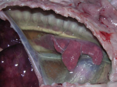
hydrothorax causing pulmonary atelectasis in a pig |
|
|
hydrothorax secondary to hepatic cirrhosis |
|
|
hemothorax (this case due to aortic rupture) |
|
What is this? Name 3 of the 6 possible causes |
chylothorax (lymph fluid) causes: neoplasia, trauma, anomalies, mycotic infection, parasites, iatrogenic (surgery) |
|
What is this? Name 3 ways this can arise |
Pleuritis 1. penetration 2. direct extension from bronchpneumonia 3. hematogenous (Haemophilus parasuis, P. multocida, Step species, E. coli) |
|
|
What type of inflammation can result in pyothorax? |
Suppurative |
|
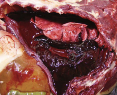
Lungs are not involved here. What is it? |
Fibrinous pleuritis (subacute and severe) |
|
|
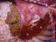
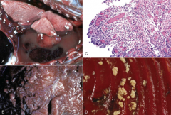
Chronic nocardiosis (sulphur granules due to Nocardia asteroides infection - bacterial) |
|
|
wet form of FIP (fibrin strands in peritoneal and pleural cavities) |