![]()
![]()
![]()
Use LEFT and RIGHT arrow keys to navigate between flashcards;
Use UP and DOWN arrow keys to flip the card;
H to show hint;
A reads text to speech;
66 Cards in this Set
- Front
- Back
|
Function of the glomerulus |
Act as a filter for the ultrafiltration of plasma
|
|
|
What is protein losing nephropathy |
Damage to the filter which results in the leakage of protein into the urine |
|
|
What are the characteristics of nephrotic syndrome |
Proteinuria Hypoalbuminaemia Hypercholesterolaemia Oedema |
|
|
What causes hypercholesterolaemia |
Increased hepatic production and defective metabolism of VLDL Pathogenesis is not well understood |
|
|
What is p:c ratio used for |
Quantify urinary protein loss in cats and dogs Does not indicate the cause of proteinuria
|
|
|
Causes of proteinuria |
Pre-glomerular or glomerular or post glomerular |
|
|
Example of preglomerular proteinuria |
Bence jones proteins in multiple myeloma |
|
|
Example of post glomerular proteinuria |
Pyelonephritis |
|
|
Example of familial renal disease |
Samoyed hereditary glomerulopathy - characterised by splitting of the BM components |
|
|
What is immune complex glomerulonephritis |
Deposition of circulating antibody - antigen complexes Followed by compliment fixation and neutrophil chemotaxis |
|
|
Causes of immune complex glomerulonephritis |
Bacterial infection Autoimmune disease Neoplasia |
|
|
What is the basis of autoimmune glomerulonephritis |
Antibodies produced against the glomerular basement membrane |
|
|
What happens when compliment is activated at the GBM |
Endothelial hypertrophy Podocyte fusion GBM thickening Inflammatory infiltration |
|
|
What is the gross pathology seen with acute GN |
Kidney is swollen and tense Individual glomeruli may be prominent at pinpoint red dots on the surface of the cortex |
|
|
What is the pathology see n with gross chronic GN |
Interstitial nephritis resemblant Shrinking and pitting of the capsule, cortical thinning and fibrosis |
|
|
Histopathology seen with gross chronic GN |
Cellularity increase in glomerular tufts Thickening of the capillary basement membrane Adhesions between the bowmans capsule and tuft Thrombi in capillaries |
|
|
What is glomerulosclerosis |
Hypocellular and nonfunctional glomerul which result from severe and prolonged glomerular damage Fibrosis of the glomeruli which may progress such that the glomeruli shrink and become hyalinised Bowmanns capsule can rupture |
|
|
How can we diagnose amyloidosis |
Immunoflourescent staining - conjugate amyloid with antibodies |
|
|
Macroscopic appearence of amyloidosis |
Swollen and pale kidneys Large with a smooth to granular capsular surface |
|
|
What are the 2 types of amyloidosis |
AL - amyloid light chain overproduction AA - chronic inflammation -acute phase proteins |
|
|
What would you see histologically with amyloidosis |
HandE - homogeneous eosinophillic distending glomerulus Congo red confirms |
|
|
Suppurative glomerulitis is also known as what |
Embolic nephritis |
|
|
What causes suppurative glomerulonephritis |
Localisation of bacteria within the glomeruli fomr bacteraemia |
|
|
What is viral glomerulonephritis |
Swelling of the glomeruli after viral attack of the endothelium |
|
|
What causes ATN |
Toxic insult to the renal tubular epithelial cells |
|
|
Why is the PCT most susceptible to injury |
High metabolic rate - susceptible to toxic injury |
|
|
How does the PCT protect itself from toxins |
P450 enzymes |
|
|
What happens if the tubular BM remains intact |
Regeneration from tubular stable cells (as long as scaffold intact) |
|
|
What happens if the BM is destroyed |
Complete loss of scaffold and loss of function Fibrosis and scarring |
|
|
What are the subtypes of ATN |
Ischaemic/Tubulorrhectic Nephrotoxic |
|
|
What causes nephrotoxic ATN |
Heavy metals Oxalates (ethylene glycol precipitation) Antibacterials |
|
|
What causes ischaemic ATN |
Hypotension Haemoglobinurea |
|
|
Tubulorrhexis |
BM damage and disruption in the tubules |
|
|
Tubulointerstitial disease |
Affecting the tubules and the interstitium |
|
|
Causes of TID in dogs |
Leptospirosis - leptospira canicola Infectious canine hepatitis virus |
|
|
Causes of TID in cattle |
E.coli septicaemia (can be incidental if not severe) "white spotted kidney" Malignant catarrhal fever |
|
|
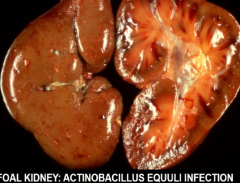
What causes TID in horses |
Equine viral arteritis Actinobacillus equuili in foal |
|
|
How does leptospira cause TID in dogd |
Localisation of bacteria in renal capillaries Migration through vascular endothelium into the renal interstitium Migration through the intercellular junctions of the renal tubular epithelial cells Tubular lumen and associated epithelial migrovilli Persistance in phagosomes Degeneration of epithelium and inflammatory reaction |
|
|
What causes granulomatous nephritis |
Chronic systemic disease sequalae e.g. FIPV |
|
|
What does granulomatous nephritis look like |
Multiple irregular cortical foci Bulge from capsular surface |
|
|
T. Canis lesions |
2-3mm granulomas Subcapsular renal cortex |
|
|
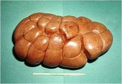
Diotophyma renale |
Giant kidney worm Renal pelvis Resulting in hydronephrosis |
|
|
Stephanurus dentatus |
Affects adult pigs in USA Encyst in perirenal fat or in kidney itself |
|
|
Pyelonephritis |
Inflammation of pelvis and parenchyma |
|
|
Pyelitis |
renal pelvis inflammation |
|
|
What causes pyelonephritis |
Ascending from lower UT or haematogenous *descending* |
|
|
Grossly seen with pyelonephritis |
Variable amounts of mucopus Medulla exhibits streaks of inflammatory debris which extend into kidney substance Bilateral Chronic deformation and scarring Can look like calculi (dryer) |
|
|
Histologically seen with pyelonephritis |
oRganisms in tubules Inflammatory reaction in interstitial tissue Degeneration and fibrosis of bowmans capsule in ascending infections Fibrosis in chronic cases |
|
|
Pyonephrosis |
Infection and obstruction (calculi in ureter) results in the kidney filling with pus |
|
|
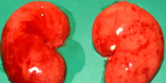
Pulpy kidney |
c perfringens type D toxin Rapid autolysis of organs such as kidney (can be hard to differentiate from PM change) widespread haemorrhage |
|
|
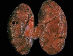
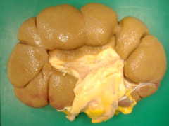
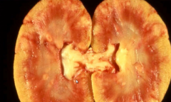
Glomerulonephritis gross |
|
|
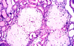
RBC Inflammatory cells and infiltrate Glomerulonephritis histo |
|
|
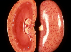
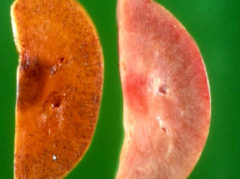
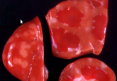
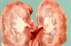
Bovine amyloid kidney Swolen, greasy, granular look like fatty change Heavy and granular Fatty would be smooth |
|
|
Iodine dip to identify presence of SAA |
|
|
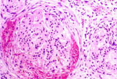
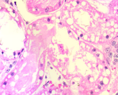
Amyloid is space occupying Doesnt elicit inflammatory response Even staining pink material is the amyloid Notice the protein in the tubulear lumen Physical presence causing ishcaemia and pressure atrophy |
|
|
Acute tubular necrosis caused by crystals - ethylene glycol |
|
|
Pathogenesis of leptospirosis |
Bacteraemia Organisms localise in the renal capillaries Interstitium MIgrate through the epithelial intercellular junctions into tubules persist - necrosis and degeneration of the tubules |
|
|
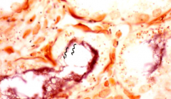
Canine leptospirosis (silver stain) |
|
|
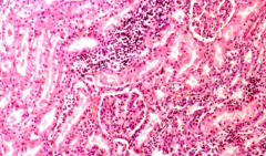
Tubulointerstitial neprhitis caused by leptospira Heavily w |
|
|
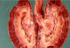
White spotted kidney sequalae to E. coli infection Chronic - previous usually incidental on pm tubulointerstitial nepritis |
|
|
Causes tubulointerstitial disease |
|
|
Granulomatous nephritis from FIP |
|
|
Suppurative pyelonephritis dog |
|
|
chronic pyelonephritis |
|
|
pyelonephrosis |
pyelonephritis and hydronephrosis |
|
|
Pulpy kidney C Perfringens type D toxin Rapid autolysis |