Reading...
![]()
Play button
![]()
Play button
![]()
Use LEFT and RIGHT arrow keys to navigate between flashcards;
Use UP and DOWN arrow keys to flip the card;
H to show hint;
A reads text to speech;
50 Cards in this Set
- Front
- Back
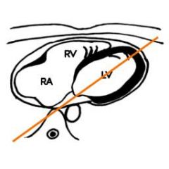
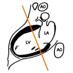
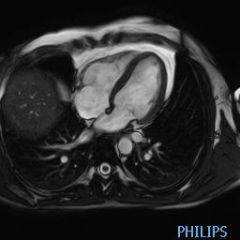
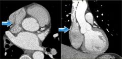
This is a transverse view. What is view does this view create.
|

Long axis view
|
|
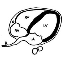
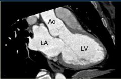
What are each of these chambers and major vessels
long axis view |

This is the long axis view. The left main pulmonary artery is below the aorta
|
|
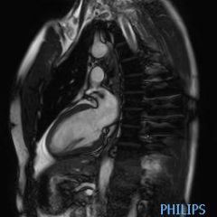
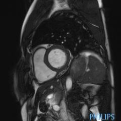
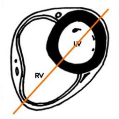
This is a long axis view. What cut does this form. This cut goes up diagnolly as it moves from the left atrium to the bottom of the pulm artery on the right
|
semi-4 chamber view
|
|
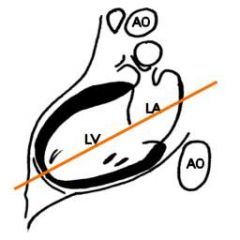
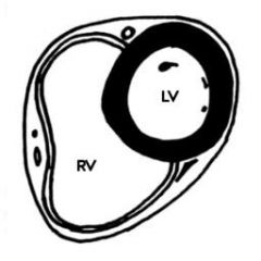
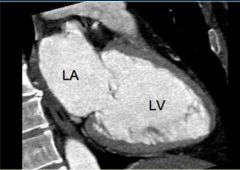
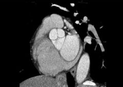
This is a long axis view. What view does this form
|

short axis view
|
|
Label this short axis view
|
|
|

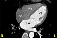
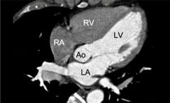
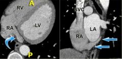
Label the chambers and major vessels of this semi 4 chambered image
|
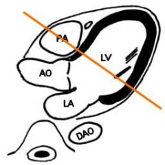
Note that the aorta loops over the pulmonary artery and connects with the descenting aorta
|
|

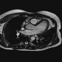
This is a short axis view. What does this cut form
|
This is a 4 chamber view (different than semi 4 chamber view)
NOTE: when attempting to get a semi 4 chamber view the line would go diagnol from the bottom of the left ventricle upwards (opposite this diagnole) so the mitral valve and the main pulm artery is seen. |
|

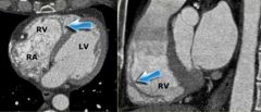
This is a 4 chamber view. What are the the chambers and vessels
|
|
|
This is the first cut to form a for chamber view what is the other
|

|
|
|
Where is the location of the right ventricle
|
anterior
|
|
|
Where is the location of the left ventricle
|
posterior
|
|
|
What does a 4 chamber view of the heart look like
|
|
|
|
What does a 3 chamber view look like
|
|
|
|
What valves are you able to see in the 4 chamber view
|
mitral
tricuspid (as long as contrast is present) |
|
|
What valves are you able to see in the 3 chamber view
|
mitral
aortic |
|
|
What does a 5 chamber view look like
|
|
|
|
How is a 5 chamber view created
|
similar to the 4-chamber view, but additionally displays the aortic valve and left ventricular outflow tract.
This view is achieved by rotating the 4-chamber view a little more cranially |
|
|
What does a 2 chamber view look like
|
|
|
|
How is a 2 chamber view created
|
rotating the images perpendicularly to the mitral valve and parallel to the cardiac septum.
This axis gives an overview of the left atrium ventricle and mitral valve. |
|
|
What is the 2 chamber view used for
|
It is a good view for analyzing ventricular function, especially that of the inferior and anterior walls.
|
|
|
What is best for obtaining functional data
|
consecutive short axes must be reconstructed making use of the 3- and 4-chamber views.
|
|
|
What vessels supply blood to the right atrium
|
the coronary sinus and superior and inferior vena cava.
|
|
|
What does the coronary sinus look like on axial CT
|
|
|
|
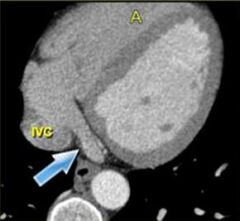
Where does the coronary sinus (carries blood back from coronary arteries) enter the right atrium
|
which enters anterior to, and just to the left of the inferior vena cava.
|
|
|
What chamber is the crista terminalis located
|
right atrium
|
|
|
What is the crista terminalis
|
In the right atrium lies the crista terminalis, a muscular ridge that runs from the entrance of the superior- to that of the inferior vena cava.
|
|
|
What does the crista terminalis seperate
|
This structure separates the smooth part of the right atrium - the sinus venosus - from the trabecularized right atrial appendage.
|
|
|
What does the crista terminalis look like on axial and coronal CT
|
|
|
|
Where does the coronary sinus run
|
It runs in the atrioventricular groove on the posterior surface of the heart and enters the right atrium in the vicinity of the tricuspid valve.
|
|
|
What does the coronary sinus look like in axial and coronal CT
|
|
|
|
the coronary sinus in the atrioventricular groove on the posterior surface of the heart.
|
|
|
|
What is the trabecularized portion of the right atrium
|
the right atrial appendage
|
|
|
What does the right atrial appendage look like on coronal and 3D reformats
|
|
|
|
What is a major difference between the mitral and tricuspid valve
|
This valve has three leaflets and three papillary muscles, which partially insert on the septum (in contrast to the papillary muscles of the mitral valve, which do not).
|
|
|
How is the right ventricle wall different then the left ventricle wall
|
The right ventricle also has a thinner wall which is more trabecularized, especially towards the apex.
|
|
|
What is the location of the moderator band
|
t runs from the septum to the lateral wall of the right ventricle, and plays a key role in the electrophysiological conduction of the right ventricle's free
|
|
|
What does the moderator band look like
|

|
|
|
What doe the moderator band look like
|
|
|
|
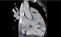
What area of the heart seperates the TV from the PV
|
the crista supraventricularis
|
|
|
How is the alignment of the valve and the right and left ventricles different
|
TV and PV seperated by crista supraventricularis which differs from the left ventricular outflow tract, where the mitral and aortic valves lie side by side
|
|
|
What does the crista supraventricularis look like
|
|
|
|
What are 4 characteristics of the RV that differ from the LV
|
moderator band
septal papillary muscle infundibulum no fibrous continuity of the AV valves |
|
|
What is the MC configuration of pulmonary veins
|
there are two pulmonary veins on the left and two on the right. (superior and inferior)
|
|
|
Where does the middle pulmonary vein (right) usually drain into
|
the superior pulmonary vein
|
|
|
What is associated with a fib
|
anomalous insertion of pulmonary vein on the right
|
|
|
Where is the left atrial appendage
|
The left atrial appendage is a finger like, trabecularized structure which originates supralaterally in the left atrium. It lies over the left atrioventricular groove, and partially covers the left coronary artery in it.
|
|
|
What is a common pitfall of the left atrial appendage
|
Its small, parallel-running muscles should not be mistaken for thrombus.
|
|
|
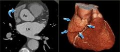
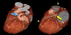
To visualize the left coronary artery what must be done to a 3D reformat
|
When assessing the coronary arteries, the left atrial appendage must be removed, so that the LCX and proximal LAD may be visualized.
|
|
|
What does a 3D reformat of the LCA look like before and after removing the left atrial appendage
|
|
|
|
What do the coronary cusp look like
|
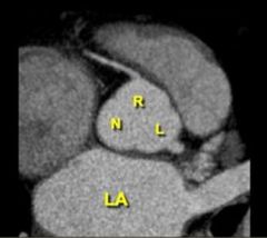
|