Reading...
![]()
Play button
![]()
Play button
![]()
Use LEFT and RIGHT arrow keys to navigate between flashcards;
Use UP and DOWN arrow keys to flip the card;
H to show hint;
A reads text to speech;
94 Cards in this Set
- Front
- Back
|
Know your fractures... Page 101 (the definition)
|
Smith, Barton's, Collie, Boxer's, Bennett's
|
|
|
A radiograph of an AP oblique elbow with medial rotation reveals that the radial head is superimposed over part of the coronoid process. What postioning error was committed?
|
Excessive medial rotation
|
|
|
What is the proper name the acute elbow flexion projection?
|
Jones
|
|
|
Which of the following best demonstrates the radial head using the trauma lateral coile method?
|
Elbow flexed 90 degrees, CR angled 45 degrees toward the shoulder.
|
|
|
With the radial head projections, what is the only difference between the four projections?
|
Position of the hand and wrist
|
|
|
What basic position of the elbow best demonstrates an elevated or visible posterior fat pad?
|
Lateral with 90 degree flexion
|
|
|
A radiograph of the elbow demonstrates the proxiaml radius directly superimposed over the ulna and the coronoid process in the profile? Which projection of the elbow has been performed?
|
Medial rotation oblique
|
|
|
How should the humeral epichondyles be positioned for a lateral projection of the elbow?
|
Perpendicular
|
|
|
Which basic projection of the elbow demonstrates the trochlear notch?
|
Lateral
|
|
|
Which basic projection of the elbow demonstrates the olecranon process in profile?
|
Lateral
|
|
|
Which basic projection of the elbow best demonstrates the radial head and tuberosity free of superimposition?
|
AP Oblique with lateral rotation.
|
|
|
What is the purpose of performing the partially flexed projection of the elbow?
|
Trauma
|
|
|
Which of the following actions leads to proximal radius crossing over the ulna?
|
Pronation of the hand
|
|
|
The nonvisible posterior fat pad on a well exposed correctly positioned lateral elbow radiograph correctly suggests...
|
Negative study of the elbow
|
|
|
The radiocarpal joint possesses a __________ type of joint movement.
|
Ellipsoidal
|
|
|
The smooth depressed center postion of a trochlea used for evaluating rotation on a lateral elbow is termed the...
|
Trochlear sulcus
|
|
|
What two bony landmarks are palpated when positioning the elbow?
|
humeral epichondyles
|
|
|
What two structures form the distal radioulnar joint?
|
Ulnar notch and head of the ulna
|
|
|
What is the name of the two small depressions found on the anterior aspect of the distal humerus?
|
Coronoid and radial fossa
|
|
|
Which two structures primarily form the hinge-like structure and movement of the elbow joint?
|
Trochlea and Olecranon process
|
|
|
Which of the following structures is considered the most distal?
|
Styloid Process
|
|
|
Which of the following structures is considered to be the most posterior?
|
Olecranon Process
|
|
|
Which of the following structures is considered to be the most lateral?
|
Capitulum
|
|
|
Which of the following structures is considered to be the most proximal?
|
Olecranon Process
|
|
|
Which of the following is not part of the ulna?
|
Ulnar notch
|
|
|
Fat pads of the elbow are normally seen on correctly postioned and correctly exposed anterior elbow projections.
|
False
|
|
|
To visualize fat pads surrounding the elbow, exposure factors must be adjusted to see both bone and soft tissue structures.
|
True
|
|
|
For the lateral projection the hand should be adjusted in the lateral position with the thumb up.
|
True
|
|
|
It's important to get the Corocoid Process in profile for the lateral view of the elbow.
|
False
|
|
|
The CR should be directed perpendicular to the forearm in a lateral projection.
|
True
|
|
|
The hand should be supinated for the lateral projection
|
False
|
|
|
The hand should be pronated for the AP projection.
|
False
|
|
|
The posterior fat pad of the elbow joint is usually not seen unless the elbow has sustained some sort of trauma.
|
True
|
|
|
The hand should be pronated for a medial oblique of the elbow.
|
True
|
|
|
The humeral epichondyles are superimposed on a lateral elbow exam.
|
True
|
|
|
When performing a lateral elbow exam, place the humeral condyles perpendicular to the film.
|
True
|
|
|
The elbow should be flexed 70 degrees to put the olecranon process in profile when doing a lateral elbow exam.
|
False
|
|
|
For a lateral forearm exam, the humeral chondyles are parallel to the IR.
|
False
|
|
|
For a lateral forearm, the radial head should be free from superimposition of the coronoid process.
|
False
|
|
|
The ulna and radius should be crossed over on an AP forearm exam.
|
False
|
|
|
The humeral epichondyles are equidistant from the IR for an AP forearm.
|
True
|
|
|
When performing and AP forearm, supinate the hand and extend the forearm.
|
True
|
|
|
When performing a forearm exam, include both joints.
|
True
|
|
|
Displacement of the superior fat pad is useful in diagnosing fractures of the radial head and neck.
|
True
|
|
|
What position should the hand be in for an AP medial rotation oblique, elbow position?
|
Pronated
|
|
|
What position should the hand be in for an AP elbow projection?
|
Supinated
|
|
|
Why should the forearm never be taken as a PA projection?
|
Crossover of radius and ulna
|
|
|
Which projection of the elbow best demonstrates the olecranon process and coronoid in profile.
|
AP medial oblique.
|
|
|
Which projection of the elbow best demonstrates the olcranon process in profile.
|
Lateral.
|
|
|
Which oblique projection of the upper limb best demostrates the radial head superimpositioned over ulna.
|
Medial oblique.
|
|
|
Which oblique projection of the upper limb best demostrates the radial head free of bony superimpositioned.
|
AP laterl oblique of the elbow.
|
|
|
With referenence to the plane of IR how should the humeral epicondral coronal plane be positioned for a lateral projection of the elbow.
|
Perpendicular.
|
|
|
Which projection of the elbow demostrates the radial head free of bony superimpositioned.
|
AP lateral oblique.
|
|
|
How much should the elbow be flexed for the lateral projection of the elbow.
|
90
|
|
|
Which projection of the forearm requires the elbow to be flexed 90 degrees.
|
Lateral.
|
|
|
Which projection of the upper limb is radial crossover a primary concern.
|
Trying to do the AP.
|
|
|
Which description best explaines how radial crossover occurs when the forearm is demonstrated...
|
AP projection with hand pronated
|
|
|
How should the hand be positioned for the AP projection and later projection of the forearm.
|
Hand supinated and lateral.
|
|
|
For AP oblique projections of the elbow how should the CR be directed.
|
Perpendicular.
|
|
|
How much medial rotation of the elbow is needed to position it for the AP oblique projection.
|
45
|
|
|
For the AP projection of the elbow how should the coronal plane through the humeral epicondyles be set?
|
Parallel
|
|
|
For the AP projection of the elbow how should the CR be directed?
|
Perpendicular.
|
|
|
For the AP projection of the forearm how should the elbow be positioned?
|
fully extended
|
|
|
When positioning for the AP projection select the a cassette long enough to include the entire forearm from the _______ of the ulna, to the __________ of the radius.
|
Olecranon process and styloid process.
|
|
|
The clinical clue to the prescence of a radial head fracture....
|
All of the above.
|
|
|
An accurately positioned lateral elbow radiograph demostrates ...
|
1- open elbow joint space
3- radial tuberosity out of profile 4-Anterior fat pad |
|
|
For a lateral forearm radiograph...
|
All of the above.
|
|
|
With reference to the plane of the IR, how should the humeral epicondral coronal plane be positioned in the AP projection of the elbow ?
|
Parallel.
|
|
|
Bartons Fracture
|
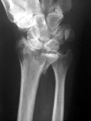
An intra-articular fracture of the distal radius with dislocation of the radiocarpal joint
|
|
|
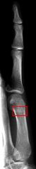
Bennets Fracture
|
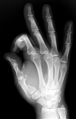
fracture at the base of the first metacarpal
|
|
|
Boxers Fracture
|
Fracture of the metacarpal neck
|
|
|
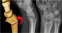
Colles Fracture
|

Fracture of the distal radius with posterior (dorsal) displacement
|
|
|
Smiths Fracture
|
Fracture of the distal radius with anterior (palmar) displacement
|
|
|
Torus or Buckle Fracture
|
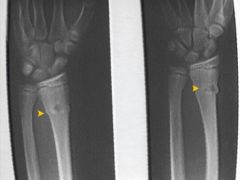
Impacted fracture with bulging of the periosteum
|