![]()
![]()
![]()
Use LEFT and RIGHT arrow keys to navigate between flashcards;
Use UP and DOWN arrow keys to flip the card;
H to show hint;
A reads text to speech;
952 Cards in this Set
- Front
- Back
|
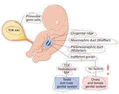
Indifferent stage of gonads (weeks) |
Weeks 4-7 |
|
|
Where do the indifferent gonads develop? |
In a longitudinal elevation or ridge of intermediate mesoderm - urogenital ridge |
|
|
Where do the primordial germ cells arise from? |
From the lining cells in the wall of the yolk sac At week 4, primordial germ cells migrate into the indifferent gonad Provide a critical inductive influence on gonad development |
|
|
Components of the indifferent gonad |
1. Primordial germ cells 2. Primary sex cords - finger-like epithelial extensions inside the gonads that are populated by migrating primordial germ cells 3. Mesonephric (Wolffian) duct - male genital truct Paramesonephric (Mullerian) duct - female genital truct |
|
|
Development of testis and ovary |
|
|
|
Factors for the development of male reproductive system (4) |
1. Sry gene - short arm of the Y chromosome (encodes testis-determining factor [TDF]) 2. Testosterone - Leydig cells 3. Mullerian-inhibiting factor (MIF) - Sertoli cells 4. Dihydrotestosterone (DHT) - external genitalia |
|
|
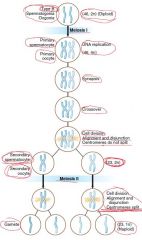
Events during meiosis I |
|
|
|
Events during meiosis II |
|
|
|
Meiosis chart |
|
|
|
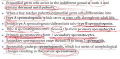
Type B spermatogonia/oogonia genetic status |
46, 2n (Diploid) |
|
|
Primary spermatocyte/oocyte genetic status |
46, 4n |
|
|
Secondary spermatocyte/oocyte genetic status |
23. 2n |
|
|
Gametes genetic status |
23, 1n (Haploid) |
|
|
Spermatogenesis |
|
|
|
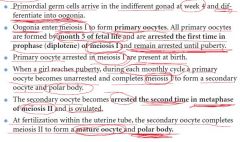
Oogenesis |
|
|
|
Week 1 of development chart |
|
|
|
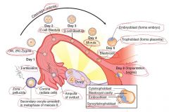
2 changes of a spermatozoa in the female genital truct |
1. Capacitation - during 7 hours in the female truct, removal of several proteins from the plasma membrane of the acrosome of the spermatozoa 2. Acrosome reaction - release of hydrolytic enzymes from the acrosome used by sperm to penetrate the zona pellucida -> cortical reaction that prevents other spermatozoa penetrating the zona pellucida |
|
|
Morula |
32-cell stage of division |
|
|
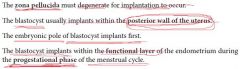
Blastocyst |
Forms as fluid develops in the morula Consists of an inner cell mass known as the embryoblast, and the outer cell mass known as the trophoblast, which becomes the placenta At the end of the first week, the trophoblast differentiates into the cytotrophoblast and syncytiotrophoblast and then implantation begins |
|
|
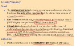
Ectopic pregnancy |
|
|
|
Risk factors for the tubal ectopic pregnancy |
-Endometriosis -Pelvic inflammatory disease (PID) -Tubular pelvic surgery -Exposure to diethylstilbestrol (DES) |
|
|
What risks in diethylstilbestrol (DES) exposure |
-clear cell carcinoma, a rare vaginal tumor in girls and women who had been exposed to this drug in utero -tubal ectopic pregnancy |
|
|
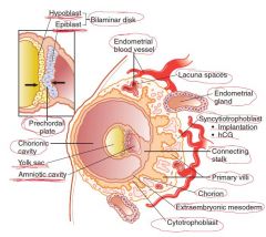
Implantation |
|
|
|
Week 2 of the development |
Formation of the bilaminar embryo |
|
|
Week 2 of development chart |
|
|
|
Into what does the embryoblast differentiates? |
Epiblast and hypoblast, forming a bilaminar embryonic disk Epiblast -> amniotic cavity Hypoblast -> primary yolk sac |
|
|
From what structure does the mouth develops? |
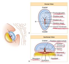
Prechordal plate (formed by fusion of epiblast and hypoblast) |
|
|
Extraembryonic mesoderm |
Is derived from the epiblast Extraembryonic somatic mesoderm lines the cytotrophoblast, forms the connecting stalk, and covers the amnion Extraembryonic visceral mesoderm covers the yolk sac |
|
|
Connecting stalk |
Suspends the conceptus within the chorionic cavity |
|
|
Chorion |
Wall of the chorionic cavity Consists of extraembryonic somatic mesoderm, cytotrophoblast, and the syncitiotrophoblast |
|
|
Is there mitosis in syncytiotrophoblast or cytotrophoblast? |
No mitosis in syncytiotrophoblast! Cytotrophoblast is mitotically active |
|
|
Locations of hematopoiesis |
1. Mesoderm surrounding the yolk sac (up to 6 weeks) 2. Fetal liver, spleen, thymus (6 weeks - 3rd trimester) 3. Bone marrow |
|
|
Where is the hCG produced? |
Syncytiotrophoblast |
|
|
Function of the hCG |
Stimulates progesterone production by the corpus luteum |
|
|
Low hCG |
-spontaneous abortion -ectopic pregnancy |
|
|
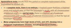
High hCG |
-multiple pregnancy -hydatidiform mole -gestational trophoblastic disease |
|
|
What replaces the blastocyst cavity? |
Primary yolk sac |
|
|
When are the extraembryonic mesoderm and chorion formed? |
2 week (formation of the bilaminar embryo) |
|
|
Embryonic period |
Weeks 3-8 |
|
|
Embryonic week 3 |
|
|
|
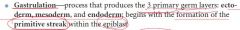
Gastrulation |
|
|
|
What does the mesoderm form? (3) |
1. Paraxial mesoderm (35 pairs of somites) 2. Intermediate mesoderm 3. Lateral mesoderm |
|
|
What happens in third week of embryonic development? |
Gastrulation and early development of nervous and cardiovascular systems; corresponds to first missed period! |
|
|
Sacrococcygeal teratoma |

|
|
|
Chordoma |

|
|
|
Hydatidiform mole. Two types. Their genotype. |
|
|
|
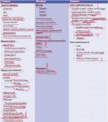
Germ layer derivatives |
|
|
|
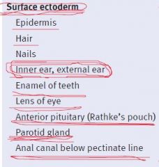
Surface ectoderm derivatives (9) |
|
|
|
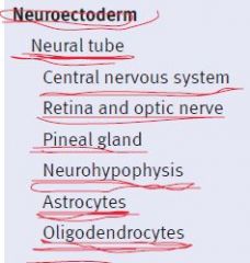
Neuroectoderm derivatives (7) |
|
|
|
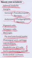
Neural crest ectoderm derivatives (13) |
|
|
|
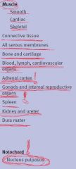
Mesoderm derivatives (13) |
|
|
|
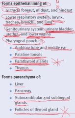
Endoderm derivatives (11) |
|
|
|
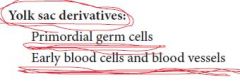
Yolk sac derivatives (2) |
|
|
|
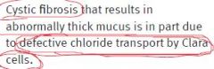
Pathogenesis of cystic fibrosis |
The apical Cl channels do not open -> NaCl and water cannot move across -> thickening of the mucus layer covering the epithelia |
|
|
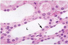
Simple cuboidal epithelium |
-renal tubules -salivary gland acini |
|
|
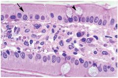
Simple columnar epithelium |
-Small intestine -Large intestine |
|
|
Simple squamous epithelium |
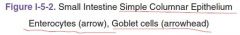
-Endothelium -Mesothelium -Epithelium lining the inside of the renal glomerular capsule |
|
|
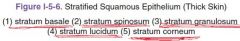
Stratified squamous epithelium |
-Nonkeratinized: oral cavity, pharynx, and esophagus -Keratinizing: skin |
|
|
Pseudostratified columnar epithelium |
-nasal cavity -trachea -bronchi -epididymis |
|
|
Transitional epithelium (urothelium) |
-ureter -bladder |
|
|
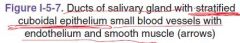
Stratified cuboidal epithelium |
Salivary gland ducts |
|
|
|
|
|
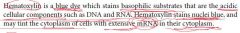
Hematoxylin |
|
|
|
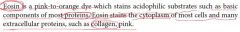
Eosin |
|
|
|
|
|
|

|
|
|
|
|
|
Bladder transitional epithelium |
|

|
|
|
|
Layers of the epidermis |

|
|
|
|
|
|

|
|
|
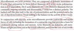
Microfilaments |
|
|
|
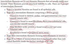
Intermediate filaments. 4 groups. |
|
|
|
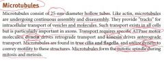
Microtubules. Function of dynein and kinesin. |
|
|
|
First step in the invasion of malignant cells through an epithelium results from ... |
... a loss of expression of cadherinsthat weakens the epithelium |
|
|
Changes in ... are evident in neurons in Alzheimer's disease and in cirrhotic liver diseases |
intermediate filaments |
|
|
Colchicine action |
Prevents microtubule polymerization and is used to prevent neutrophil migration in gout |
|
|
Vinblastine and vincristine action |
Inhibit the formation of the mitotic spindle Used in cancer therapy |
|
|
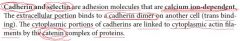
Cadherin and selectin |
|
|
|
Catenin complex of proteins |
Links cytoplasmic portions of cadherins to cytoplasmic actin filaments |
|
|
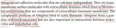
Integrins |
|
|
|
Basement membrane structure |
1. Basal lamina (type IV collagen, glycoproteins [laminin], and proteoglycans [heparin sulfate]) 2. Reticular lamina (reticular fibers) - under the basal lamina |
|
|
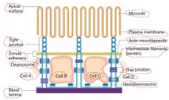
Cell junctions chart |
|
|
|
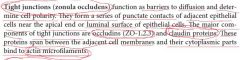
Tight junctions (zona occludens) |
|
|
|
Proteins associated with tight junctions |
-occludins (ZO-1,2,3) -claudin proteins These proteins bind to actin microfilaments |
|
|
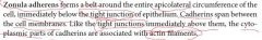
Zonula adherens |
|
|
|
Proteins of zonula adherens |
Cadherins Bind to actin microfilaments |
|
|
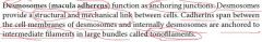
Desmosomes (macula adherens) |
|
|
|
Proteins of desmosomes |
Cadherins Bind to intermediate filaments in large bundles (tonofilaments) |
|
|
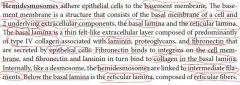
Hemidesmosomes |
|
|
|
Proteins of hemidesmosomes |
Integrins on the cell bind to fibronectin and fibronectin and laminin in turn bind to collagen (type IV) in the basal lamina Internally, like a desmosome, the hemidesmosomes are linked to intermediate filaments |
|
|
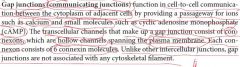
Gap junctions (communicating junctions) |
|
|
|
Connexon structure |
6 connexin molecules - gap junction |
|
|
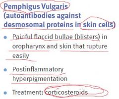
Pemphigus vulgaris |
|
|
|
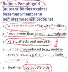
Bullous pemphigoid |
|
|
|
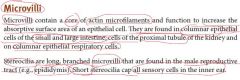
Microvilli |
|
|
|
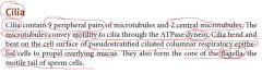
Cilia |
|
|
|
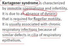
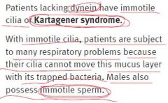
Kartagener syndrome |
|
|
|
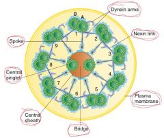
Structure of the axoneme of a cilium |
|
|
|
Embryology of vertebrae |
During week 4, sclerotome cells of the somites (mesoderm) migrate medially to surround the spinal cord and notochord. After proliferation of the caudal portion of the sclerotomes, the vertebrae are formed, each consisting of the caudal part of one sclerotome and the cephalic part of the next |
|
|
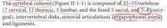
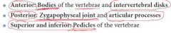
Composition of the vertebral column |
|
|
|
Each intervertebral disk is numbered by the vertebral body ... the disk |
Above |
|
|
Notochord derivative |
Nucleus pulposus |
|
|
Anulus fibrosus function |
Connects the adjacent bodies |
|
|
Nucleus pulposus function |
Shock absorber |
|
|
Anterior longitudinal ligament function |
Prevents hyperextension of the vertebrae Often involved in "whiplash" accidents |
|
|
Posterior longitudinal ligament function |
Limits flexion of the vertebral column Causes the herniation of a disk to be positioned posterolaterally |
|
|
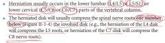
Most common direction of disk herniation |
Posterolateral |
|
|
Disk herniation |
|
|
|
Boundaries of the intervertebral foramina |
|
|
|
Termination of spinal cord in adults |
L1-L2 |
|
|
2 specializations of the pia mater |
1. Denticulate ligaments 2. Filum terminale |
|
|
Termination of the dural mater/dural sac |
S2 |
|
|
Arachnoid termination |
Like dural mater - S2 |
|
|
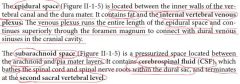
Epidural and subarachnoid spaces |
|
|
|
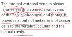
Significance of the internal vertebral venous plexus |
|
|
|
How many spinal nerves are there? |
31 pairs: 8 cervical, 12 thoracic, 5 lumbar, 5 sacral, and 1 coccygeal |
|
|
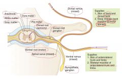
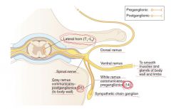
Cross section of spinal cord and parts of spinal nerve |
|
|
|
Where are sensory ganglions located? |
Dorsal roots |
|
|
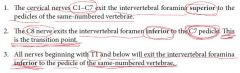
Relationship of exit of spinal nerves and vertebral levels |
|
|
|
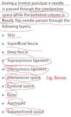
On what level is the lumbar puncture performed? |
L4-L5 interspace |
|
|
A horizontal line drawn at the top of the iliac crest marks the level of the ... |
L4 vertebra |
|
|
The interlaminar spaces are covered by the ... |
Ligamentum flava |
|
|
Layers passed during lumbar puncture |
|
|
|
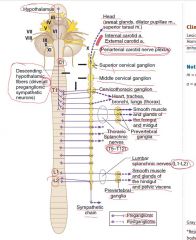
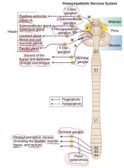
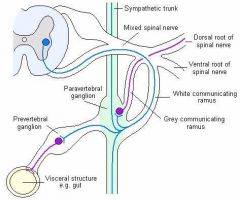
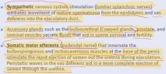
Preganglionic cell bodies of the SNS |
Lateral horn gray matter of spinal cord segments T1-L2 (14 segments) |
|
|
Postganglionic cell bodies of the SNS |
2 types of motor ganglia in the PNS: 1. Chain or paravertebral 2. Collateral or prevertebral (found only in abdomen or pelvis) |
|
|
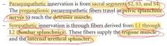
Thoracic splanchnic nerves |

|
|
|
Lumbar splanchic nerves |

|
|
|
Sympathetic (thoracolumbar) outflow |
|
|
|
Sympathetic outflow chart |
|
|
|
Cross section of spinal cord's sympathetic outflow |
|
|
|
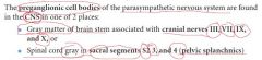
Preganglionic cell bodies of the PsNS |
|
|
|
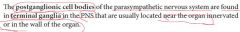
Postganglionic cell bodies of the PsNS |
|
|
|
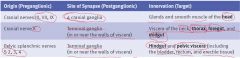
Parasympathetic (craniosacral) outflow |
|
|
|
PsNS ganglions |
1. CN III - ciliary ganglion: -pupillary sphincter -ciliary m. 2. CN VII - submandibular ganglion: -submandibular gland -sublingual gland CN VII - pterygopalatine ganglion: -lacrimal gland -nasal and oral mucosal glands 3. CN IX - otic ganglion: -parotid gland 4. CN X and S2-S4 - terminal ganglia |
|
|
PsNS outflow chart |
|
|
|
Cooper ligaments |
Suspensory ligaments that attach the mammary gland to the skin and run from the skin to the deep fascia |
|
|
Arterial supply to the mammary glands |
1. Internal thoracic artery - a branch of subclavian artery which supplies the medial aspect of the gland 2. Lateral thoracic artery - a branch of the axillary artery, supplies the lateral part of the gland. Courses with the long thoracic nerve, superficial to the serratus anterior muscle |
|
|
Orange-peel appearance of the mammary glands |
The presence of a tumor within the breast can distort Cooper ligaments. which results in dimpling of the skin |
|
|
What nerves can be lesioned during the radical mastectomy? Symptoms? |
1. Long thoracic nerve (serratus anterior muscle) during ligation of the lateral thoracic artery. Few weeks after surgery: -winged scapula -weakness in abduction of the arm above 90 degrees 2. Thoracodorsal nerve (latissimus dorsi): -weakness in extension -weakness in medial rotation of the arm |
|
|
The lymphatic drainage of the breast |
1. Laterally, most of the lymphatic flow (75%) drains from the nipple, superior, lateral, and inferior quadrants of the breast -> pectoral nodes -> axillary nodes 2. Medial quadrant -> parasternal nodes, which accompany the internal thoracic vessels -> opposite breast |
|
|
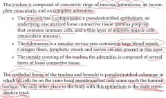
Embryology of lower respiratory system |
Week 4, lower respiatory tract (trachea, bronchi, and lungs) -> single respiratory (laryngotracheal) diverticulum of endoderm from the ventral wall of the foregut -respiratory epithelium develops from endoderm -muscles, connective tissues, and cartilages develop from mesoderm *Respiratory diverticulum -> lung bud *Diverticulum and lung bud -> bifurcation -> 2 bronchial buds -> series of divisions -> main, secondary, and tertiary bronchi (by the sixth month) *Tracheoesophageal septum forms to separate the esophagus from the trachea |
|
|
What structure is related to the bronchopulmonary segments of the lungs? |
Tertiary segmental bronchi |
|
|
A critical timeline in lung development. Why? |
25-28th weeks. Surfactant production. |
|
|
Tracheoesophageal fistula |
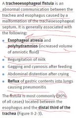
|
|
|
Tracheoesophageal fistula chart |
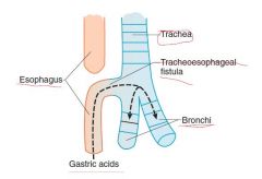
|
|
|
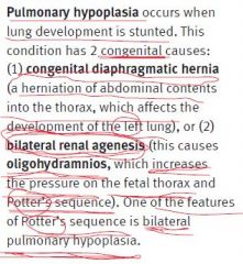
Pulmonary hypoplasia. 2 causes. |
|
|
|
How many intercostal spaces are there? With what are they filled? |
11 intercostal spaces Filled by the 3 layers of intercostal muscles and their related fasciae |
|
|
Costal groove. Correct abbreviation for the structures in it. |
VAN (superior to inferior) |
|
|
Intercostal arteries. Source. |
Anteriorly: -branches of internal thoracic artery << subclavian artery Posteriorly: -branches of the thoracic aorta Thus, collateral circulation between the subclavian artery and the thoracic aorta |
|
|
Where is the intercostal nerve block done? |
Upper portion of the intercostal space |
|
|
Where is the reflection poiny between parietal and visceral pleura? |
Hilum of the lungs |
|
|
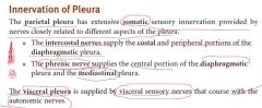
Innervation of pleura |
|
|
|
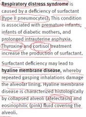
Respiratory distress syndrome and hyaline membrane disease |
|
|
|
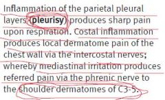
Differential diagnosis: pleurisy, costal inflammation, mediastinal irritation |
|
|
|
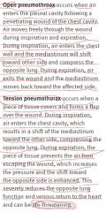
Open and tension pneumothorax |
|
|
|
Sympathetic outflow from the thoraco-lumbar spinal cord chart |
|
|
|
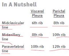
Sternal line of reflection (where the costal pleura is continuous with the mediastinal pleura posterior to sternum) |
Costal cartilages 2-4 The pleural margin then passes inferiorly to the level of the sixth costal cartilage |
|
|
What occupies the costomediastinal recess? |
Lingula of the left lung (during inspiration) |
|
|
Visceral and parietal pleuras borderline |
|
|
|
Diaphragmatic surface of what lung is more superior? |
Right lung (owing to the liver) |
|
|
Pancoast tumor |
A tumor at the apex of the lung, may result in thoracic outlet syndrome |
|
|
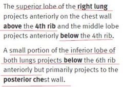
Lobes and fissures of the right lung |
3 lobes: superior, middle, inferior 2 fissures: *horizontal - separates the superior from the middle lobe *oblique - separates the middlefrom inferior lobe |
|
|
Lobes and fissures of the left lung |
2 lobes: superior and inferior 1 fissure: oblique The lingula of the left upper lobe corresponds to the middle lobe of the right lung |
|
|
Projection of the horizontal fissure |
Follows the curvature of the 4th rib, ending medially at the 4th costal cartilage |
|
|
Projection of the horizontal fissure |
Approximately 5th intercostal space in the midclavicular line, ending medially deep to the 6th costal cartilage |
|
|
Projection of the lungs' lobes on the chest wall |
|
|
|
Locations for the auscultation of lungs |
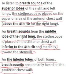
|
|
|
Aspiration of a foreign body. Where it will more probably fall? |
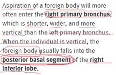
|
|
|
2 major lymphatic vessels |
|
|
|
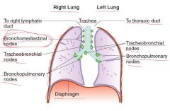
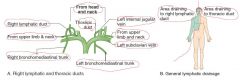
Lymphatic drainage of the lungs |
-Superficial plexus (immediately deep to the visceral pleura) -Deep plexus - begins deeply in the lungs and drains through pulmonary nodes which follow the bronchial tree toward the hilum Plexuses -> bronchopulmonary (hilar) nodes -> tracheobronchial nodes -> bronchomediastinal nodes and trunk -> right lymphatic duct or thoracic duct |
|
|
Lymphatic drainage of the lungs chart |
|
|
|
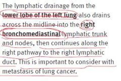
The lymphatic drainage from the lower lobe of the left lung |
|
|
|
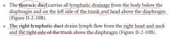
Right lymphatic and thoracic ducts |
|
|
|
Metabolic functions of pulmonary endothelium |
-transformation of lipoproteins and prostaglandins -production of the enzyme that converts angiotensin I to angiotensin II |
|
|
Clinical correlate |
|
|
|
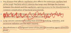
Main causes of lung cancer |
-smoking -asbestos -excessive radiation |
|
|
Mesothelioma |
Malignant tumor of the pleura Causative agent: asbestos dust |
|
|
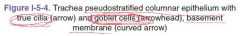
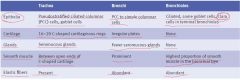
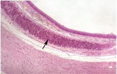
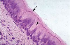
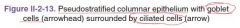
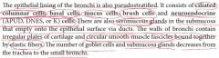
Histologic features of trachea, bronchi, and bronchioles |
|
|
|

|
|
|
Histological layers of the trachea |
|
|
|
|
|
|
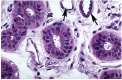
Tracheal epithelial cell types |
-columnar cells -goblet cells -pulmonary neuroendocrine (PNE) cells (APUD cells/DNES cells - diffuse neuro endocrine system/K [Kulchitsky] cells) -brush cells - may represent goblet cells that have secreted their products or intermediate stages in the formation of goblet or the tall ciliated cells; may be sensory receptors -basal cells - stem cells for the ciliated and goblet cells; stem cells lie on the basal membrane but do not extend to the lumen of the trachea; along with epithelial neuroendocrine cells are responsible for the pseudostratified appearance of the trachea |
|
|
Kartagener syndrome |
|
|
|
What cells give the pseudostratified appearance to the trachea? |
Basal cells (stem cells), not goblet cells! Along with epithelial neuroendocrine cells. |
|
|
Bronchial tree |
Trachea Primary bronchi Secondary (lobar) bronchi - 3 in right and 2 in left Tertiary (segmental) bronchi - 10 in each lung |
|
|

|
|
|
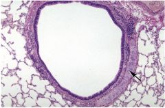
Histology of bronchi |
|
|
|
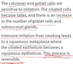
Reaction of columnar and goblet cells on irritation |
|
|
|
From what cells do the bronchial metastatic tumors arise? |
Kulchitsky cells |
|
|
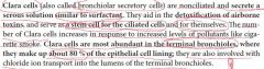
Cystic fibrosis and Clara cells |
|
|
|
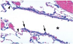
Is there cartilage or glands in the bronchioles? |
No |
|
|
Epithelial type of bronchioles |
Still ciliated but is a simple columnar or cuboidal rather than pseudostratified Epithelial lining is composed of ciliated cells (goblet and basal cells are absent in the terminal bronchioles) and an additional type called the Clara cell |
|
|
Clara cells |
|
|
|

|
|
|
Histology of terminal and respiratory bronchioles |
|
|
|
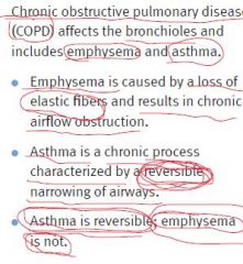
COPD |
|
|
|
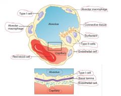
Alveolus and blood-air barrier |
|
|
|
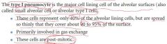
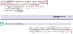
Type I pneumocyte |
|
|
|
Type II pneumocyte |
|
|
|

|
|
|
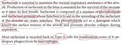
Surfactant |
|
|
|
Types of collagen in the alveolar wall |
Type I and II collagens, as well as elastic fibers, are in the septa Type I collagen is present primarily in the walls of the bronchi and bronchioles |
|
|
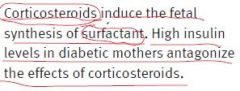
Why infants of diabetec mothers have a higher incidence of respiratory distress syndrome? |
|
|
|
Blood-gas barrier |
-Surfactant -Squamous Type I pneumocyte -Shared basal lamina -Capillary endothelium |
|
|
What's the distance between the lumen of the capillary and the lumen of the alveolus? |
0.1 microns |
|
|
Pores of Kohn |
Openings in the wall of alveoli Important in collateral ventilation d = 10-15 microns |
|
|
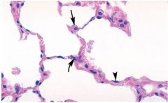
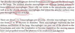
Alveolar macrophages |
|
|
|
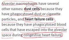
Other names of the alveolar macrophages |
|
|
|
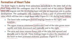
When the heart begins to develop? From what germ layer? |
Latter half of the third week From splanchnic mesoderm + neural crest cells |
|
|
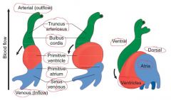
Formation of heart tube |
|
|
|
Development of the heart tube chart |
|
|
|
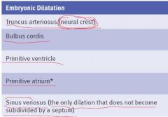
Germ layer for the truncus arteriosus (aorta, pulmonary trunk, semilunar valves) |
Neural crest |
|
|

|
|
|
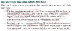
Development of the veins of liver (sinusoids, hepatic portal vein, hepatic vein) and part of the inferior vena cava |
From viteline (omphalomesenteric) veins |
|
|
Development of some of the major veins of the body (brachiocephalic, superior vena cava, inferior vena cava, azygos, renal) |
From cardinal veins |
|
|
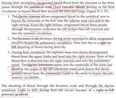
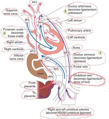
Venous systems associated with the fetal heart |
|
|
|
Arterial systems associated with the fetal heart |
|
|
|
Fetal circulation and shunts |
|
|
|
What substances facilitate closure of the ductus arteriosus? |
-release of bradykinin -immediate drop of prostaglandin E |
|
|
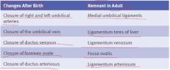
Adult vestiges derived from the fetal circulatory system |
|
|
|
When does the septation of atria and ventricles occur? |
Week 4 - week 8 |
|
|
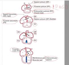
Formation of atrial septum |
|
|
|
Where is the foramen primum located? |
Between the inferior edge of the septum primum and the endocardial cushion; it is the only one foramen obliterated (when the septum primum later fuses with the endocardial cushions) |
|
|
Where is the foramen secundum located? |
Within the upper part of the SEPTUM PRIMUM (!) just before the FP closes |
|
|
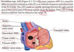
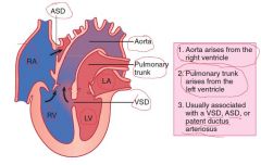
Two clinically important types of ASD |
1. Secundum-type - most common ASD 2. Primum-type |
|
|
Secundum-type ASD |
|
|
|
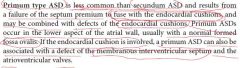
Primum-type ASD |
|
|
|
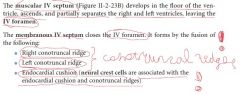
Ventricular septation |
|
|
|
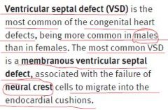
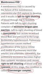
Ventricular septal defect |
|
|
|
Membranous VSD |
|
|
|
In what conditions PDA is common? |
-premature infants -maternal rubella infection |
|
|
Machine-like murmur |
PDA |
|
|
What substances sustain PDA? |
-Prostaglandin E (PGE) -Low oxygen tension |
|
|
I what conditions PGE is used to keep PDA? |
Transposition of great vessels |
|
|
What substances promote closure of the ductus arteriosus in a premature birth? |
-PGE inhibitors (eg, indomethacin) -acetylcholine -histamine -catecholamines |
|
|
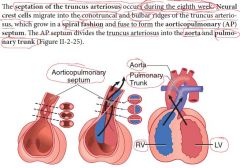
When does the septation of the truncus arteriosus occur? |
During the 8th week |
|
|
Septation of the truncus arteriosus |
|
|
|
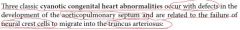
What is the defect in classic cyanotic congenital heart abnormalities? |
|
|
|
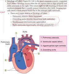
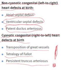
Most common cyanotic congenital heart deffect |
Tetralogy of Fallot |
|
|
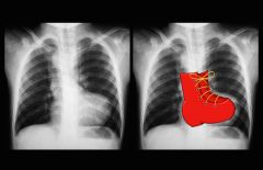
Boot-shaped heart |
Tetralogy of Fallot (due to enlarged right ventricle) |
|
|
Boot-shaped heart.Tetralogy of Fallot (due to enlarged right ventricle). |
|
|
Tetralogy of Fallot |
|
|
|
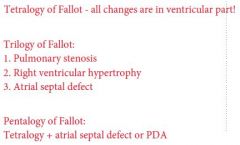
Trilogy vs. tetralogy vs. pentalogy of Fallot |
|
|
|
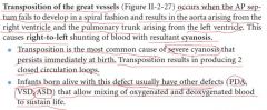
Transposition of the great vessels |
|
|
|
The most common cause of severe cyanosis that persists immediately at birth |
Transposition of the great vessels |
|
|
Transposition of the great vessels chart |
|
|
|
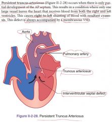
What defect is ALWAYS accompanied by a membranous VSD? |
Persistent truncus arteriosus |
|
|
Persistent truncus arteriosus |
|
|
|
Non-cyanotic and cyanotic congenital heart defects at birth (3 and 3) |
|
|
|
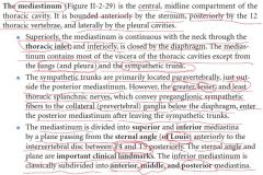
Mediastinum |
|
|
|
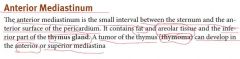
Anterior mediastinum |
|
|
|
Point of division of the mediastinum on superior and inferior |
Plane through the sternal angle of Louis and intervertebral disk between T4 and T5 |
|
|
Aortic hiatus level. What contains? |
T12 Thoracic descending aorta and thoracic duct |
|
|
Location of the esophagus in the posterior mediastinum |
Immediately posterior to the left primary bronchus and the left atrium |
|
|
Level of the esophageal hiatus. What contains? |
T10 Esophagus and vagal nerve trunks |
|
|
Constrictions of the esophagus |
1. at its origin from the pharynx 2. posterior to the arch of the aorta 3. posterior to the left primary bronchus 4. at the esophageal hiatus of the diaphragm |
|
|
Location of the thoracic duct. Its origin. |
Lies posterior to the esophagus and between the thoracic aorta and azygos vein Arises from the cisterna chyli in the abdomen (at vertebral level L1) and enters the mediastinum through the aortic hiatus of the diaphragm |
|
|
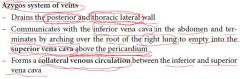
Azygos system of veins |
|
|
|
4 structures of the posterior mediastinum |
1. Thoracic (descending) aorta 2. Esophagus 3. Thoracic duct 4. Azygos system of veins |
|
|
Middle mediastinum |
Contains the heart, parts of the great vessels, phrenic nerves, and the pericardium |
|
|
In what mediastinum are the pulmonary trunk and pulmonary arteries located? |
Completely in the middle mediastinum! |
|
|
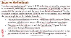
Superior mediastinum |
|
|
|
Where the superior vena cava is formed? |
2 brachiocephalic veins join posterior to the right first costal carilage |
|
|
On what level the superior vena cava drains into RA? |
Deep to the right third costal cartilage |
|
|
Where are aortic arch and its 3 branches located? |
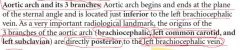
|
|
|
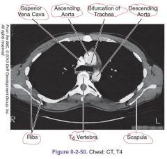
Location of the bifurcation of the trachea |
T4 level - border of the superior and inferior mediastinum |
|
|
Location of the esophagus |
Lies posterior to the trachea and courses posterior to the left primary bronchus to enter the posterior mediastinum |
|
|
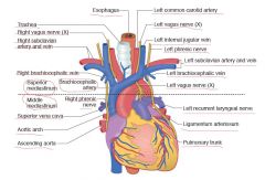
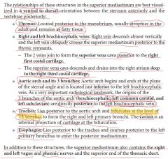
Superior mediastinum structures, from ventral to dorsal |
|
|
|
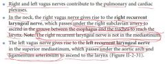
Is the right recurrent laryngeal nerve in the mediastinum? |
No |
|
|
Vagus nerves |
|
|
|
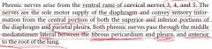
Phrenic nerves |
|
|
|
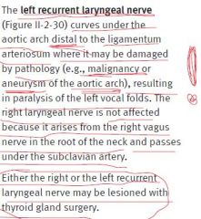
Recurrent laryngeal nerves |
|
|
|
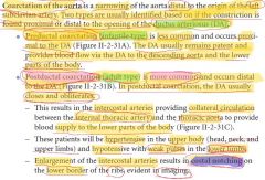
Coarctation of the aorta |
|
|
|
Infantile and adult types of the coarctation of the aorta |
-infantile type - preductal - less common, occurs proximal to the DA, which remains open! -adult type - postductal - more common, occurs distal to the DA, which usually closes and obliterates -> costal notching |
|
|
Coarctation of the aorta pictures |
|
|
|
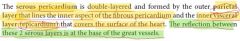
Layers of pericardium |
3 layers: an outer fobrous layer and a double-layered parietal and visceral (epicardium) serous layers |
|
|
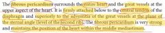
Fibrous pericardium |
|
|
|
Serous pericardium |
|
|
|
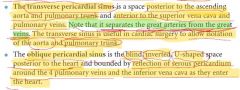
Sinuses of the heart |
|
|
|
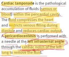
Cardiac tamponade |
|
|
|
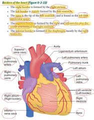
Borders of the heart |
|
|
|
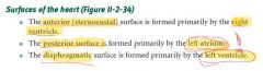
Surfaces of the heart |
|
|
|
Sulci of the heart |
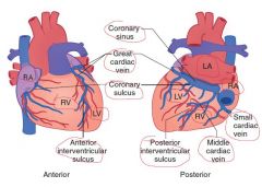
1. Coronary 2. Anterior interventricular 3. Posterior interventricular |
|
|
Surface projections of the heart: -upper right aspect -lower right aspect -upper left aspect -apex -right border -left border -inferior border -superior border |

|
|
|
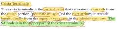
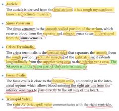
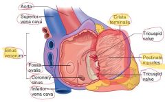
What is crista terminalis? |
|
|
|
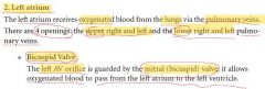
Right atrium structures |
|
|
|
Where is the SA node located? |
In the upper part of the crista terminalis |
|
|
Inside the right atrium picture |
|
|
|
Left atrium structures |
|
|
|
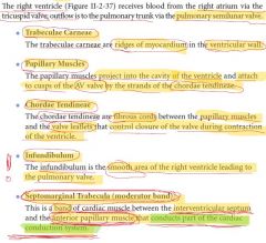
What is infundibulum (the heart)? |
The smooth area of the right ventricle leading to the pulmonary valve |
|
|
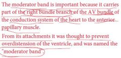
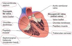
What is septomarginal trabecula (moderator band)? |
This is a band of cardiac muscle between the interventricular septum and the anterior papillary muscle that constitutes a part of the cardiac conduction system |
|
|
Right ventricle structures |
|
|
|
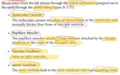
What is aortic vestibule? |
The aortic vestibule leads to the aortic semilunar valve and ascending aorta |
|
|
Left ventricle structures |
|
|
|
Right and left ventricles picture |
|
|
|
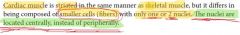
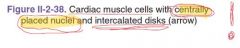
Histology: cardiac muscle vs. skeletal muscle |
|
|
|
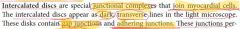
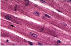
Intercalated disks (heart) |
|
|
|
|
|
|
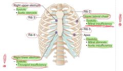
Points of auscultation of the heart valves |

|
|
|
How many cusps are in the aortic and pulmonary valves? |
3 cusps |
|
|
Auscultation of heart murmurs chart ("A heart murmur is heard downstream from the valve") |
|
|
|
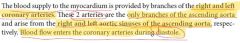
Arterial supply of the heart |
|
|
|
Arterial supply to the heart chart |
|
|
|
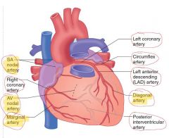
Right coronary artery branches |

|
|
|
Left coronary artery branches |

|
|
|
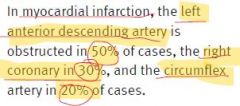
Epidemiology of vessels obstruction leading to the myocardial infarction |
|
|
|
From what does the coronary sinus develop? |
Left sinus venosus |
|
|
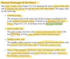
Main vein of the coronary circulation |
Coronary sinus |
|
|
Main tributary of the coronary sinus |
Great cardiac vein (lies in the anterior interventricular sulcus) |
|
|
Venous drainage of the heart |
|
|
|
Venous drainage of the heart picture |
|
|
|
Where is the AV node located? |
In the interatrial septum near the opening of the coronary sinus |
|
|
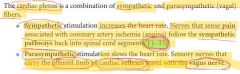
Cardiac plexus |
|
|
|
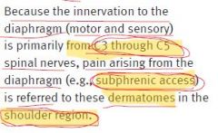
Phrenic nerve segments |
C3-C5 |
|
|
Caval hiatus |
T8, right of the midline, within the central tendon Inferior vena cava and some branches of the right phrenic nerve |
|
|
Esophageal hiatus |
T10, left of the midline, within the muscle of the right crus Esophagus and the anterior and posterior vagus trunks |
|
|
Aortic hiatus |
T12, lLocated in the midline behind the two crura Aorta and the thoracic duct |
|
|
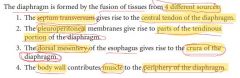
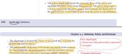
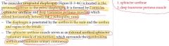
Development of the diaphragm: 1. central tendon of the diaphragm 2. parts of the tendinous portion of the diaphragm 3. crura of the diaphragm 4. muscle periphery |
|
|
|
Pain referral from the phrenic nerve |
|
|
|
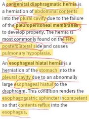
A congenital diaphragmatic and esophageal hiatal hernia |
|
|
|
|
|
|
|
|
|
|
|
|
|
|
|
|
|
|
|
|
|
|
|
|
|
|
|
Development of the alveolar macrophages |
They are derived from monocytes |
|
|
From what does the primitive heart tube develop? |
From the lateral plate mesoderm |
|
|
Where is the coronary sinus orifice located? |
Between the inferior vena cava and the right atrioventricular orifice or tricuspid valve in the right atrium |
|
|
Linea semilunaris |
Is a curved line defining the lateral border of the rectus abdominis, a bilateral feature |
|
|
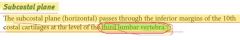
Subcostal plane |
|
|
|
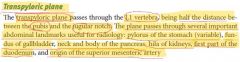
Transpyloric plane |
|
|
|
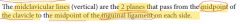
Midclavicular lines |
|
|
|
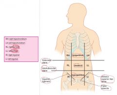
Regions and plains of the abdomen |
|
|
|
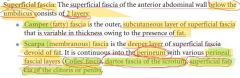
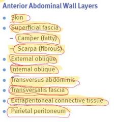
Superficial fascia of the anterior abdominal wall |
|
|
|
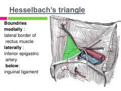
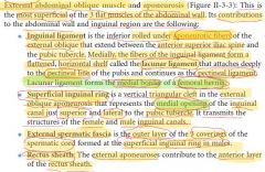
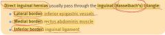
Hesselbach's triangle |
|
|
|
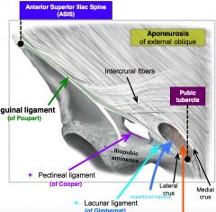
Lacunar and pectineal ligaments |
|
|
|
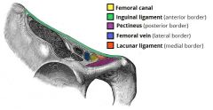
Femoral canal |
|
|
|
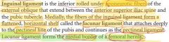
Inguinal ligament |
|
|
|
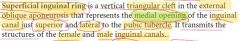
Superficial inguinal ring |
|
|
|
Anteror abdominal wall layers |
|
|
|
External abdominal oblique muscle |
|
|
|
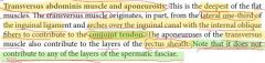
External spermatic fascia derives from ... |
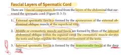
External abdominal oblique muscle Is the outer layer of 3 coverings of the spermatic cord formed at the superficial inguinal ring in males |
|
|
Cremasteric muscle and fascia derive from ... |
Internal abdominal oblique muscle Is the middle layer of the spermatic fascia covering the spermatic cord and testis in the male. It forms in the inguinal canal. |
|
|
Internal abdominal oblique muscle |

|
|
|
Transversus abdominis muscle and aponeurosis |
|
|
|
Internal spermatic fascia derives from ... |
Transversalis fascia Deepest of the coverings of the spermatic cord formed at the deep ring in the male |
|
|
Superficial inguinal ring is formed by ... |
External oblique aponeurosis |
|
|
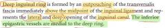
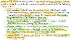
Deep inguinal ring |
|
|
|
Deep inguinal ring is formed by ... |
Outpouching of the transversalis fascia immediately above the midpoint of the inguinal ligament The inferior epigastric vessels are medial to the deep ring |
|
|
Transversalis fascia |
|
|
|
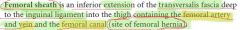
Femoral sheath |
|
|
|
Where do the gonads develop? |
Within the extraperitoneal connective tissue! |
|
|
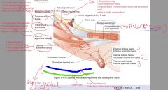
Layers of anterolateral abdominal wall and inguinal canal picture |
|
|
|
Innervation of the skin and muskulature of the anterior abdominal wall |
-Ventral primary rami of T7-T12 (includes the subcostal nerve) -Iliohypogastric and ilioinguinal branches of the ventral primary rami of L1 |
|
|
Arterial blood supply to the anterior abdominal wall |
-superior epigastric branch << internal thoracic artery -inferior epigastric and DEEP circumflex iliac << external iliac artery |
|
|
Venous drainage from the anterior abdominal wall |
Superiorly: -superficial epigastric -lateral thoracic veins Inferiorly: -great saphenous vein |
|
|
Lymph drainage from the anterior abdominal wall |
Superiorly - axillary nodes Inferiorly - superficial inguinal |
|
|
Inguinal canal |
|
|
|
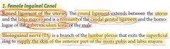
Remnant of the caudal genital ligament (females) |
Round ligament of the uterus |
|
|
Homologue of the gubernaculum testis of the male |
Round ligament of the uterus |
|
|
Ilioinguinal nerve innervation in females |
Anterior part of the mons pubis and labia majora |
|
|
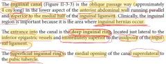
Female inguinal canal contents |
|
|
|
Ilioinguinal nerve innervation in males |
Skin of the lateral and anterior scrotum |
|
|
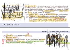
Contents of the spermatic cord (6) |
|
|
|
Male inguinal canal contents |
1. Ilioinguinal nerve 2. The spermatic cord |
|
|
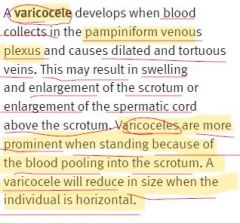
Varicocele |
|
|
|
Fascial layers of the spermatic cord |
|
|
|
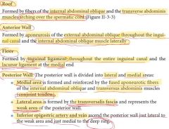
Boundaries of the inguinal canal |
|
|
|
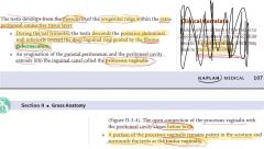
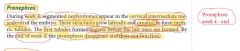
When does the descent of testes occur? |
During the last trimester |
|
|
When does the processus vaginalis close? |
Before birth |
|
|
What is gubernaculum? |
Fibers that guide the testis during its descent to the scrotum |
|
|
Descent of the testes |
|
|
|
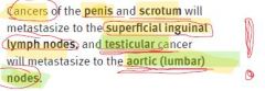
Where do the cancers of penis and scrotum will metastasize? Testicular cancer? |
|
|
|
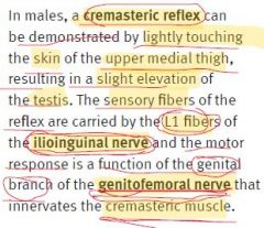
Cremasteric reflex limbs |
|
|
|
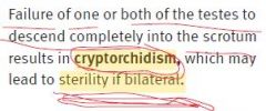
Cryptorchidism |
|
|
|
Inguinal hernias occur ... to the inguinal ligament |
superior |
|
|
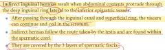
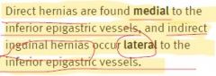
Indirect inguinal hernias |
|
|
|
Direct inguinal hernias |

|
|
|
Cuase of the congenital indirect inguinal hernia |
Persistent process vaginalis |
|
|
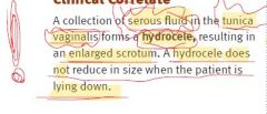
Hydrocele |
|
|
|
Direct vs. indirect hernias |
|
|
|
Hasselbach's triangle |
|
|
|
Inguinal hernias most often occur in ... Femoral hernias most offen occur in ... |
Inguinal - men Femoral - women (Femoral-Female) |
|
|
What does the femoral sheath contain? |
Femoral artery, vein, and canal |
|
|
Femoral hernias occur ... to the inguinal ligament |
inferior |
|
|
Femoral artery, nerve, and vein - correct order (medial to lateral!) |
VAN |
|
|
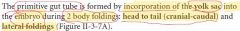
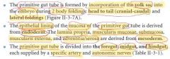
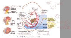
How is the primitive gut tube formed? |
|
|
|
Primitive gut tube |
|
|
|
Adult structures derived from each of the 3 divisions of the primitive gut tube |
|
|
|
Development of gastrointestinal tract |
|
|
|
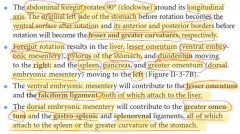
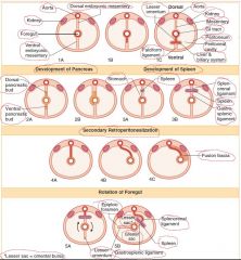
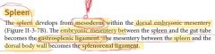
Where does the liver develop? Spleen and dorsal pancreatic bud? |
Liver - ventral embryonic mesentery Spleen and dorsal pancreatic bud - dorsal embryonic mesentery |
|
|
Rotation of the foregut |
|
|
|
From what do the lower respiratory tract, liver and biliary system, and pancreas develop? |
Endodermal outgrowth of the foregut |
|
|
Cross-sectional view of foregut development and rotation |
|
|
|
From what does the midgut develop? |
From the cranial and caudal intestinal loops |
|
|
Development and rotation of the midgut |
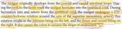
|
|
|
Innervation of the parietal peritoneum |
-lower intercostal nerves -ilioinguinal and the iliohypogastric nerves of the lumbar plexus |
|
|
What are mesenteries? |
Double-layered peritoneal membranes that suspend parts of the GI tract from the body wall |
|
|
Postnatal remnants of mesenteries |
|
|
|
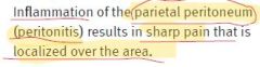
Peritonitis |
|
|
|
lesser sac |
Omental bursa Is a cul-de-sac formed posterior to the stomach and the lesser omentum |
|
|
Greater sac |
Formed by the larger area of the remaining peritoneal cavity |
|
|
The only communication between the lesser sac and the greater sac |
Epiploic foramen (of Winslow) |
|
|
Intraperitoneal vs. retroperitoneal organs |
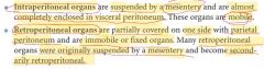
|
|
|
Intraperitoneal vs. retroperitoneal organs table |
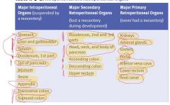
|
|
|
Boundaries of the epiploic foramen (of Winslow) |
|
|
|
Where is the ligamentum teres of liver located? |
Falciform ligament |
|
|
What does the splenorenal ligament contain? |
Tail of pancreas and distal splenic vessels |
|
|
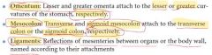
Composition of the lesser omentum |
-Hepatogastric ligament -Hepatoduodenal ligament |
|
|
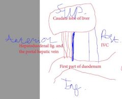
What does the hepatoduodenal ligament contain? |
-common bile duct -proper hepatic artery -hepatic portal vein |
|
|
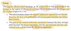
Development of the liver |
|
|
|
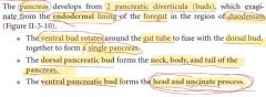
Development of the pancreas |
|
|
|
What does the dorsal pancreatic bud form? |
Neck, body, and tail of the pancreas |
|
|
What does the ventral pancreatic bud form? |
Head and uncinate process |
|
|
Development of the spleen |
|
|
|
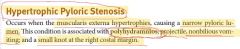
Hypertrophic pyloric stenosis |
|
|
|
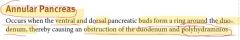
Annular pancreas |
|
|
|
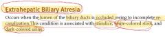
Extrahepatic biliary atresia |
|
|
|
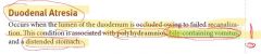
Duodenal atresia |
|
|
|
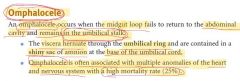
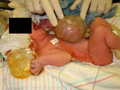
Omphalocele |
|
|
|
Omphalocele (with membrane) |
|
|
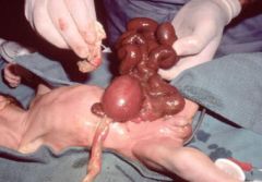
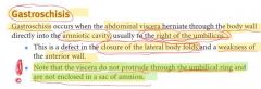
Gastroschisis |
|
|
The amnion stems from the ... on the outer side and the ... on the inner side |
The amnion stems from the extraembryonic somatic mesoderm on the outer side and the extraembryonic ectoderm on the inner side |
|
|
Gastroschisis |
|
|
|
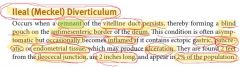
Ileal (Meckel) diverticulum |
|
|
|
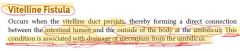
Vitelline fistula |
|
|
|
Drainage of meconium from the umbilicus |
Vitelline fistula |
|
|
Volvulus |
Twisting of intestines |
|
|
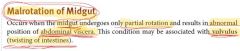
Malrotation of midgut |
|
|
|
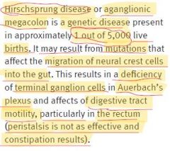
Hirschprung disease |
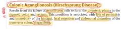
|
|
|
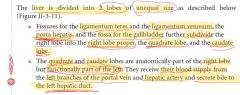
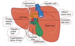
Division of the liver |
|
|
|
Visceral surface of the liver |
|
|
|
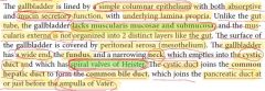
Where does the gallbladder lie? |
In a fossa on the visceral surface of the liver to the right of the quadrate lobe |
|
|
Biliary ducts |
|
|
|
By what is the uncinate process of pancreas crossed? |
Superior mesenteric vessels |
|
|
Site of formation of the hepatic portal vein |
Posterior to the neck of pancreas |
|
|
The only part of the pancreas that is intraperitoneal |
Tail |
|
|
Blood supply to the pancreas |
Head: -superior pancreaticoduodenal branch << gastroduodenal artery << celiac trunk -inferior pancreaticoduodenal branch << superior mesenteric artery Collateral circulation between celiac trunk and superior mesenteric artery! Neck, body, and tail: splenic artery |
|
|
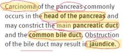
Carcinoma of the pancreas |
|
|
|
The splenic artery and vein reach the hilus of the spleen by traversing the ... |
splenorenal ligament |
|
|
Border between foregut and midgut |
2 part of duodenum - at the point of entry of the common bile duct |
|
|
What is mesoappendix? |
Own mesentery of the appendix |
|
|
What's longer - jejunum or ileum? |
Jejunum - 2/5 Ileum - 3/5! |
|
|
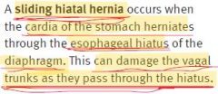
Sliding hiatal hernia |
|
|
|
What is transverse mesocolon? |
Own mesentery of the transverse colon |
|
|
Border between midgut and hindgut |
Junction of the proximal two-thirds and distal one-third of the transverse colon |
|
|
What is sigmoid mesocolon? |
Own mesentery of the sigmoid colon |
|
|
Junction point between descending colon and sigmoid colon |
Pelvic brim |
|
|
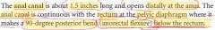
Anal canal |
|
|
|
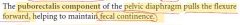
Function of the puborectalis component of the pelvic diaphragm |
|
|
|
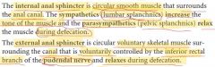
Internal and external anal sphincter |
|
|
|
Innervation of the external anal sphincter |
Inferior rectal branch of the pudendal nerve |
|
|
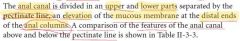
Pectinate line (rectum) |
|
|
|
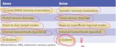
Comparison of features above and below the pectinate line (table) |
|
|
|

|
|
|
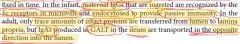
What is located in the lamina propria of GI tract? |
-Capillary networks -Lacteals! -GALT (IgA production)! |
|
|
Meissner's plexus |
An interconnected network of ganglia and nerves located in the submucosa |
|
|
Auerbach's plexus |
Located between the 2 muscle layers of the muscularis externa |
|
|
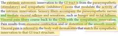
Extrinsic autonomic innervation of the GI tract |
|
|
|
Hirschprung disease |
|
|
|
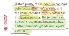
Brunner glands |
Discharge alkaline secretion in duodenum Located in submucosa! |
|
|
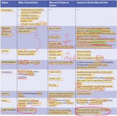
Histology of GI tract table |
|
|
|
Function of M cells in ileum |

|
|
|
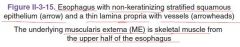
Muscularis externa of the esophagus |

|
|
|
|
|
|
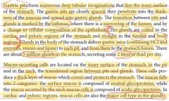
Gastric pits |
|
|
|
|
|
|
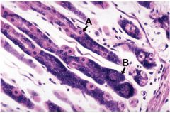
Where are the parietal cells located? |
Upper regions of the gastric glands (under the isthmus!) |
|
|
Where are the mucus-secreting cells located in the stomach? |

|
|
|
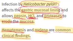
H. pylori infection |
|
|
|
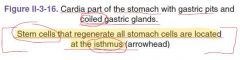
Where are the stem cells responsible for the regeneration of all types of cells located in the stomach? |
Isthmus! |
|
|
Where are chief cells located? |
Deep within the glands, thus they can have life span greater than 190 days (not acidic environment) |
|
|
|
|
|
The glands are ... in the cardiac and pyloric regions of the stomach and ... in the fundus and body regions |
The glands are coiled in the cardiac and pyloric regions of the stomach and straight in the fundus and body regions |
|
|
Valves of Kerckring |
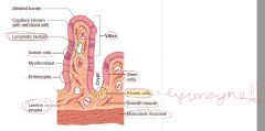
Plicae circulares (circular folds) in the small intestine Involve both mucosa and sub-mucosa Increase the surface area by factor of 3 |
|
|
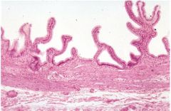
Villi of the small intestine |

|
|
|
Microvilli of the small intestine |
|
|
|
Function of glycocalix |

|
|
|
Crypts of Lieberkuhn |
|
|
|
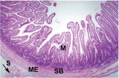
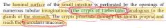
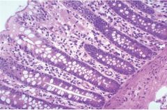
Small intestine mucosal histology |
|
|
|
What cells produce mucus in the GI tract? |
Duodenum - Brunner glands Intestine - goblet cells |
|
|
|
|
|
Maternal IgGs ingestion and IgAs secretion |
|
|
|
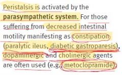
Constipation |
|
|
|
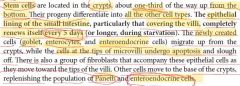
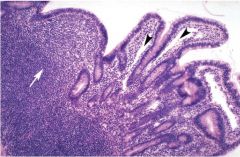
Where are the Peyer's patches located? |
|
|
|
Goblet cells |

|
|
|
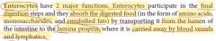
Enterocytes |
|
|
|
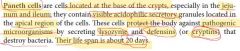
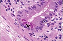
Paneth cells |
|
|
|
|
|
|
Stain for the enteroendocrine cells |
Silver-based stains |
|
|
Where are the enteroendocrine cells located? |
|
|
|
Stem cells of the intestinal epithelia |
|
|
|
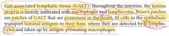
GALT |
|
|
|

|
|
|
Does the large intestine have plicae, villi, and crypts of Lieberkuhn? |
No plicae, villi, but has short crypts of Lieberkuhn |
|
|
Why are there haustra in the colon? |
Because the colon is longer than teniae coli |
|
|
Difference in location between stem cells in stomach and small and large intestine |
Stomach - isthmus Small and large intestine - lower part of the crypts |
|
|

|
|
|
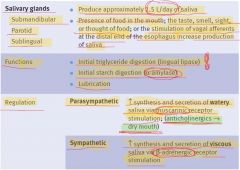
Salivary glands |
|
|
|
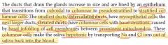
Intercalated and striated ducts of the salivary glands |
|
|
|
|
|
|
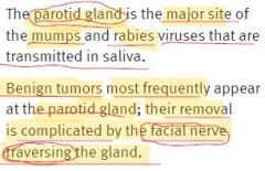
Parotid gland pathology |
|
|
|
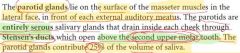
Stensen's duct |
Parotid gland's duct, opens above the second upper molar tooth |
|
|
Parotid gland |
|
|
|
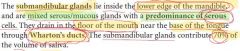
Wharton's duct |
Duct of the submandibular gland, drain in the floor of the mouth near the base of the tongue |
|
|
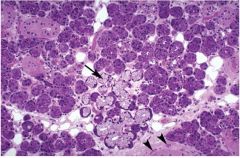
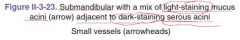
Submandibular gland |
|
|
|
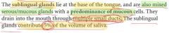
Sublingual gland |
|
|
|
|
|
|
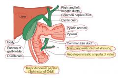
Duct of Wirsung |
Main pancreatic duct |
|
|
Duct of Santorini |
Accessory pancreatic duct |
|
|
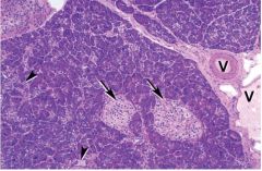
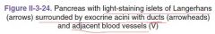
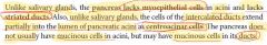
Histological difference between pancreas and salivary glands |
|
|
|
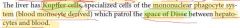
Kupffer cells |
|
|
|
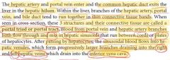
Blood flow into the liver |
Blood flow into the liver is dual (75% from portal vein, 25% from hepatic artery) |
|
|
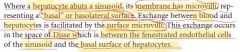
Space of Disse |
|
|
|
How many hepatic veins are there? |
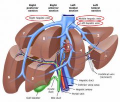
The hepatic veins are three large veins which drain the hepatic parenchyma into the inferior vena cava (IVC), named the right hepatic vein, middle hepatic vein and left hepatic vein |
|
|
Portal triad (portal tract) |
|
|
|
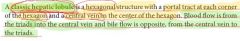
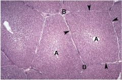
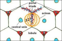
Classic hepatic lobule |
|
|
|
|
|
|
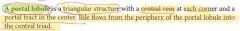
Portal lobule |
|
|
|
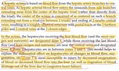
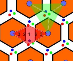
Hepatic acinus |
|
|
|
Hepatic acinus picture |
|
|
|
Classic lobule vs. portal lobule vs. hepatic acinus |
|
|
|
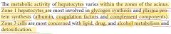
Metabolic activity of hepatocytes within the zones of the acinus |
|
|
|
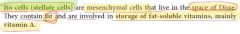
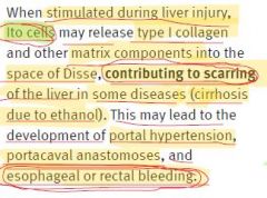
Ito cells (stellate cells) |
|
|
|
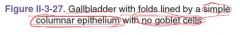
Epithelium of the gallbladder |
|
|
|
Spiral valves of Heister |
Valves in the cystic duct |
|
|
Mechanism of the development of the portal hypertension |
|
|
|
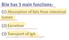
3 main functions of bile |
|
|
|
|
|
|
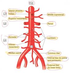
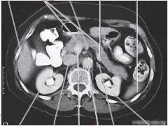
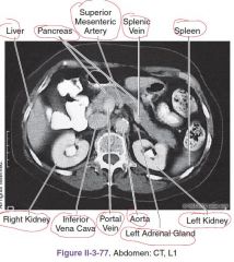
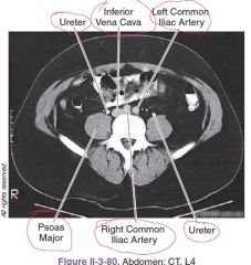
Level of the aortal bifurcation |
L4 |
|
|
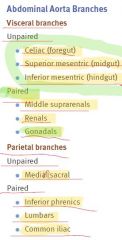
3 groups of the aortal branches |
-3 unpaired visceral branches -3 paired visceral branches -several parietal branches to the body wall |
|
|
Abdominal aorta branches |
|
|
|
Visceral and parietal branches of the abdominal aorta picture |
|
|
|
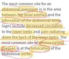
The most common site for an abdominal aneurism |
|
|
|
Celiac artery (trunk) branches |
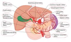
Arises at T12-L1 level Supplies the structures derived from the foregut 3 branches: 1. left gastric artery (supplies most of the lesser curvature) -esophageal branch 2. splenic artery -branches to the neck, body, and tail of the pancreas -left gastroepiploic artery (supplies the left side of the greater curvature of the stomach) -short gastric branches (supply the fundus of the stomach) 3. common hepatic artery -proper hepatic artery *right gastric artery -left hepatic artery -right hepatic artery *cystic artery -gastroduodenal artery *right gastroepiploic artery *superior pancreaticoduodenal arteries (anastomoses with inferior pancreaticoduodenal branches of the superior mesenteric artery) |
|
|
Celiac artery branches |
|
|
|
Superior mesenteric artery |
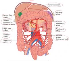
Arises at the L1 level just below the celiac artery Supplies the midgut The superior mesenteric vein is to the right of the artery Branches: 1. inferior pancreaticoduodenal arteries - anastomose with the superior pancreaticoduodenal branches of the gastroduodenal artery 2. intestinal arteries - 12-15 branches from the left side of SMA 3. ileocolic artery 4. right colic artery 5. middle colic artery |
|
|
SMA picture |
|
|
|
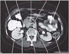
Inferior mesenteric artery |
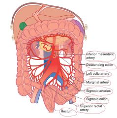
Supplies hindgut Arises at the level of L3 Branches: 1. Left colic artery 2. Sigmoid arteries 3. Superior rectal artery |
|
|
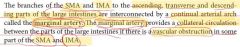
Marginal artery |
|
|
|
IMA picture |
|
|
|
The most common site of bowel ischemia |
Splenic flexure |
|
|
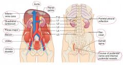
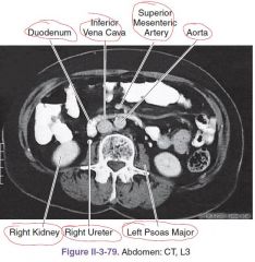
Renal arteries' level |
Upper border of the L2 vertebra |
|
|
Difference between the right and left renal arteries |
The right renal artery is longer and passes posterior to the IVC |
|
|
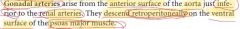
Gonadal arteries |
|
|
|
Level of the IVC origin |
To the right of the lumbar vertebrae at the L5 level |
|
|
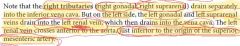
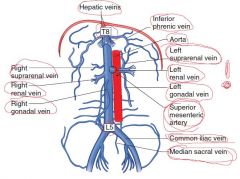
Where do the right and left gonadal and suprarenal veins drain? |
|
|
|
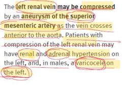
How does the left renal vein pass? |
Crosses anterior to the aorta, just inferior to the origin of the superior mesenteric artery |
|
|
IVC picture |
|
|
|
Aneurysm of the superior mesenteric artery |
|
|
|
Hepatic portal system |
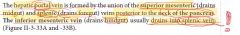
|
|
|
Hepatic portal system picture |
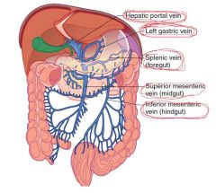
|
|
|
Are there valves in the portal system? |
No! |
|
|
Palm tree sign |
Caput medusae |
|
|
Portacaval anastomoses |
|
|
|
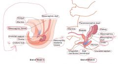
Embryology of kidneys and ureter |
|
|
|
Pronephros derivatives |
Nothing! |
|
|
Mesonephros derivatives |

Bowman's capsule into which a tuft of capillaries, or glomerulus, invaginates |
|
|
Metanephros derivatives |
1. Ureteric bud - calyces, pelvis, ureter 2. Metanephric mass - proximal convoluted tubule, the loop of Henle, and the distal convoluted tubule |
|
|
Positional change of the kidneys |
|
|
|
Development of the urinary system pictures |
|
|
|
Embryology of bladder and urethra |
|
|
|
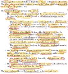
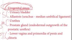
Urogenital sinus |
|
|
|
Potter sequence |
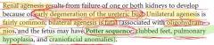
-clubbed feet -pulmonary hypoplasia -craniofacial anomalies Seen in cases of oligohydramnios (eg, bilateral renal agenesis) |
|
|
Renal agenesis |
|
|
|
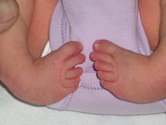
Club foot or Congenital Talipes Equinovarus (CTEV) |
|
|
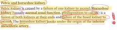
Pelvic and horseshoe kidney |
|
|
|
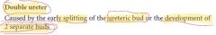
Double ureter |
|
|
|
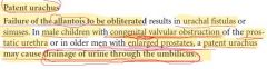
Patent urachus |
|
|
|
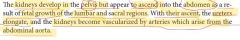
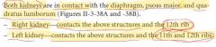
Right kidney vs. left kidney position |
T12-L3 The right kidney is positioned slightly lower than the left because of the mass of the liver |
|
|
Kidney's relation to the posterior abdominal wall |
|
|
|
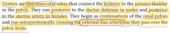
Ureters |
|
|
|
On what muscle does the ureter lie? |
Psoas major muscle |
|
|
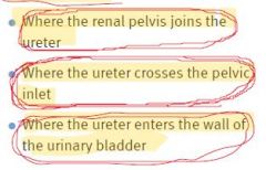
Critical points of the ureter for the blockage by renal calculi |
|
|
|
Muscles and bony landmarks of the posterior abdominal wall |
|
|
|
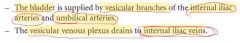
Blood supply to the urinary bladder |
|
|
|
Lymphatic drainage of the urinary bladder |
Drain to the external and internal iliac nodes |
|
|
Innervation of the urinary bladder |
|
|
|
Innervation of the external urethral sphincter |
Perineal branches of the pudendal nerve |
|
|
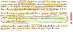
Urethra |
|
|
|
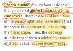
Spastic bladder |
|
|
|
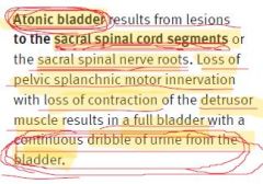
Atonic bladder |
|
|
|
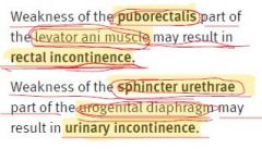
Weakness of the puborectalis and sphincter uretrae |
|
|
|
Column of Bertin |
Renal column |
|
|
Endocrine functions of kidneys |
-renin -erythropoietin -prostaglandins - vasodilators |
|
|
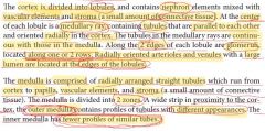
Collections of straight tubules form the medullary rays, which run up the center axis of a renal lobule. The border of the renal lobule is defined by the interlobular blood vessels. Between the interlobular blood vessels and the medullary ray are the renal corpuscles and convoluted tubules, together constituting the cortical labyrinth. |
|
|
Organization of the kidney |
|
|
|
Blood circulation in kidneys |
|
|
|
Uriniferous tubule |
Nephron + collecting duct |
|
|
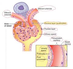
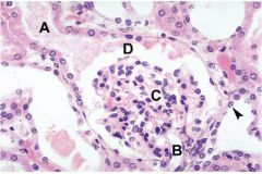
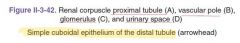
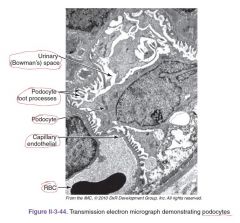
Renal corpuscle and Bowman's capsule picture |
|
|
|
|
|
|

|
|
|
|
|
|
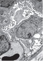
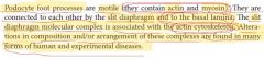
Podocyte foot processes |
|
|
|
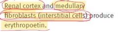
Erythropoietin production |
|
|
|
Polkissen or Lacis cells |
|
|
|
Renal corpuscle and juxtaglomerular apparatus picture |
|
|
|
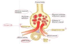
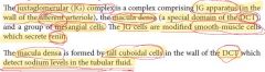
Juxtaglomerular complex |
|
|
|
Juxtaglomerular cells. Location and function. |
Modified smooth-muscle cells! Location: in the wall of the afferent arteriole Function: secrete renin |
|
|
Macula densa. Location and function. |
Formed by tall cuboidal cells Location: in the wall of the distal convoluted tubule Function: detect sodium levels in the tubular fluid |
|
|
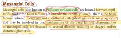
Intraglomerular mesangial cell |
|
|
|
Extraglomerular mesangial cell |
|
|
|
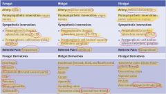
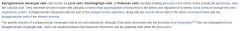
Adult male and female reproductive structures derived from precursors of the indifferent embryo |
|
|
|
Gonads derivatives in male and female |
Male (+TDF!): testes, seminiferous tubules, rete testes Female: ovary, follicles, rete ovarii |
|
|
Paramesonephric ducts derivatives in male and female |
Male (-MIF!): Appendix of testes (hydatid of Morgagni = remnant) Female: Uterine tubes, uterus, cervix, and upper part of vagina |
|
|
Mesonephric ducts derivatives in males and females |
Males (+Testosterone!): Epididymis, ductus deferens, seminal vesicle, ejaculatory duct FEmales: Duct of Gartner = remnant |
|
|
Genital tubercle derivatives in males and females |
Males (+DHT!): Glans and body of penis Females: Clitoris |
|
|
Urogenital folds derivatives in males and females |
Male (+DHT!): ventral aspect of penis Female: Labia minora |
|
|
Labioscrotal swellings derivatives in males and females |
Male: Scrotum Female: Labia majora |
|
|
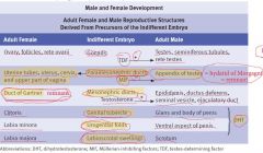
Hydatid of Morgagni |
|
|
|
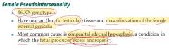
Female pseudointersexuality |
|
|
|
Male pseudointersexuality |
|
|
|
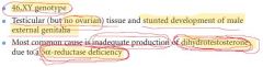
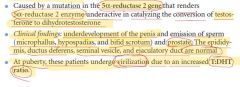
5α-reductase 2 deficiency |
|
|
|
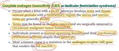
CAIS |
|
|
|
Chordee |
A condition in which the head of the penis curves downward or upward, at the junction of the head and shaft of the penis. The curvature is usually most obvious during erection, but resistance to straightening is often apparent in the flaccid state as well. |
|
|
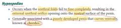
Hypospadias |
|
|
|
Bladder exstrophy |
Also known as "Ectopia vesicae" A congenital anomaly that exists along the spectrum of the exstrophy-epispadias complex and most notably involves protrusion of the urinary bladder through a defect in the abdominal wall. Its presentation is variable, often including abnormalities of the bony pelvis, pelvic floor, and genitalia. The underlying embryologic mechanism leading to bladder exstrophy is unknown, though it is thought to be in part due to failed reinforcement of the cloacal membrane by underlying mesoderm. |
|
|
Epispadias |
|
|
|
Cryptorchidism |
|
|
|
When does the descend of testes normally occur? |
Within 3 months after birth |
|
|
Vaginal process |
|
|
|
Canal of Nuck |

|
|
|
Hydrocele of the testes |
|
|
|
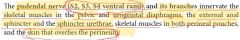
Innervation of the pelvic and urogenital diaphragms |
Branches of the pudendal nerve |
|
|
Pelvic diaphragm |
|
|
|
Urogenital diaphragm |
|
|
|
Kegel exercise |
|
|
|
Parts of the levator ani muscle |
1. Pubococcygeus - main, can be damaged in parturition 2. Puborectalis 3. Ileococcygeus |
|
|
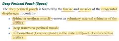
Where is the bulbourethral gland located? |
Deep perineal pouch |
|
|
Where is the Bartholin gland located? |
Superficial perineal pouch |
|
|
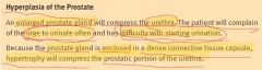
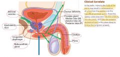
Hyperplasia of the prostate |
|
|
|
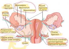
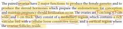
Where are the ovarian vessels located? |
In the suspensory ligament of ovary |
|
|
Ureter vs. suspensory ligament of the ovary location |

|
|
|
Ligaments of the female reproductive tract |
|
|
|
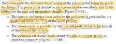
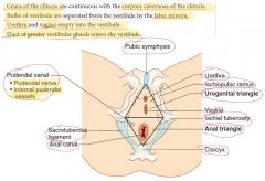
Perineum |
|
|
|
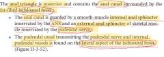
Anal triangle |
|
|
|
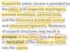
Supporting structures of the pelvic viscera |
|
|
|
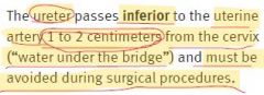
"Water under the bridge" |
|
|
|
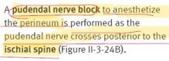
Pudendal nerve block |
|
|
|
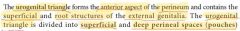
Urogenital triangle |
|
|
|
Cowper glands |
Bulbourethral Located in the deep perineal pouch |
|
|
Bartholin glands |
Greater vestibular glands Located in the superficial perineal pouch |
|
|
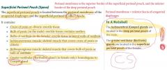
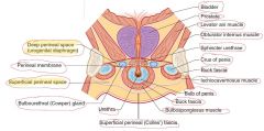
Superficial perineal pouch |
|
|
|
Deep perineal pouch |
|
|
|
Colles' fascia |
Superficial perineal = Scarpa fascia in abdomen = dartos fascia of scrotum = superficial fascia of clitoris or penis |
|
|
Superficial and deep perineal pouches of male picture |
|
|
|
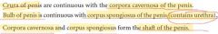
Shaft of the penis |
|
|
|
Male reproductive system |
|
|
|
Perineum of female picture |
|
|
|
Pelvic and perineal innervation |
|
|
|
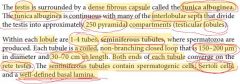
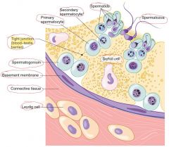
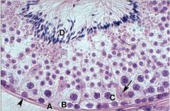
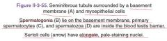
Testicular histology |
|
|
|
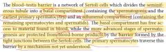
Blood-testis barrier |
|
|
|
Seminiferous tubule diagram |
|
|
|
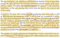
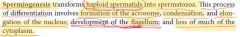
Spermatogenesis |
|
|
|
|
|
|
Spermiogenesis |
|
|
|
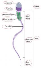
Spermatozoan diagram |
|
|
|
The acrosome is derived from the ... |
Golgi complex |
|
|
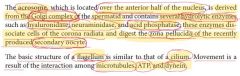
Acrosome |
|
|
|
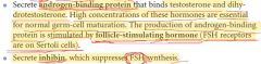
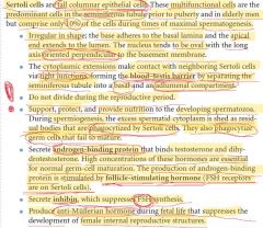
Androgen binding protein and inhibin |
|
|
|
Sertoli cells |
|
|
|
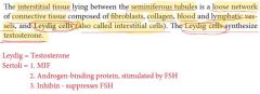
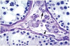
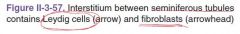
Leydig cells |
|
|
|
|
|
|

|
|
|
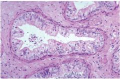
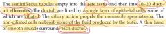
Rete testis |
|
|
|
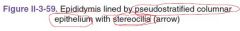
Epithelial lining of the epididymis |
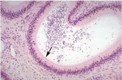
Pseudostratified columnar epithelium wich contains stereocilia (tall microvilli) |
|
|
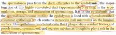
Where do the spermatozoa become motile? |
Epididymis |
|
|
Epididymis |
|
|
|
|
|
|
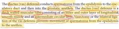
Ductus (vas) deferens |
|
|
|

|
|
|
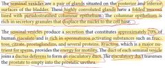
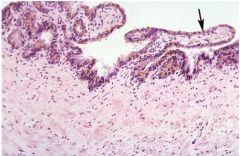
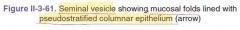
Epithelial lining of the seminal vesicles |
Pseudostratified columnar epithelium |
|
|
How much (in %) do the seminal vesicles constitute to the human ejaculate |
70% |
|
|
Seminal vesicles |
|
|
|
What substance provides energy for the spermatozoa's motility? |
Fructose |
|
|
|
|
|
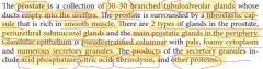
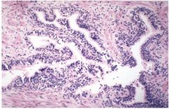
Prostate histology |
|
|
|
Epithelial lining of the prostate |
Pseudostratified columnar epithelium |
|
|

|
|
|
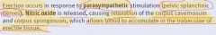
Penile erection |
|
|
|
Ejaculation |
|
|
|
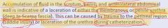
Accumilation of fluid in the scrotum, penis, and anterolateral abdominal wall |
|
|
|
Ovarian histology |
|
|
|
Female reproductive system diagram |
|
|
|
Follicular development diagram |
|
|
|
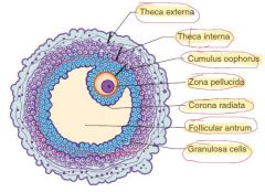
Graafian follicle diagram |
|
|
|
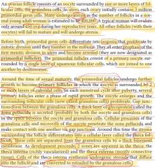
Primordial follicles |
Primary oocyte surrounded by a single layer of squamous follicular cells, which are joined to one another by desmosomes |
|
|
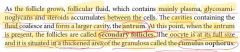
Primary follicles |
Develop from the primordial follicles Oocyte is surrounded now by 2 or more layers of cuboidal cells |
|
|
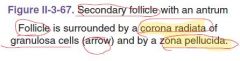
What is zona pellucida? |
A thick layer of glycoprotein synthesized by both granulosa cells and oocyte |
|
|
Difference between the theca interna and theca externa (follicle) |
Theca interna - richly vascularized, synthesize androgenic steroids, that diffuse into follicle and are converted to estradiol by the granulosa cells |
|
|
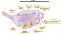
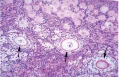
Ovarian follicles |
|
|
|

|
|
|
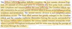
Secondary follicle |
|
|
|
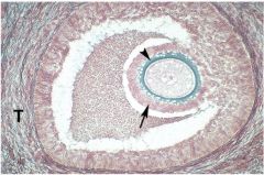
Graafian follicle |
|
|
|
|
|
|
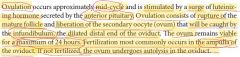
Ovulation |
|
|
|
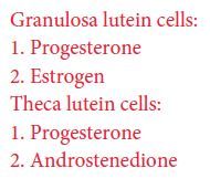
What do the granulosa lutein cells and theca lutein cells produce? |
From theca interna! |
|
|
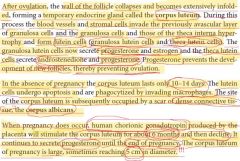
Corpus luteum |
|
|
|
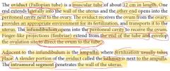
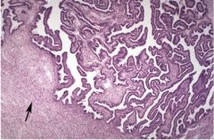
Oviduct |
|
|
|
|
|
|
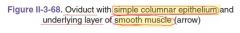
Epithelial lining of the oviducts |
Simple columnar! Some cells are ciliated! |
|
|
Layers of the oviduct |
No submucosa! 1. Mucosa 2. Muscularis 3. Serosa |
|
|
Will the Kartagener's syndrome cause impairment of the tubular transport in women? |
No |
|
|
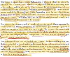
Source of the stem cells for the functional layer of the myometrium |
The bases of the uterine glands, which lie deep in the basalis |
|
|
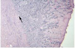
Epithelial lining of the uterus |
Simple columnar cells (ciliated and secretory) |
|
|
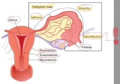
Uterus |
|
|
|
|
|
|
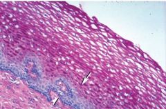
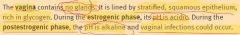
Does the vagina have glands? |
No! |
|
|
Epithelial lining of the vagina |
Stratified sqamous |
|
|
|
|
|
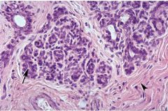
Mammary glands' histology |
|
|
|

|
|
|
|
|
|
|
|
|
|
|
|
|
|
|
|
|
|
|
|
|
|
|
|
|
|
|
|
|
|
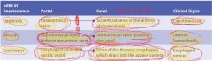
4 collateral portacaval anastomoses |
1. Esophageal 2. Rectal 3. Umbilical 4. Retroperitoneal |
|
|
How does the gallbladder concentrate bile? |
It removes water through active transport of sodium and chloride ions (especially chloride!) |
|
|
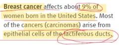
Breast cancer epidemiology |
|
|
|
Epithelial lining of the urethra |
Prostatic urethra - transitional epithelium Distal urethra - stratified epithelium |
|
|
Corpora CAVERNOSA are surrounded by ... |
Tunica albuginea |
|
|
Why there is breakdown of the functional level? |
Constriction of the spiral arteries |
|
|
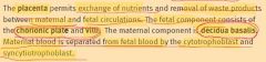
Fetal and maternal components of the placenta |
|
|
|
pH in the vagina during estrogenic and postestrogenic phases |
|
|
|
Brachial plexus segments |
C5-T1 |
|
|
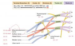
Brachial plexus diagram |
|
|
|
Musculocutaneous nerve Segments? Muscles innervated? Primary actions? |

|
|
|
Median nerve Segments? Muscles innervated? Primary actions? |

|
|
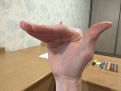
What muscles are in action? |
Lumbricals 1-4 |
|
|
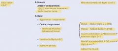
Ulnar nerve Segments? Muscles innervated? Primary actions? |
|
|
|
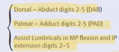
Palmar and dorsal interossei muscles mnemonics |
|
|
|
Axillary nerve Segments? Muscles innervated? Primary actions? |

|
|
|
Teres minor and teres major functions |
Teres major - medial rotation (we are stronger in medial rotation, thus, teres MAJOR) Teres minor - lateral rotation |
|
|
Radial nerve Segments? Muscles innervated? Primary actions? |

|
|
|
Nerves of the upper limbs that provide supination and pronation |
Supination: -Musculocutaneous nerve (biceps brachii) -Radial nerve (supinator muscle) Pronation: -Median nerve |
|
|
Is the biceps brachii the most powerful flexor of the forearm? |
No! Brachialis is. Biceps is powerful supinator. |
|
|
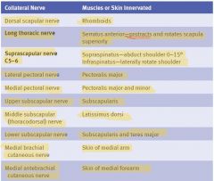
Collateral nerves of the brachial plexus |
|
|
|
Dorsal scapular nerve innervation |
Rhomboids |
|
|
Long thoracic nerve innervation |
Serratus anterior - protracts and rotates scapula superiorly |
|
|
Suprascapular nerve innervation |
Supraspinatus - abduct shoulder 0-15 degrees Infraspinatus - LATERALLY rotate shoulder |
|
|
Lateral pectoral nerve innervation |
Pectoralis major |
|
|
Medial pectoral nerve innervation |
Pectoralis major and minor |
|
|
Upper subscapular nerve innervation |
Subscapularis |
|
|
Middle subscapular (thoracodorsal) nerve innervation |
Latissimus dorsi |
|
|
Lower subscapular nerve innervation |
Subscapularis and teres major |
|
|
Medial brachial cutaneous nerve innervation |
Skin of medial arm |
|
|
Medial antebrachial cutaneous nerve innervation |
Skin of medial forearm |
|
|
Segmental inervation to muscles of upper limbs |
|
|
|
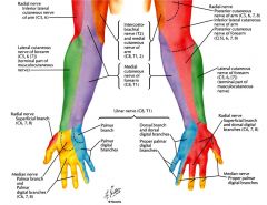
Sensory innervation of the hand |
|
|
|
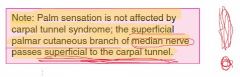
Is the palm sensation affected by the carpal tunnel syndrome? |
|
|
|
Sensory innervation of the hand and forearm diagram |
|
|
|
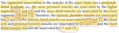
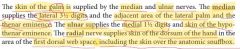
Sensory innervation of the lateral forearm |
Musculocutaneous nerve (C5-C6) -> lateral cutaneous nerve of forearm |
|
|
Sensory innervation of the medial forearm |
Medial cutaneous nerve of forearm (C8-T1) |
|
|
Sensory innervation of the lateral arm |
Radial nerve -> lateral cutaneous nerve of arm |
|
|
Sensory innervation of the medial arm |
Medial cutaneous nerve of arm (C8-T1) |
|
|
Sensory innervation of the lateral dorsal 3.5 distal phalangs |
Median nerve |
|
|
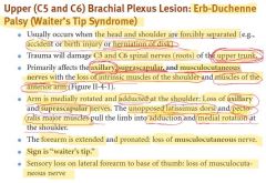
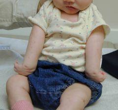
Erb-Duchenne palsy |
|
|
|
What nerves are affected in Erb-Duchenne palsy? |
-axillary -suprascapular -musculocutaneous |
|
|
Erb-Duchenne palsy |
|
|
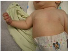
Klumpke's paralysis |
|
|
|
Klumpke's paralysis |
|
|
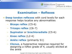
Deep tendon reflexes levels |
|
|
|
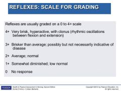
Deep tendon reflexes scale |
|
|
|
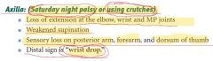
Radial nerve injury in axilla |
Also, honeymoon palsy |
|
|
Elbow - lateral epicondyle or radial head dislocation |
Radial nerve injury |
|
|
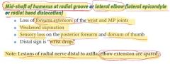
Radial nerve injury in mid-shaft of humerus at radial groove or lateral elbow (lateral epicondyle or radial head dislocation) |
|
|
|
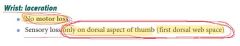
Radial nerve injury in wrist laceration |
|
|
|
Waiter's tip sign |
Erb-Duchenne palsy |
|
|
Combination of "claw hand" and "ape hand" signs |
Klumpke's paralysis |
|
|
"Wrist drop" |
Radial nerve injury |
|
|
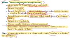
Median nerve injury - elbow (supracondylar fracture of humerus) |
|
|
|
Combination of the "hand of benediction" and "ape hand" |
Median nerve injury - elbow (supracondylar fracture of humerus) |
|
|
Median nerve injury - wrist laceration or carpal tunnel |
|
|
|
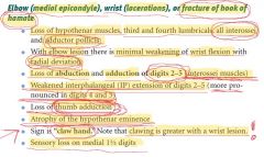
Ulnar nerve injury |
|
|
|
Fracture of hook of hamate |
Ulnar nerve injury |
|
|
Axillary nerve injury |
|
|
|
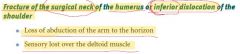
Fracture of the surgical neck of the humerus or inferior dislocation of the shoulder |
Axillary nerve injury |
|
|
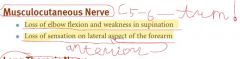
Musculocutaneous nerve injury |

|
|
|
Long thoracic nerve injury |
|
|
|
"Winged scapula" |
Long thoracic nerve injury (radical mastectome or stab wound to lateral chest) |
|
|
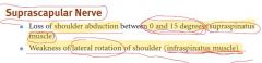
Suprascapular nerve injury |
|
|
|
"Ulnar claw" vs. "Hand of benediction" |

|
|
|
"Dupuytren's contracture" vs. "ulnar claw" |

|
|
|
Dupuytren's contracture |
|
|
"Ulnar claw" |
|
|
Spinster's claw |
"Ulnar claw" |
|
|
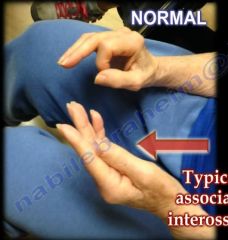
Ulnar paradox |

|
|
|
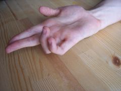
Pathogenesis of the "ulnar claw" |

|
|
Unable to make the O.K. sign |
Anterior interosseus nerve injury |
|
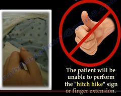
Unable to perform the "hitch hike" sign |
Radial nerve injury |
|
|
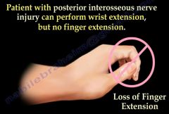
Posterior interosseus nerve injury |
|
|
|
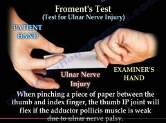
Froment's test |
|
|
|
Pronator teres syndrome |
Median nerve injury |
|
|
Supracondylar fracture of humerus |
Median nerve injury |
|
|
Fracture of medial epicondyle of humerus |
Ulnar nerve injury |
|
|
What nerve may be damaged in fracture of clavicle? |
Ulnar nerve |
|
|
Effects of lesions to branches of the brachial plexus |
|
|
|
Axillary artery margins |
From the first rib to the posterior edge of the teres major muscle |
|
|
What nerve runs with the lateral thoracic artery? |
Long thoracic nerve |
|
|
Shoulder arterial collateral |
-Subscapular artery << axillary artery -Suprascapular artery << subclavian artery |
|
|
What artery runs with axillary nerve? |
Posterior humeral circumflex artery - at surgical neck |
|
|
What artery runs with radial nerve in radial groove at midshaft of humerus? |
Profunda brachii artery |
|
|
Deep palmar arch is from ... |
Radial artery |
|
|
Superficial palmar arch is from ... |
Ulnar artery |
|
|
Common interosseus artery is from ... |
Ulnar artery |
|
|
Arterial supply to the upper limb picture |
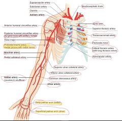
|
|
|
Collateral circulation in the hand |
Superficial and deep palmar arches (ulnar and radial arteries) |
|
|
Boundaries of the carpal tunnel |
Anteriorly - flexor retinaculum Posteriorly - proximal row of carpal bones (lunate) |
|
|
Carpal tunnel |
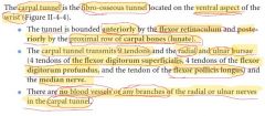
|
|
|
Carpal tunnel syndrome |
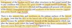
|
|
|
Rotator cuff muscles mnemonics |
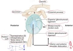
Counterclockwise: SITS! |
|
|
Humeral head dislocation |
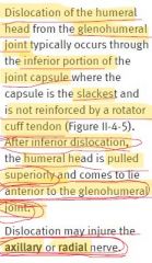
|
|
|
Most frequently damaged muscle of the rotator cuff |
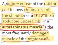
|
|
|
Humeral surgical neck fracture |
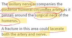
|
|
|
Mid-shaft (radial groove) humeral fracture |
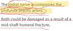
|
|
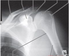
|
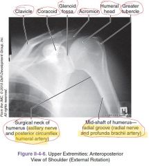
|
|
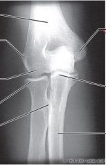
|
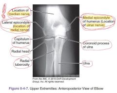
|
|
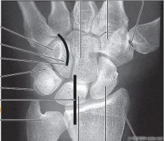
|
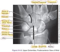
|
|
|
Most frequently fractured and most frequently dislocated carpal bones |
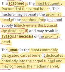
|
|
|
Hook of the hamate fracture |
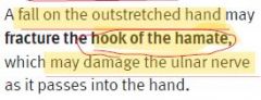
|
|
|
Lumbosacral plexus levels |
L2-S3 |
|
|
Division. Level. -Femoral nerve -Obturator nerve -Tibial nerve -Common fibular nerve -Superior gluteal nerve -Inferior gluteal nerve |
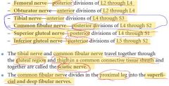
|
|
|
Lumbosacral plexus chart |
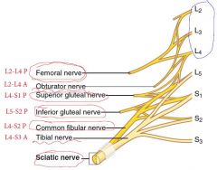
|
|
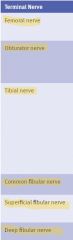
Origin. Muscles innervated. Primary actions. |
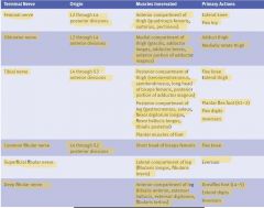
|
|
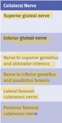
Origin. Muscles or skin innervated. Primary actions. |
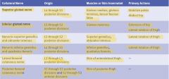
|
|
|
Segmental innervation to muscles of lower limb |
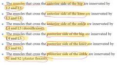
|
|
|
Trendelenburg gait |
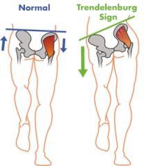
Gluteus medius (weakness in abduction) - superior gluteal nerve |
|
|
Superior gluteal nerve injury |
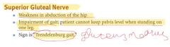
|
|
|
Inferior gluteal nerve injury |

|
|
|
Femoral nerve injury |
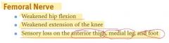
|
|
|
Obturator nerve injury |

|
|
|
Sciatic nerve injury |
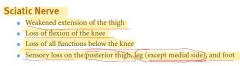
|
|
|
Tibial nerve injury |
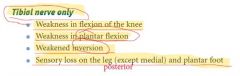
|
|
|
Common fibular nerve injury (neck of fibula) |
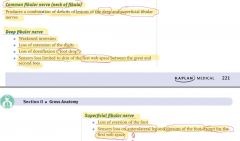
|
|
|
Deep fibular nerve injury |
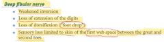
|
|
|
Superficial fibular nerve injury |

|
|
|
Most frequently damaged nerve of the lower limb |
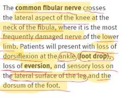
|
|
|
Piriformis syndrome |
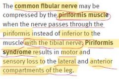
|
|
|
What nerve may be lesioned in posterior hip dislocation? |
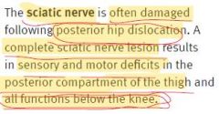
|
|
|
Sensory innervation of the lower leg and foot |
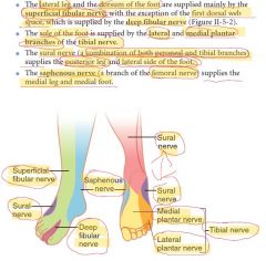
|
|
|
Sural nerve |
It is made up of collateral branches of the tibial nerve and common fibular nerve. The medial cutaneous branch arises from the tibial nerve, and the lateral branch arises from the common fibular nerve. |
|
|
Arterial supply to the legs |
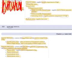
|
|
|
What artery supplies head of femur? |
Medial circumflex femoral artery |
|
|
Avascular necrosis of the head of the femur |
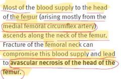
|
|
|
What artery is responsible for pulse on dorsum of foot? |
Dorsalis pedis artery |
|
|
Arterial supply to lower limb picture |
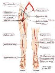
|
|
|
Tibial shaft fractures |
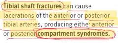
|
|
|
Femoral triangle |
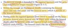
|
|
|
Femoral sheath contents |
Femoral artery anv vein and canal No femoral nerve! |
|
|
Mnemonics for the structures in the femoral triangle |
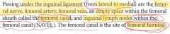
|
|
|
Ligaments of the hip joint |
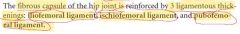
|
|
|
Structures of the knee picture |
|
|
|
Most frequently torn ligament at the knee |
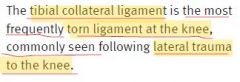
|
|
|
Tibial collateral ligament |
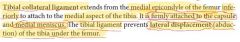
|
|
|
The collateral ligaments are taut with knee flexion or extension? |
Extension |
|
|
Fibular collateral ligament |
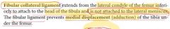
|
|
|
Anterior and posterior cruciate ligaments |
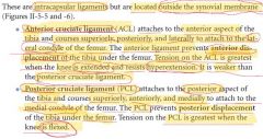
|
|
|
Anterior and posterior cruciate ligaments chart |

|
|
|
Medial and lateral menisci |
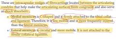
|
|
|
Anterior and posterior drawer signs |
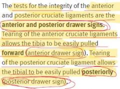
|
|
|
Pain when the leg is medially rotated at the knee |
Medial meniscus tear |
|
|
3 most commonly injured structures at the knee |
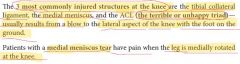
|
|
|
Lateral (collateral) ligament of ankle parts |
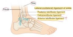
|
|
|
Medial (deltoid) ligament of ankle parts |
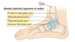
|
|
|
Most common injuries in ankle joint |
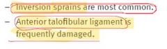
|
|
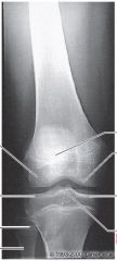
|
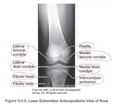
|
|
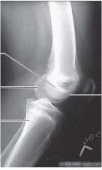
|
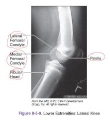
|
|
|
Thoracic outlet. Scalene triangle. |
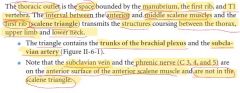
|
|
|
Thoracic outlet syndrome |
|
|
|
Scalene triangle picture |
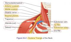
|
|
|
Internal (2) and external (8) carotid arteries branches |
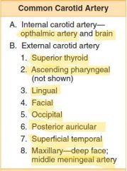
|
|
|
Subclavian artery branches (7) |
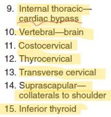
|
|
|
The most significant artery of the external carotid system |
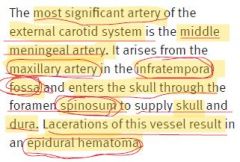
|
|
|
Pharyngeal apparatus |
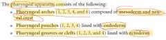
|
|
|
Fetal pharyngeal apparatus picture |
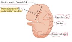
|
|
|
Section through the developing pharynx (picture) |
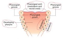
|
|

Pharyngeal arch 1 |
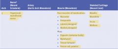
|
|

Pharyngeal arch 2 |

|
|

Pharyngeal arch 3 |

|
|

Pharyngeal arch 4 |

|
|

Pharyngeal arch 6 |

|
|
|
Where do the ocular muscles (III, IV, VI) and the tongue muscles (XII) derive from? |
Mesoderm of the occipital somites (somitomeres) Not from the pharyngeal arch mesoderm! |
|
|
Fetal pharyngeal pouches picture |
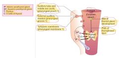
|
|
|
Adult structures derived from the fetal pharyngeal pouches |
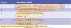
|
|
|
Adult structures derived from the fetal pharyngeal grooves (clefts) |
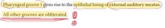
|
|
|
Branchial cyst (lateral cervical cyst) |
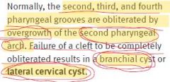
|
|
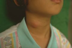
|
Branchial cyst (lateral cervical cyst) |
|
|
DiGeorge syndrome. Contemporary name. |
22q11.1 deletion syndrome Pharyngeal pouches 3 and 4 fail to differentiate into the parathyroid glands and thymus. Neural crest cells are involved. -Immunologic problems -Hypocalcemia May be combined with: -Cardiovascular defects (persistent truncus arteriosus) -Abnormal ears -Micrognathia |
|
|
Thyroid gland development |
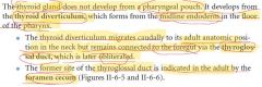
|
|
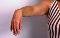
|
"Wrist drop" - radial nerve palsy |
|
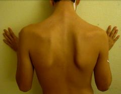
|
"Winged scapula" - serratus anterior - long thoracic nerve |
|
|
Treacher Collins syndrome |
|
|
Pierre Robin sequence |
|
|
Treacher Collins syndrome |
Abnormal formation of pharyngeal arch 1 because of faulty migration of neural crest cells. -Mandibular hypoplasia -Zygomatic hypoplasia -Down-slanted palpebral fissures -Colobomas -Malformed ears |
|
|
Pierre Robin sequence |
Abnormal formation of pharyngeal arch 1 because of faulty migration of neural crest cells. Triad: 1. Poor mandibular growth 2. Cleft palate 3. Posteriorly placed tongue Normal ears (differentiation with DiGeorge) |
|
|
What is foramen cecum (tongue)? |
Former site of the thyroglossal duct |
|
|
Development of the tongue |

|
|
|
Innervation of the tongue |
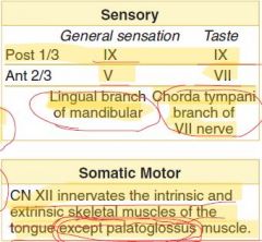
|
|
|
Paltoglossus muscle innervation |
Vagus nerve (via pharyngeal branch to pharyngeal plexus) All other intrinsic and extrinsic muscles of tongue - CN XII |
|
|
Development of the face and palate |
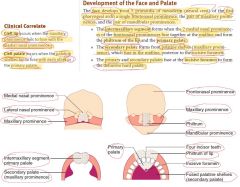
|
|
|
Cleft lip and cleft palate pathogenesis |
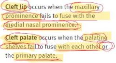
|
|
|
First arch syndrome |
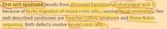
|
|
|
Pharyngeal fistula and pharyngeal cyst |
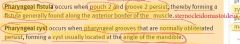
|
|
|
Ectopic thyroid, parathyroid, or thymus |
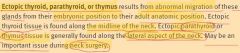
|
|
|
Thyroglossal duct cyst or fistula |
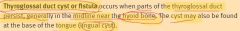
|
|
|
Cribriform plate fractures |
May result in dysosmia and rhinorrhea (CSF) |
|
|
Foramen lacerum function |
-The foramen lacerum fills with cartilage after birth. -The artery of pterygoid canal, the nerve of pterygoid canal and some venous drainage pass through the foramen lacerum. -Some emissary veins pass through the foramen lacerum. These connect the extracranial pterygoid plexus with the intracranial cavernous sinus and present an unopposed route for infection. -The Foramen lacerum is a potential route for nasopharyngeal carcinoma to gain access to the cavernous sinus and affect cranial nerves. |
|
|
What do the cranial foramens contain? 1. Cribriform plate 2. Optic canal 3. Superior orbital fissure 4. Foramen rotundum 5. Foramen ovale 6. Foramen spinosum 7. Foramen lacerum 8. Internal auditory meatus 9. Jugular foramen 10. Hypoglossal canal 11. Foramen magnum |
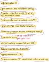
|
|
|
Jugular foramen syndrome |
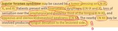
|
|
|
Where the tongue will deviate in CN XII lesion? |
Towards the lesioned side! |
|
|
What does the stylomastoid foramen contain? |
CN VII |
|
|
What does the carotid canal contain? |
Internal carotid artery and carotid sympathetic nerve |
|
|
What do the supraorbital, infraorbital, and mental foramens contain? |
Supraorbital, infraorbital, and mental VANs, respectively |
|
|
What does theinferior orbital fissure contain? |
-zygomatic branch of the maxillary nerve -ascending branches from the pterygopalatine ganglion -inferior division of ophthalmic vein |
|
|
Where does the CSF returns back to the systemic venous circulation? |
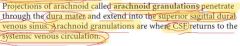
|
|
|
Coronal section of the dural sinuses picture |
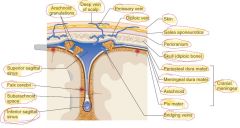
|
|
|
What does the jugular foramen contain? |
CN IX, X, XI and internal jugular vein |
|
|
3 duplicatons of meningeal dura mater |
-falx cerebri -tentorium cerebelli -diaphragma sellae |
|
|
Primary tributaries that flow into the dural sinuses |
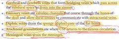
|
|
|
Dural venous sinuses picture |
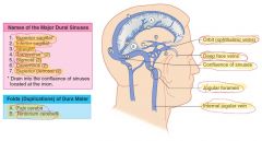
|
|
|
How is the inferior sagittal sinus formed? |
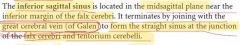
|
|
|
Dural venous sinuses |
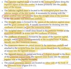
|
|
|
Cavernous sinus |
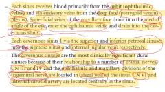
|
|
|
Cavernous sinus picture |

|
|
|
What does the cavernous sinus contain? |
-Lateral wall of the sinus: CN III, IV, V1, and V2 -Centrally: CN VI and internal carotid artery |
|
|
Cavernous sinus thrombosis |
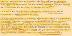
|
|
|
Initial symptom of cavernous sinus thrombosis |
Internal strabismus (CN VI lesion) |
|
|
Epidural hematoma |
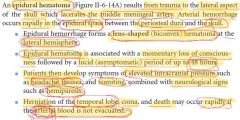
|
|
|
Lens-shaped (biconvex) hematoma at the lateral hemisphere |
Epidural hematoma (middle meningeal artery) |
|
|
Initial symptoms of uncal herniation |
The uncus can squeeze the oculomotor nerve (a.k.a. CN III), which may affect the parasympathetic input to the eye on the side of the affected nerve, causing the pupil of the affected eye to dilate and fail to constrict in response to light as it should. Pupillary dilation often precedes the somatic motor effects of CN III compression called oculomotor nerve palsy or third nerve palsy. This palsy presents as deviation of the eye to a "down and out" position due to loss of innervation to all ocular motility muscles except for the lateral rectus (innervated by abducens nerve (a.k.a. CN VI) and the superior oblique (innervated by trochlear nerve a.k.a. CN IV). The symptoms occur in this order because the parasympathetic fibers surround the motor fibers of CN III and are hence compressed first. |
|
|
Subdural hematoma |
|
|
|
Crescent-shaped hematoma at the lateral hemisphere |
Subdural hematoma |
|
|
Epidural hematoma |
|
|
Subdural hematoma |
|
|
Subarachnoid hemorrhage |
|
|
|
Xanthochromia |
Yellowish appearance of cerebrospinal fluid that occurs several hours after bleeding into the subarachnoid space caused by certain medical conditions, most commonly subarachnoid hemorrhage. Its presence can be determined by either by spectrophotometry (measuring the absorption of particular wavelengths of light) or simple visual examination. |
|
|
Orbital muscles and their innervation |
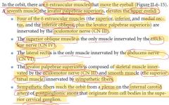
|
|
|
Clinical testing of superior oblique muscle |
Ask patient to look medially and downward! We can asses only the function of moving the eye downward. |
|
|
Function of thesuperior oblique muscle |
Abducts, depresses and internally rotates the eye |
|
|
Function of the inferior oblique muscle |
Primary action is extorsion (external rotation); secondary action is elevation; tertiary action is abduction (i.e. it extorts the eye and moves it upward and outwards). The inferior oblique muscle is the only muscle that is capable of elevating the eye when it is in a fully adducted position. |
|
|
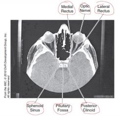
|
|
|
Muscles of the tongue developments |
|