Reading...
![]()
Play button
![]()
Play button
![]()
Use LEFT and RIGHT arrow keys to navigate between flashcards;
Use UP and DOWN arrow keys to flip the card;
H to show hint;
A reads text to speech;
95 Cards in this Set
- Front
- Back
|
Hemianopia
|
Loss of vision on the opposite side
i.e. RHS brain removal, LHS loss of vision |
|
|
Hemiplegia
|
Paralysis of one side of the body.
Due to damage to contralateral motor cortex. Paralysis is worse at the extremitites (finger movements may be lost, but some movement of the arm maintained). |
|
|
Association areas
|
Don't show fixed sensory mapping, implicated in higher mental functions. Can't point to a particular location and say its the organizing centre for "planning"- higher tasks depend on control exerted from many locations
|
|
|
Agnosia
|
Visual Agnosia - cannot identify familiar objects. If you give the object to the person to hold they would be able to identify it.
Caused by damage to occipital cortex, parts of temporal and parietal lobes. Can identify separate details of a picture but cannot identify the picture as a whole. |
|
|
Prosopagnosia
|
Damage to the association area on the temporal/occipital lobes
Difficulty recognising faces. Some can't recognise familiar faces, some can't recognise a face as a face. |
|
|
How the brain knows WHAT things are:
|
Visual stimuli (after following an intricate pathway) finally ends up at the occipital lobe, & then is directed to the temporal lobe: "what" system, ventral stream, memory.
So if you have damage to the "what" system- you can locate objects in space, and shape your hand to pick things up, but you don't know what it is. Can use other cues |
|
|
How the brain knows WHERE things are:
|
The "where" system - dorsal stream
Visual stimuli (after following an intricate pathway) finally ends up at the occipital lobe and then is directed to the parietal lobe So if you have damage to the "where" system - you can't locate objects in space, and shape your hand to pick things up, but you know what it is. |
|
|
Symptoms of damage to prefrontal cortex and an example of a person this happened to:
|
Phineas gage.
Deficiency in response inhibition Inability to plan Appear uninvolved, depressed and apathetic Some may appear "psychopathic", acting flagrantly and crudely, being sexually promiscuous and may engage in criminal conduct. Frontal lobes not fully understood |
|
|
Describe prefrontal lobotomy and a cite an example case.
|
1940's-1950's surgery disconnected prefrontal areas. Helped some, used indiscriminately.
Patients - docile but cognitively disabled. No longer widely used after the 1950's due to drugs. |
|
|
Describe Apraxia
|
Damage in the frontal lobe. Serious disturbances in initiation or organisation of voluntary action
Unable to perform well known actions Actions become fragmented and disorganised. |
|
|
Describe Aphasia
|
Language disorder. Results from left-hemisphere damage. Middle-cerebral artery (MCA) runs close to language areas.
|
|
|
Functions of Broca's area
|
speech production and syntax
|
|
|
Functions of Wernicke's area
|
speech comprehension
|
|
|
Function of Arcuate fasciculus
|
Connects Broca and Wernickes areas. (i.e. connects speech production and speech comprehension)
|
|
|
Function of Angular Gyrus
|
Reading
|
|
|
Broca's aphasia: describe and give an example of
|
expressive, motor, production, nonfluent
Paul Broca. Can move tongue, vocalize, swear, but suffered from: Agrammatism - understanding generally alright, but complex syntax not well understood. Lack of grammar. eg. Monday ah Dad and Paul and Dad...hospital etc. |
|
|
Describe and give an example of Wernicke;s aphasia
|
Sensory, receptive, fluent
Carl Wernicke Lack of comprehension. Speaking is fluent and grammatical but meaningless (jargon aphasia) The words may not actually exist |
|
|
Sign Language Aphasia
|
Occurs with language and involves the same areas.
Sign language has all the essential properties of spoken language. |
|
|
Characteristics of Dyslexia
|
Any reading disability not associated with obvious problems such as bad eyesight.
Occurs more commonly in LHers Inability to name letters, read words or sentences, or recognise words directly although they can be sounded out |
|
|
The two types of dyslexia and their characteristics
|
Acquired: reading impairment due to left brain damage (including angular gyrus) in those with previously normal language. Deep (inability to read), phonological (inability to read non words), surface (inability to tell some letters apart)
Developmental: difficulty in learning to read despite adequate intelligence and appropriate educational opportunity. |
|
|
Characteristics of neglect syndrome
|
People with right sided parietal damage tend to neglect the left side of space (seldom vice versa, if it does occur usually transient)
Can be visual, auditory or tactual. Problem on attention - sometimes called hemi-inattention an emotional component - denial of any deficit (anosognosia) |
|
|
Why is neglect asymmetrical?
|
Right side of brain (parietal lobe) controls attention to both sides of space.
Left side controls attention to right side of space only. Therefore damage to the right side causes neglect of the left side. |
|
|
The asymmetrical brain: Right side dominant for:
|
Spacial attention: neglect
Melody - amusia Facial recognition - prosopagnosia Recognition of natural objects - agnosia |
|
|
The asymmetrical brain: Left side dominant for:
|
Language- aphasia
Recognition of manufactured objects- agnosia Voluntary action -apraxia |
|
|
explain split brain surgery and its use
|
Relief of intractable, multifocal epilepsy
Separates left and right hemispheres and prevents seizures from spreading through the brain. In the 60's all forebrain commissures were sectioned - commissurotomy From the 70's onwards only the corpus callosum was sectioned -callosotomy Done in two stages. Usually anterior (front), and if this doesn't work, followed by posterior (back) section |
|
|
Corpus Callosum
|
Largest cerebral commissure (something that communicates with both sides), over 200 million axons
|
|
|
Effects of split-brain
|
Patients seem quite normal.
Everyday evidence of a split mind is rare, but sometimes occurs - "alien hand" - right hand takes on a life of its own. Left hemisphere function in split brain: -Cant name objects or words presented in the LEFT visual field. -Cant name objects held in LEFT hand BUT can understand words in the left visual field (demonstrated by pointing) -Suggests that the right brain can understand but cannot speak- does't have access to language. |
|
|
Callosal agenesis
|
In some people the corpus callosum does not develop. These people are "naturally" split brained.
Sometimes accompanied by Probst's bundles with are remnants of a corpus callosum that failed to cross the midline. Callosal agenesis is often accompanied by other neurological problems, although many of these people seem otherwise normal. Less evidence of disconnection in callosal agenesis objects and words usually easily named in either visual field So there is some form of compensation, evidence of neural plasticity, probably occurring very early. |
|
|
Neurons
|
Communicating elements of the brain
Perform computations inside the brain that we call "thought". |
|
|
Dendrites
|

Receive nerve impulses from other neurons.
Short- few hundred microns |
|
|
Axons
|

Transmit nerve impulses
|
|
|
Cell bodies/ soma
|

5-100 microns in diameter
|
|
|
Myelinated axons and unmyelinated axons
|
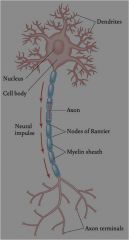
Myelinated axons= white matter
Unmyelinated axons: Grey matter |
|
|
Axon terminals
|

Secrete neurotransmitters in synapse
|
|
|
Name the 3 types of neurons and their locations
|
Motorneurons: Begin in CNS, exit through the spinal cord, end on muscle fibre.
Sensory neurons: Begin at sense organ (retina, skin, tongue) convey information to brain via spinal cord. Interneurons: interposed between other neurons, do much of the computation of the brain. |
|
|
Glial Cells
|
Make up about 90% of cells in brain
act as guidewires for growing neurons Later in development provide supportive scaffolding for mature neurons, assist in repair process when tissue is damaged |
|
|
What is the resting potential and action potential of an axon?
|
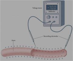
In the resting state, the inside of an axon is negative with respect to the surface, by about -70 millivolts.
If a pulse is applied the exceeds the excitation threshold (about -55 millivolts), then an action potential occurs. This causes the inside to swing positive relative to the outside. How it works: Resting potential depends on positive sodium ions (Na+) on the outside of the cell membrane. Ion channels let charged ions in and out of the cell. In resting state, Na+ ions are kept on the outside by a sodium pump, but in-going ion channels remain closed. When a pulse is applied in going channels open, Na+ ions pour into the cell, reversing the voltage difference. Na+ channels then close (potassium) K+ then leaves the cell to restore the original voltage difference. |
|
|
The Synapse
|
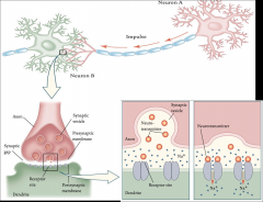
Where one neuron meets another. Transmission of an action potential along one neuron may cause the next neuron to fire (excitation) or it may inhibit firing (inhibition). Governed by the release of neurotransmitters.
|
|
|
Explain the Lock and Key model
|

Neurotransmitter molecules will only affect the postsynaptic membrane if the molecules shape fits into certain synaptic receptors
|
|
|
Explain synaptic reuptake
|
Neurotransmitters don't sit in the synapse: Inactivated by "cleanup" enzymes. Reused in synpatic reuptake
|
|
|
Define the two drug effects
|
Antagonists: Drugs that block or inhibit postsynaptic effects.
Agonists: Drugs that facilitate postsynaptic effects. |
|
|
What are the three ways in which drugs effect the synapse? *potential exam question (give me a drug example of each of these three points)
|

1. Stimulate or inhibit neurotransmitter release. (Venom of Black Widow spider: Acetylcholine agonist)
2. Stimulate or block postsynaptic receptor molecules (curare) 3. Inhibit reuptake |
|
|
Drug that stimulates the neurotransmitter release
|
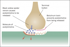
Stimulate terminal buttons to release neurotransmitters continuously even when axon is not firing.
Venom of Black widow spider: Acetylcholine agonist -releases lots of acetylcholine. Exhausts victims supply of acetylcholine. For small prey leads to paralysis and death |
|
|
Drug that inhibits neurotransmitter release
|

Botulinum toxin: present in improperly canned food (bulging cans). Acetylcholine antagonist - prevents the release of acetylcholine. Even a small amount of this toxin- less than one millionth of a gram- victim paralyzed and suffocates to death. Used in botox in very small amounts
|
|
|
Drug that stimulates postsynaptic receptor molecules
|
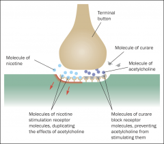
Drugs can duplicate the effects of some neurotransmitters by directly stimulating particular kinds of receptor molecules. Nicotine stimulates acetylcholine receptors - mimics acetylcholine.
Low doses - Pleasurable and addictive, excitatory effect. High doses - convulsions and death. |
|
|
Drug that blocks postsynaptic receptor molecules
|

Other drugs block receptors, making them inaccessible to the neurotransmitter and inhibit synaptic transmission. Curare- used on darts of blowguns. Blocks acetylcholine receptors located on muscle fibres. Victim can't breathe and suffocates to death.
|
|
|
Drug that inhibits reuptake
|
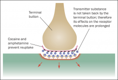
Some drugs inhibit the process of reuptake so that molecules of the neurotransmitter continue to stimulate the postsynaptic receptors for a long time.
Inhibition of reuptake increases the effect of the neurotransmitter. Amphetamines/cocaine: Dopamine, norepinephrine, ephinephrine agonists. Affects autonomic arousal, resulting in restlessness, insomnia, loss of appetite, euphoria. |
|
|
Antidepressants
|
Prozac. serotonin agonist, blocks reuptake, relieves depression
|
|
|
Benzodiazepines
|
Valium, GABA agonist, reduces anxiety, facilitates sleep
|
|
|
Heroin
|
Affects endorphins, agonist, mimics at postsynaptic receptor, analgesia, sedation, euphoria.
|
|
|
Neuropsychology
|
the study of how brain injury affects mental processes
|
|
|
Neuroimaging
|
pictures of the brain's anatomy or function, in relation to mental processes.
|
|
|
Cognitive neuroscience
|
modern term for both neuropsychology and neuroimaging
|
|
|
Lesioning
|
Experimental brain lesions, usually an injury to brain tissue, in animals.
Use stereotaxic apparatus to insert a fine wire into a particular location in the brain |
|
|
Describe Tan
|
Paul broca studied patients with left frontal lobe damage. Patient called "Tan" - only articulate sound he could make. Hence we know Broca's area in left hemisphere responsible for articulate speech
|
|
|
Henry Molaison
|
Surgery for intractable epilepsy in 1953. Removed much of the temporal lobe including the hippocampus.
Could no longer make new memories. |
|
|
MRI
|
Magnetic resonance imaging. Safer than CT scan- no xrays. Magnetic field overhead, reverberations produced by hydrogen molecules picked up by scanner. Computer examines signals for subtle differences that identify the tissue that generated them (blood, nerve fibres, or membranes).
Creates a 3D anatomical picture of the brain. Very detailed picture, shows tumours, tissue degeneration, blood clots and leaks that may signal strokes |
|
|
Neuroimaging view of brains anatomy methods
|
structure. Computerised Tomography (CT), and Magnetic resonance imaging (MRI)
|
|
|
Neuroimaging view of brains physiology
|
function.
Positron emission tomography (PET) Magneto-ence-phal-ography (MEG) Electro-ence-phal-ography (EEG) fMRI Transcranial magnetic stimulation (TMS) |
|
|
EEG
|
Electroencephalography - detects electrical currents generated by neurons on brain surface by affixing metal electrodes to scalp.
Poor spatial resolution, excellent temporal resolution (picks up fast changes). Resolution improved with high density arrays. |
|
|
Event- related potentials
|
Average signals across many trials to deal with noise. Stimulus repeated - each EEG response averaged to produce a clearer signal -ERP.
Some measure sensory responses to stimuli- N100- appearing 100ms after the onset of a stimulus Some associated with more cognitive functions - P300 and N400 |
|
|
N400
|
Linked with language processing. Elicited in sequences where the last word is surprising/inappropriate although linguistically legal. The more difficult the task, the greater the amplitude of the N400.
|
|
|
fMRI
|
Detects fast changing aspects of brain physiology (blood flow and oxygen use) without any radioactivity. Blood contains iron, changes in magnetic fields can be detected. 3D image of brain at work shows which parts are active. Superimposes activity patterns onto the MRI image.
|
|
|
TMS
|
Transcranial magnetic stimulation: Create temporary brain dysfunction, perform experiments that wouldn't be possible otherwise. Series of strong magnetic pulses on scalp, causes temporary disruption in region directly below the area,
|
|
|
The development of the Nervous System
|
3rd week of embryonic life. Small thickening on top of embryo running from head to (nearly) tail.
After a few days left and right edges of the neural plate zip together and fuse lengthwise to form the neural tube. Spina Bifida - neural tube defects. At 1 month the head end of the neural tube develops three thickenings -develop into: hindbrain- nearest the tail Forebrain- nearest the head Midbrain- between the two Enclosed by cranial bones. Lower end of hindbrain marks beginning of spinal cord. Brain and Spinal cord form the CNS. |
|
|
Peripheral nervous system:
|
1. Somatic nervous system -afferent, efferent, and cranial nerves.
2. Autonomic nervous system. Regulation of viscera- heart, lungs, blood vessels, digestion, sex organs. |
|
|
Afferent Nerves and Efferent Nerves
|
Afferent nerves=Transmit information FROM sense organs TO brain & spinal cord.
Efferent nerves= Transmit information from CNS to effectors (muscles/glands that are the organs of action). Eg. reaction when touching something hot |
|
|
Cranial Nerves
(subject: Branches of Nervous System) |
12 pairs- enter/ exit from hindbrain (pons/medulla). Poke though holes in skull- afferent/efferent functions.
Control movements of and carry sensations from head and neck. Regulate glandular secretions in head. Control visceral functions (digestion and excretion) |
|
|
Olfaction
|
smell, cranial nerves produce tears, saliva, mucus and control visceral functions,
|
|
|
Number of neurons in brain
|
10 to 100 billion neurons
|
|
|
Pons
|
Located in the hindbrain.
Responsible for arousal. Relays sensory information between cerebellum and cerebrum and other parts of the brain. Regulates respiration, involved in deep sleep and dreaming |
|
|
Locked in syndrome
|
Awake and aware but cannot move or speak due to paralysis of all voluntary muscles in the body.
Journalist Jean-Dominique Bauby had a stroke in 1995, and when he awoke 20 days later found that he way completely paralysed except for his left eyelid. He used this method of communication to write 'The Diving Bell and the Butterfly' |
|
|
Medulla
|
Part of the hindbrain.
Regulation of heart rate, blood pressure, rate of respiration. Also involved in vomiting, defecations, reflexes and swallowing. Simpler animals -crawling or swimming motions. |
|
|
Cerebellum
|
The hindbrain
Knows what each part of the body is doing. Receives information from frontal lobes, knows what movements this lobe intends to accomplish. Monitors information about posture/balance, produces eye movements that compensate for changes in head position. MAY play a role in learning of new movements and movement skills. Very well developed in humans and primates (1) Controls overall bodily balance- Damage (injury, disease or alcohol) results in wide stance and staggering gait. (2) Sequencing and timing of precise skilled movements: Damage- tremors during movement and inability to perform rapidly alternating movements. Influences thinking - damage impairs performance of tasks requiring exact sequencing. |
|
|
The Midbrain
|
Auditory and visual stimuli - eye movement. Control movements used in sexual behaviour and fighting, decrease sensitivity to pain.
|
|
|
Blind sight
|
Report not being able to see an object but when asked to grasp it, will do so 99 percent of the time, but report that they have no idea where it is and are just guessing randomly.
Ancient visual system in midbrain that guides reaching intact, but not connected with higher area of the brain that guides conscious awareness as to object location. Patient TN - blinded by strokes in both hemispheres. Essentially "blind"- no activity in any areas of the brain which control vision. Reacts to facial expressions - scanning shows he can recognise facial emotions (joy/anger/fear). |
|
|
The Forebrain
|
Everything above the midbrain.
Mammals (primates) have the largest forebrains. In humans - it's so large that it surrounds and hides from view all the midbrain and half the hindbrain. Most obvious part is the part that is wrinkled- the cortex. The cortex is the outer shell of the brain and is most important for psychological functions. |
|
|
The Cortex
|
Primates/complex mammals it accounts for more than 1/2 the brain's volume (80% in humans).
Possibly the part of the brain that makes us human. Humans gain flexibility of behaviour by having a large cortex. Cortex is 2-3mm thick - made possible by convolutions - if flat would occupy about 2 feet. Tasks performed by nonmammals in subcortical regions (limbic system/midbrain) performed by cortex in mammals. As cortex enlarged in mammalian brain evolution subcortical structures (midbrain) act more as relay stations/ middle managers. |
|
|
Gyrus
|
A gyrus (pl. gyri) is a ridge. It has psychological function
|
|
|
Sulcus
|
A sulcus (pl. sulci) is the groove between ridges, also known as a fissure. Sulci don't have psychological function, they're just land marks.
|
|
|
Thalamus
|
Subcortical structure of the Forebrain.
Receiving and relay station for sensory input. Receives sensory information from the sense organs, performs simple analyses and passes results on to primary sensory cortex. The sense of smell doesn't go to the thalamus first. |
|
|
Hypothalamus
|
Subcortical structure of the forebrain.
Hypothalamus performs homeostasis and species typical behaviours. Feeding, drinking, body temperature, sex. Controls much of the activity of the Autonomic Nervous System. Hypothalamus controls the pituitary gland. Functions of hypothlamus: Feeding, fleeing, fighting, fornicating, |
|
|
Basal Ganglia
|
Subcortical structure of the forebrain
Regulation of smoothing of movement - beside thalamus. Tourettes, tardive dyskensia (damage to BG from drugs), cerebral palsy (occurs at birth from oxygen deprivation) |
|
|
Amygdala
|
subcortical structure of the forebrain - Limbic system
Expression of emotions- especially fear. |
|
|
Hippocampus
|
Subcortical structure of the forebrain - limbic system
Memory |
|
|
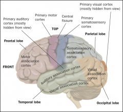
Name the 4 lobes of the cortex
|
Occipital lobe, temporal lobe, frontal lobe, parietal lobe
|
|
|
Occipital lobe
|
One of the 4 lobes of the cortex.
Is at the back of the brain. receives input from the eyes (at the front of the head!) via the thalamus. |
|
|
Parietal Lobe
|
One of the 4 lobes of the cortex.
Important for spatial perception. e.g. mapping way home and knowledge of where limbs are. Postcentral gyrus is at the front of parietal lobe. Postcentral gyrus: receiving area for skin senses (touch, cold, warmth, movement) |
|
|
Postcentral Gyrus
|
At the front of the parietal lobe. Its the receiving area for skin senses (touch, cold, warmth, movement).
|
|
|
Temporal Lobe:
|
One of the four lobes of the cortex.
Is the receiving area for auditory information. Sound goes to the thalamus then is sent to the temporal lobe, smell goes directly to the temporal lobe. Memory and smell associations with strong emotional links |
|
|
Frontal Lobe
|
One of the four lobes of the cortex.
Responsible for motor output and motor planning. Precentral gyrus is at the back of the frontal lobe. Precentral gyrus: Maps onto movelments of different parts of the body. |
|
|
Difference between primary areas and association areas
|
Primary areas - basic input (sensory) and output (motor)
Associations areas- elaboration, or 'higher functions', acting out, planning, memory |
|
|
Primary Motor Area
|
Discovered in 1860's. Mild electric currents applied to parts of the cortex in animals. Very specific effects. Evidence of contralateral control- operates in all nervous systems.
Canadian Neurosurgeon Wilder Penfield - worked with epileptics needing surgery to remove diseased cells. 400 fully conscious people helped confirm that the primary motor area lies in the frontal lobe and stimulation there led to movement of specific parts of the body. |
|
|
The homunculus
|
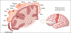
Is the primary motor projection area. Mapping body surface onto the motor cortex. It is mapped upside down. The finer (or more controlled) the movement in the body the more area it takes up in the motor cortex.
Equal sized areas of the body do not receive equal amounts of cortical space. Parts that move with greater precision receive more cortical space than those over which we have less control. |
|
|
Primary Sensory areas
|
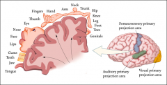
Stimulation at particular point- report tingling somewhere in the opposite side of the body. Less frequently experiences of cold, warmth or movement. Each [art of the body surface sends its sensory information to a particular part of the somatosensory cortex. Sensation is also contralateral.
Parts of the body that are the most sensitive to touch receive more cortical space. |