![]()
![]()
![]()
Use LEFT and RIGHT arrow keys to navigate between flashcards;
Use UP and DOWN arrow keys to flip the card;
H to show hint;
A reads text to speech;
118 Cards in this Set
- Front
- Back
|
Define "Biopsychology" |
Study of the brain and its relationship with the NS as a determinant of behaviour |
|
|
What are the 3 dimensions of biopsych research? |
1) Human vs. Non-Humans 2) Experimental vs. Non-experimental 3) Applied vs. Basic |
|
|
What are the 6 divisions of biopsychology? |
1) Physiological psychology 2) Psychopharmacology 3) Neuropsychology 4) Psychophysiology 5) Cognitive Neuroscience 6) Comparative psychology |
|
|
What is physiological psychology? |
Looks at: How direct manipulation of the NS causes an outcome (Pure, lab animals) |
|
|
What is psychopharmacology? |
Looks at: How pharmacological manipulation of the NS affects the brain (Pure/applied, animals/humans) |
|
|
What is neuropsychology? |
Looks at: What are the behavioural effects of brain damage (without experimentation) (Applied, humans -- With damage) |
|
|
What is psychophysiology? |
Looks at: Relating physiological and psychological processes, to determine the cognitive functional abilities of the organism (Applied, humans) |
|
|
What is cognitive neuroscience? |
Looks at: What is the neural basis of cognitive function? (I.e. Learning, memory, attention, etc.) (Applied [mostly] or pure, humans) |
|
|
What is comparative psychology? |
Looks at: Drawing parallels between different species to understand the evolutionary biological basis for behaviour - why is it similar, how did it evolve? (Pure and applied, animals and humans) |
|
|
What are the 3 methods of visualizing the human brain (IMAGING)? |
Contrast X-rays: Inject a material, uptake occurs more in certain areas, and x-rays create a colourful image (Limited to injection site) X-ray computed tomography: 3D X-ray of anywhere, low image resolution MRI: Displace hydrogen atoms with a magnetic field, and measure the resultant waves being emitted - high resolution |
|
|
What are 2 methods of visualizing ACTIVITY in the human brain? |
PET scan: Radioactive substance (Glucose-like) is injected and absorbed by ACTIVE neurons, so we can scan during an activity to observe usage of substance - LOW TEMPORAL RESOLUTION fMRI: Analyze O-blood and DeO-blood, as they react differently to different magnetic fields - we can see where activity is occuring...HIGH TEMPORAL RESOLUTION, but expensive |
|
|
What is a method of stimulating the brain (for research)? |
TMS: Magnetic field is used to disrupt or produce neuron activity (temporarily) ---CLUFF STUDY |
|
|
What are 3 methods of recording psychophysiological activity? |
Scalp EEG: Electrodes measure the activity of neurons (in the cortex) - Just tells us if activity is present or not MEG: Measures changes in magnetic field across the scalp Muscle tension, eye movement, skin conductance, CV activity: Electrodes measure physiological changes in response to an event (e.g. Lie detectors) |
|
|
What is stereoaxic surgery? |
Invasive manipulation method: A permanent implant is put in the brain |
|
|
What are 2 of the invasive inactivation/activation methods of biopsych research? (1 of each) |
Inactivation: Lesion studies -- Take out/disrupt part of the tissue, permanently or temporarily Activation: Electrical stimulation -- Used to activate certain areas of the brain |
|
|
What is electrophysiological recording? Invasive? |
Invasive recording method - small electrodes are used to record the activity of a single neuron |
|
|
What is autoradiography? |
A radioactive isotope targets a chemical of interest, and accumulated levels are measured after the animal engages in a certain behaviour |
|
|
What is cerebral dialysis? |
A sample tube is placed in the brain, and certain chemicals diffuse in (and then measured) |
|
|
What are 2 methods of measuring neurotransmitter & receptor locations? |
Immunofluoresence: Dye binds to a protein of interest, and is then imaged In Situ hybridization: Labeled RNA is used to locate neurons with complimentary mRNA |
|
|
What is pavlovian conditioning vs. operant conditioning? |
Pavlovian: Benign stimulus paired with a stimulus (that produces a behavioural outcome), and eventually the benign is able to produce the outcome Operant: Reinforcement & punishment are used to produce a response |
|
|
What does the Radial Arm Maze test? |
Assesses foraging behaviours and memory processes (multi-arm maze, food is only in some areas |
|
|
What composes the CNS? |
Brain and Spinal cord |
|
|
What are the two systems under the peripheral NS? |
Somatic and Autonomic |
|
|
In the somatic NS, explain the difference between afferent nerves and efferent nerves |
Afferent: Carries sensory info to the CNS (Approach) Efferent: Carries motor signals from CNS to muscles (Exit) |
|
|
What kind of neurons are in the somatic NS? |
Somatic neurons: Cell body in the CNS, one long cell running through PNS to effector organ |
|
|
What 2 types of efferent nerves are found in the autonomic NS? |
Sympathetic: Cell body to ganglion, then a long distance to effector organ -- Or directly on adrenal medulla Parasympathetic: Synapse on ganglion, then a short distance to effector organ |
|
|
What kind of nerves do the somatic and autonomic NS both contain? |
Afferent and efferent |
|
|
Purpose of the somatic NS? |
Interaction with the external environment |
|
|
Purpose of the autonomic NS? |
Regulation of the bodies internal environment |
|
|
What are the 3(+1) meninge layers, from innermost to outermost? |
Pia mater: Adheres to surface of the CNS Arachnoid: Web-like membrane **Sub-arachnoid space: Allows for CSF circulation Dura mater: Toughest |
|
|
What is the function of the meninges? |
Protection for the CNS |
|
|
Where is the CSF produced? How much is produced, and where does excess go? |
Choroid plexuses: Capillary networks that protrude into the ventricles from the pia mater Excess enters the dural sinuses to go to jugular veins (0.5L Made, but only 100-150ml used) |
|
|
What is hydrocephalus? |
A blockage of CSF -- the ventricles and brain expand to allow for excess fluid drainage |
|
|
Where does CSF circulate? |
Two lateral ventricles, 3rd ventricle, 4th ventricle Cerebral aqueduct: Connects 3rd/4th ventricle Central canal: Small channel that runs length of spinal cord |
|
|
What is the blood brain barrier? Is it present EVERYWHERE in the brain? |
Impedes passage of toxic substances from blood to the brain -- the cells of the blood vessel structure are packed extremely tightly No, places like the posterior pituitary gland must be able to send stuff into the bloodstream, for example |
|
|
Main difference between neurons and glial cells? |
Neurons process and transmit information Glial cells support the nervous system |
|
|
Ribosome purpose? |
Protein synthesis site |
|
|
Golgi complex function? |
Membrane that packs molecules into vesicles |
|
|
Smooth vs. rough ER? |
Smooth: Helps with fat synthesis Rough: Helps with protein synthesis (Ribosomes) |
|
|
What are the 4 classes of neurons? |
Multipolar: More than 2 processes extending from body Unipolar: One process extending from body Bipolar: 2 Processes extending Interneurons: Short axon (or none at all) |
|
|
What is the purpose of interneurons? |
To integrate neural activity within a brain structure |
|
|
Explain neuron clusters in the CNS vs. the PNS for: a) Cell bodies b) Axons |
Cell body clusters: Nuclei (CNS), Ganglia (PNS) Axons: Tracts (CNS), Nerves (PNS) |
|
|
What is the present ratio of glial cells to neurons? |
Glial : Neuron 10 : 1 |
|
|
4 Types of glial cells? |
Oligodendrocytes: Myelin sheaths in CNS Schwann Cells: Myelin sheaths in PNS (Only PNS Glial cells) Microglia: Trigger inflammatory response to respond to injury/disease Astrocytes: Form a supportive matrix for neurons, provide nourishment |
|
|
Golgi stain vs. Nissl stain? |
Golgi: Looks at shape of neurons (Portrays a silhouette image) Nissl: Stains JUST cell bodies, but shows number of neurons in an area by penetrating them with a dye |
|
|
What is immunofluorescent neural staining? |
Tags different parts of each neuron a different colour |
|
|
What is electron microscopy? Scanning electron microscopy? |
Neural tissue is coated with an electron-absorbing substance, and electrons then passed through tissue onto film to create a detailed 2D image - Provides explicit detail about neuronal structure Scanning EM: Same process, but in 3D, and with far less magnification |
|
|
What are the 2 neuroanatomical tracing techniques? |
Anterograde tracing: Trace where the information is going to Retrograde tracing: Trace where the information is coming from |
|
|
What are the 3 axes of direction? |
Anterior/posterior: Nose/tail-end (Rostral/Caudal) Dorsal-ventral: Back surface, chest surface Medial-lateral: Midline, away from midlin |
|
|
What are the 3 planes? |
Horizontal Frontal/Coronal Sagittal (Or midsagittal, if in the middle) |
|
|
What is the difference between gray matter and white matter of the spinal cord? |
Gray matter: H-Core, unmyelinated interneurons - Contains dorsal horns (back), and ventral horns (front) White matter: Surrounding tissue, myelinated axons |
|
|
Dorsal root vs. Ventral root? |
Dorsal: Afferent (sensory), unipolar neurons - cell bodies group outside cord to form dorsal root ganglia Ventral: Efferent (motor), multipolar neurons - cell bodies in the ventral horn |
|
|
Where do ventral root neurons project to in the somatic NS vs. autonomic NS? |
Somatic: Project to skeletal muscle Autonomic NS: Project to Ganglia, then synapse on neurons that project to internal organs |
|
|
What 5 divisions of the brain are composed in the forebrain, midbrain, hindbrain? |
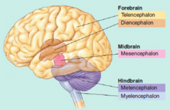
Forebrain: Telencephalon, Diencephalon Midbrain: Mesencephalon Hindbrain: Metencephalon, Myelencephalon (Medulla) |
|
|
What does the brain stem refer to? |
Everything BUT the telencephalon |
|
|
Myelencephalon: What does it do? What part of brain? |
Involved in the most basic of functions that keep us alive (sleep, attention, respiration, cardiac function, muscle tone) Contains the reticular formation (Hindbrain) |
|
|
What is the reticular formation? Composes core of what areas of the brain? |
100 Nuclei that occupy the central core of the brain stem...involved in sleep, attention, movement, cardiac/circulatory/respiratory reflexes (Basically an activation system) Hindbrain: Myenecephalon, metencephalon Mibrain: Mesencephalon |
|
|
Metencephalon: What are the 2 main areas? Function? What part of brain? |
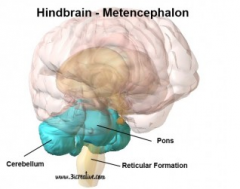
Houses part of reticular formation 1) Pons 2) Cerebellum - Sensorimotor function (Fine motor movements) (Hindbrain) |
|
|
Mesencephalon: What part of brain? a) Tectum: 2 Structures + Function? b) Tegmentum: 3 Structures and function? |
(Midbrain) Tectum: Dorsal surface 1) Inferior colliculi (Posterior): Auditory function 2) Superior collliculi (Anterior): Visual function Tegmentum: Ventral aspect 1) Periaqueductal gray: Gray matter around cerebral aqueduct, sends signals of "release pain-reducing stuff" 2) Cerebral aqueduct: Connects 3rd/4th ventricles 3a) Substania nigra: Dopamine productino 3b) Red nucleus: Motor coordination while crawling, but vestigial once we stand |
|
|
Diencephalon: What part of brain? a) Thalamus: What joins them? What covers surface? Purpose? b) Hypothalamus: Purpose? 3 Inferior structures? |
(Forebrain) a) Massa intermedia (Runs through ventricles), covered by white lamina (myelinated axons) -- contains many nuclei to project to cortex b) Involved in motivated behaviours 1) Pituitary gland: Releases hormons 2) Optic chiasm: Optic nerve location 3) Mammilary bodies: Recollective memory |
|
|
Telencephalon: What part of brain? Main functions? |
(Forebrain) Initiates voluntary movement, interprets sensory input, involved in cognitive processes |
|
|
What is the cerebral cortex? |
Gray matter (Unmyelinated neurons) that covers the entire brain |
|
|
3 Main fissures in the brain? |
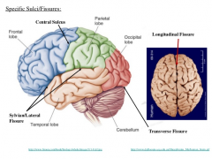
Longitudinal fissure (separates cerebral hemispheres) Central fissure Lateral fissure |
|
|
3 Main gyri found in the brain, and locations? |
Precentral gyri (Frontal) Postcentral gyri (Parietal) Superior temporal gyri (Temporal) |
|
|
Function of the occipital lobe region? |
Aids in analysis of visual input |
|
|
Function of the two areas of the parietal lobe? |
Post-central gyrus: Analyzes body sensations Remainder: Involved in spatial location of objects and own body |
|
|
Function of the 3 areas of the temporal lobe? |
Superior temporal gyrus: Involved in hearing and language Inferior temporal gyrus: Complex visual patterns Medial portion: Memory |
|
|
Function of the 2 areas of the frontal lobe? |
Precentral gyrus: Motor function Front cortex: Cognitive function |
|
|
Neocortex: a) How many layers? What is special about top 3 layers? b) 2 Categories of neocortical neurons? |
a) 6 Layers - Top 3 contain stellate cells and interneurons b) Pyramidal cells: Large multipolar neurons with very large axons b2) Stellate cells: Interneurons |
|
|
Hippocampus: Major role? Is it neocortex? How many layers? |
Memory and spatial location, not neocortex, only contains 3 layers |
|
|
Limbic system: What does it do? |
Involved in regulation of motivated behaviours: Fleeing, feeding, fighting, f-cking |
|
|
List the 6 structures in the limbic system |
Mammillary bodies: Recollective memory Amygdala: Intense emotional memory Hippocampus: Memory/spatial navigation Cingulate cortex: Large strip of cortex on medial cerebral hemisphere Fornix: Fibers connecting hippocamp/mamillary bodies/thalamus Septum: Completes the limbic ring |
|
|
Basal Ganglia: Main function? List the structures within |
Amygdala: Intense emotional memory Striatum: Dopamine release site Tail of caudate: Encircles putamen Globus pallidus: Center of circle of caudate, medial to the putamen -- Fine-tuning of movements |
|
|
What is a membrane potential? How to record? |
Difference in elec. charge between the inside and outside of a cell Recorded by microelectrode on inside and outside of cell |
|
|
What is the resting potential of a cell? Is it polarized? |
Polarized, -70mV |
|
|
At rest, what are the positive and negative ions inside and outside the cell? |
Outside: Cl-, Na+ Inside: K+, Negative protein ions |
|
|
Factors responsible for sodium gradient at resting? |
Wants to move into cell, but: - Membrane resistant to passive diffusion (into cell) - Pumped out by Na-K pump at the same rate they move in |
|
|
Factors responsible for potassium gradient at resting? |
Wants to move out of cell, but: - Internal negative potential keeps most in - When they move out (via passive diffusion), Na-K pumps bring them in at same rate |
|
|
Factors responsible for chlorine gradient at resting? |
Wants to stay out of cell:
- Negative internal potential keeps them out, so at equilibrium, there are far more on outside |
|
|
Factors responsible for protein ion gradient at resting? |
Negatively charged and trapped in cell |
|
|
What are the 2 characteristics of PSPs? |
1) Rapid transmission - Fast enough to assume instantaneous 2) Decremental - Further away it travels, the weaker the signal |
|
|
What is the threshold of excitation? |
-65mV to -55mV |
|
|
What are the 2 types of summations of signal that form an AP? |
Spatial summation: IPSPs and EPSPs sum together over the same space Temporal summation: PSPs produced in rapid succession before the original signal dies completely, eventually elicits an AP |
|
|
What are the steps of an AP? |
1) Membrane potential reaches threshold of activation 2) Sodium channels open, sodium rushes in 3) Potassium channels open, potassium rushes out 4) Sodium pushed back out fast, as the channels close at +50mV 5) Potassium channels close slower, so they travel in later, neuron reaches hyperpolarized state of -90mV |
|
|
Difference between absolute and relative refractory period? |
Absolute: Impossible to elicit a 2nd AP Relative: Possible to elicit, but must apply higher-than-normal stimulation (With overstimulation, we can fire the neurons at a faster than normal rate) |
|
|
What are the 2 directions of AP conduction? |
Antidromic conduction: From terminal end towards cell body Orthodromic conduction: From cell body towards terminal buttons |
|
|
What is saltatory conduction? |
As an AP travels, it jumps from node to node (it runs through the myelinated areas, but passively, so the process is basically instantaneous) |
|
|
Velocity of conduction in myelinated vs nonmyelinated axons? |
Myelinated is way faster, up to 100m/second in some mammals Unmyelinated is only 1m/second |
|
|
What is unique about axoaxonic synapses? |
Since they can synapse on a site just before a terminal button, they can mediate the presynaptic facilitation or inhibition of the neuron they act on |
|
|
What is neurotransmitter coexistence |
Typically, most neurons contain 2 neurotransmitters, a large (Neuopeptide) and small one |
|
|
Difference between ionotropic receptors and metabotropic receptors |
Ionotropic - Ligand-activated (Effects are immediate) Metabotropic - Associated with signal and G proteins (Effects are way slower to develop |
|
|
What is an autoreceptor? |
Metabotropic receptor 1) Attached to presynaptic membrane 2) Binds to neurotransmitter Function: Triggers uptake or release when levels are too low or high |
|
|
How do Glial cells communicate with each other? |
Gap junctions |
|
|
What are the 2 important amino acid neurotransmitters? |
Glutamate - Excitatory CNS GABA - Inhibitory CNS |
|
|
What are the 4 monoamine neurotransmitters? |
Catecholamines: Dopamine, norepinephrine, epinephrine (All synthesized from Tyrosine) Indolaimes: Serotonin |
|
|
What are the 7 steps common to most neurotransmitters? |
1) Synthesis 2) Storage (in vesicles) 3) Breakdown in cytoplasm 4) Exocytosis 5) Inhibitory feedback (via autoreceptors) 6) Activation of postsynaptic receptors 7) Deactivation |
|
|
What are the 6 causes of brain damage? |
Brain tumours Strokes Closed-head injuries Infections of the brain Neurotoxins Genetic factors |
|
|
Difference between encapsulated tumors and infiltrating tumours? |
Encapsulated: Grow within their own membrane, exert pressure on tissue Infiltrating: Grow diffusely through the tissue, very difficult to destroy |
|
|
2 Major types of causes of strokes? |
Cerebral hemorrhage: Blood vessel bursts, and the blood damages surrounding neural tissue Cerebral ischemia: Blood supply to brain is disrupted |
|
|
What is the link between cerebral ischemia and glutamate? |
Damage results in excessive glutamate release: - Allows lots of Na and Ca into the postsynaptic neurons 1) Triggers more glutamate release surrounding neurons 2) Triggers a sequence of self-death for the post-synaptic neurons |
|
|
What is a coup-contrecoup? |
Brain injury from the blow, and the resultant hit of the brain on the other side of the skull |
|
|
What is punch-drunk syndrome? |
Dementia (intellectual deterioration) and cerebral scarring from repeated concussions |
|
|
What is encephalitis? |
Inflammation resulting from microorganism-caused brain infection |
|
|
What is tardive dyskinesia (TD)? |
A motor disorder that arose from treatment with antipsychotic drugs (that contained neurotoxins) |
|
|
What are the two types of cell death? |
Apoptosis: Programmed death, slow process where the cells shrink and get packed up -- minimal surrounding damage Necrosis: Cell death from injury - swelling and breakage of cells leads to inflammation and damages nearby cells -- very quick |
|
|
What are the 5 diseases associated with brain damage? |
Epilepsy: Seizures Parkinson's: Progressive movement disease Huntington's: Progressive movement disease (Genetic) Multiple Sclerosis: Autoimmune disorder on myelin Alzheimer's: Cognitive deterioration |
|
|
2 Types of partial seizures? |
Simple partial: Symptoms are sensory/motor, can spread to rest of body Complex partial: Restricted to temporal lobes -- results in engaging in behaviour repetitively (e.g. Buttoning a button) Partial seizures do not involve entire brain |
|
|
What are the 2 types of generalized seizures? |
Grand mal: Violent tonic-clonic convulsions, loss of consciousness, and hypoxia (leads to brain damage) can occur Petit mal: 3spike/second EEG, and petit mal absence occurs - disruption of consciousness (Kids diagnosed as "frequent daydreamers") |
|
|
What are 2 methods of alleviating parkinson's symptoms? |
L-Dopa: Precursor to dopamine, and is able to cross the BBB, whereas dopamine cannot Deep brain stimulation - stimulates the subthalamic nucleus at low-intensity |
|
|
Huntington's Disease: 2 Differences from Parkinson's? |
1) Strong genetic basis (Dominant gene), but doesn't appear until after having kids 2) Associated with dementia |
|
|
Alzheimer's: Autopsy is performed to find 3 things? |
1) Neurofibrillary tangles: Threadlike tangles of protein in the neural cytoplasm 2) Amyloid plaques: Clumps of scar tissue 3) Substantial neural loss |
|
|
Where does Alzheimer's affect in the brain, mainly? |
Medial temporal lobe structure: Amygdala, hippocampus, entorhinal cortex Inferior temporal cortex, posterior parietal cortex, prefrontal cortex (I.e. Memory and complex cognitive function areas) |
|
|
Kindling epilepsy model - what is it? |
Stimulations 1x/day eventually result in twitches, which progress to full convulsive seizures in animals Neurplastic changes that occur are permanent, even if subject is left unstimulated for months Used to study interictal behaviour - periods between seizures |
|
|
What is the issue with most transgenic mouse models of alzheimer's disease? |
Amyloid synthesis acceleration genes are implemented - this is an issue if the neurofibrillary tangle hypothesis ends up being the primary symptom |
|
|
What is MPTP? Benefit to science? |
Drug that results in substantial cell loss in the substantia nigra (and resultant ability to produce dopamine) - basically produces the symptoms of parkinson's Good because otherwise we'd have to wait 50 years in our models to study the disease |
|
|
What are the 3 outcomes for PNS axonal regeneration in mammals? |
1) If schwann cells remain intact, regrowth occurs 2) If small separation of nerve ends occurs, crossing of threads will occur and grow into incorrect sheaths to the wrong targets 3) Substantial separation will lead to proximal ends of the nerves tangling and growing into a stump....they die |
|
|
Which neurons have regenerative capacities? |
Mammals: CNS cannot, but PNS can (schwann cells promote regeneration, but oligodendroglia (CNS) prevent it) Lower vertebrates: Extremely accurate CNS and PNS regeneration (e.g. entire limbs can regrow) |
|
|
What are 4 major methods of treating neuro damage? |
1) Blocking neurodegeneration: Drugs 2) Promoting regeneration in CNS: Induced by transplanting PNS cells (or olfactory ensheathing cells) 3) Neurotransplantation: Stem cells or fetal tissue 4) Rehabilitative training: Cognitive/Physical exercise |