Reading...
![]()
Play button
![]()
Play button
![]()
Use LEFT and RIGHT arrow keys to navigate between flashcards;
Use UP and DOWN arrow keys to flip the card;
H to show hint;
A reads text to speech;
94 Cards in this Set
- Front
- Back
|
intervertebral foramina are spaces or openings between ________ when two vertebrae are stacked on each other.
|
pedicles
|
|
|
The intervertebral foramina in the lumbar region are best demonstrated on a ________ radiographic image
|
lateral
|
|
|
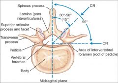
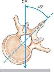
The intervertebral foramen are situated ______ degrees relative to the midsagittal plane
|
90 degrees
|
|
|
When vertebra are stacked, the superior and inferior vertebral notches line up and form a single opening called the _________
|
intervertebral foramina
|
|
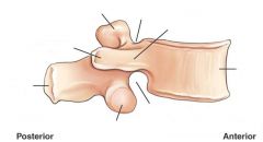
Label
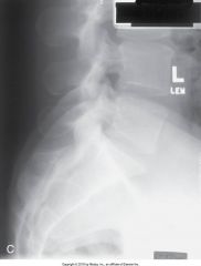
Lateral Lumbar Vertebra |
|
|
|
How many articular processes does each typical lumbar vertebra have?
|
Four
|
|
|
The zygapophyseal joints form an angle open from ____ to ____ degrees to the midsagittal plane.
|
30 to 50 degrees
|
|
|
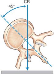
Demonstration of the zygapophyseal joints is achieved by rotating the patients body an average of _____ degrees
|
45 degrees
|
|
|
The portion of each lamina between the superior and inferior articular processes is the ____________
|
pars interarticularis
|
|
|
in which position is the pars interarticularis demonstrated?
|
oblique lumbar image
|
|
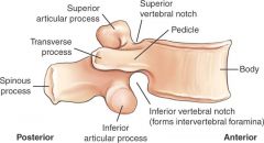
Label
Lumbar superior view |
|
|
|
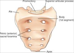
______ are the large masses of bone lateral to the 1st sacral segment
|
Alae (wings)
|
|

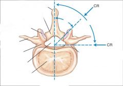
The CR projection labeled A in this drawing would best demonstrate the ________________
|

Zygapophyseal joints
|
|
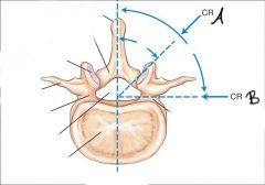
The CR projection labeled B would best demonstrate the _____________
|

Intervertebral foramina
|
|
|
The upper or proximal lumbar vertebrae are nearer to a ______ degree angle while the lower or distal vertebra are nearer to a ______ degree angle.
|
The upper or proximal lumbar vertebrae are nearer to a 50 degree angle while the lower or distal vertebra are nearer to a 30 degree angle.
|
|
Label the sacrum
|
|
|
|
The anterior ridge of the body of the first sacral segment helps to form the posterior wall of the inlet of the true pelvis and is termed the ____________
|
promontory
|
|
|
This is a continuation of the vertebral canal and contains certain sacral nerves.
|
sacral canal
|
|
|
Another term for sacral horns
|
cornua
|
|
|
This is formed by fused spinous processes of the sacral vertebrae
|
median sacral crest
|
|
|
What does the sacrum articulate with?
|
ilium of the pelvis
|
|
|
What is the surface called where the sacrum articulates with the ilium of the pelvis
|
auricular surface
|
|
|
Each sacroiliac joint opens obliquely and posteriorly at an angle of _______
|
30 degrees
|
|
|
Whats the formal name for the tail bone?
|
coccyx
|
|
|
Whats the name for the upper broad aspect of the coccyx?
|
Base
|
|
|
Whats the most distal portion of the vertebral column?
|
coccyx
|
|
|
An average of _____ coccygeal segments have fused in the adult to form the single coccyx.
|
four
|
|
|
The distal pointed tip of the coccyx is termed the _______
|
apex
|
|

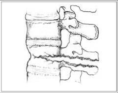
This photograph shows the vertebra in a __________ oblique position and demonstrates the _________ of the zygapophyseal joint
|
This photograph shoes the vertebra in a posterior oblique position and demonstrates the downside of the zygapophyseal joint
|
|
This figure represents a ________ oblique projections and demonstrates the _______ zygapophyseal joints.
|

This figure represents a anterior oblique projections and demonstrates the upside zygapophyseal joints.
|
|
|
The zygapophyseal joints of the L-spine are classified as _________, have a _________ mobility type, and a ________ movement
|
The zygapophyseal joints of the L-spine are classified as synovial, have a diarthrodial mobility type, and a plane (gliding) movement
|
|
|
The intervertebral joints of the L-spine are classified as _________, and have an ________ mobility type.
|
The intervertebral joints of the L-spine are classified as cartlaginous and have an amphiarthodial mobility type.
|
|
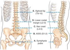
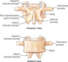
Name the vertebral level of each landmark
|
|
|
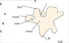
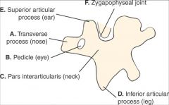
Identify what each part of the scottie dog represents on the vertebra
|
|
|
|
What does the eye of the scottie dog represent on a oblique lumbar vertebrae?
|
pedicle
|
|
|
Which zygapophyseal joint is demonstrated with an LAO position of the lumbar spine?
|
Right upside joints
|
|
|
What does the nose of the scottie dog represent on a oblique lumbar vertebrae ?
|
transverse process
|
|
|
What does the neck of the scottie dog represent on a oblique lumbar vertebrae?
|
pars interarticularis
|
|
|
What does the leg of the scottie dog represent on a oblique lumbar vertebrae?
|
inferior articular process
|
|
|
What does the ear of the scottie dog represent on a oblique lumbar vertebrae?
|
superior articular process
|
|
|
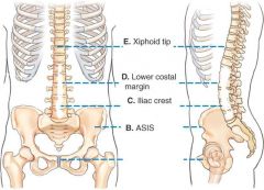
The ASIS is at the level of _______
|
S1-2
|
|
|
The xiphoid tip is at the level of ______
|
T9-T10
|
|
|
The lower costal margin is at the level of ______
|
L2-3
|
|
|
The Iliac crest is at the level of _______
|
L4-L5
|
|
|
The Symphysis pubis is at the level of
_________ |
tip of coccyx
|
|
|
Does the AP or PA projection of the lumbar spine open the intervertebral joint spaces better?
|
PA
|
|
|
Should the knees be extended or slightly flexed for a AP projection of the lumbar spine?
|
slightly flexed
|
|
|
True of False
It would be advantageous to increase the SID to 44 or 46 inches for AP and lateral lumbar projections. |
true
|
|
|
Where is the CR centered for AP lumbar projection?
|
CR centered to level of iliac crest
|
|
|
What should the collimation width be for AP lumbar projection
|
collimation width to the SI joints
|
|
|
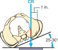
Where is the CR for oblique lumbar spine
|
CR is 1½ inches (4 cm) above iliac crest
2 inches (5 cm) medial to upside ASIS |
|
|
Which two structures can be evaluated to determine whether rotation is present on a radiograph of an AP projection of the lumbar spine
|
1.) sacroiliac joints should be equidistant from the spine
2.) spinous process should be midline to the vertebral column |
|
|
Where should the pedicle (which is the eye of the scottie dog) be on a well positioned oblique lumbar spine
|
center of the vertebral body
|
|
|
If the eye of the scottie dog is projected too far posterior on a obl lumbar spine what positioning error has occurred?
|
excessive rotation
|
|
|
Which position or projection of the lumbar spine best demonstrates a compression fracture?
|
lateral
|
|
|
A patient with a wide pelvis and narrow thorax may require a central ray of?
|
5-8 degrees caudal
|
|
|
How should the spine of a patient with scoliosis be positioned for a lateral position of the lumbar spine?
|
with the convexity of the spine closest to the IR
|
|
|
Where is the CR for the L5-S1 lateral L-spine projection
|
CR 1½ inches (4 cm) inferior to iliac crest and 2 inches (5 cm) posterior to ASIS
|
|
What is this picture pointing to?
|
zygapophyseal joint
|
|
|
What type of angulation is required for an AP axial L5-S1 projection of a male patient
|
30 degrees cephalad
|
|
|
When performing a L5-S1 projection, if the abdomen is blocked it helps placed the interiliac line _______ to the IR
|
perpendicular
|
|
|
When performing the L5-S1 projection it may be necessary to angle _________ for a patient with a large waist
|
cephalad
|
|
|
the coccyx curves ________and _________ from articulation with sacrum
|
the coccyx curves inferiorly and anteriorly from articulation with sacrum
|
|
|
Where should the lower margin of the IR be for a scoliosis series
|
1 to 2 inches below iliac crests
|
|
|
Which side of the spine should be elevated for the second exposure for the Ferguson scoliosis series.
|
convex side of the spine by having patient stand on a block with one foot.
|
|
|
How much CR angulation is required for an AP projection of the sacrum for a typical male patient?
|
15 degrees cephalad
|
|
|
where is the CR for an AP of the coccyx?
|
2 inches superior to the symphysis pubis
|
|
|
The _______ projection of the sacrum and coccyx can be combined to decrease gonadal dose
|
lateral
(AP cannot be combined because different CR angles are required) |
|
|
Patients should be asked to empty the bladder before performing which projection of the vertebral column?
|
AP of sacrum and coccyx
|
|
|
Which sacroiliac joint is demonstrated with a RPO position
|
Left joint (always upside)
|
|
|
How much rotation of the body is required for oblique positions of the SI joints
|
25-30 degrees
|
|
|
Which sacroiliac joint is demonstrated for a LPO position?
|
right side (always the side that's up)
|
|
|
Where is the CR for posterior OBL SI joints?
|
CR perpendicular 1 inch medial to upside ASIS
|
|
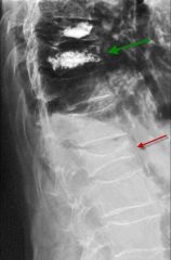
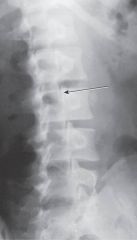
What type of fracture is in this picture?? described as anterior wedging of the vertebrae with loss of body height.
|
Compression fracture
|
|
|
What can cause a compression fracture?
|
trauma, osteoporosis or metastatic disease.
|
|
Name the fracture that results from hyperflexion force that causes fx through the vertebral body and posterior elements.
|
Chance Fracture
|
|
|
for which fracture are pts at risk when wearing lap type seat belts acting as a fulcrum during sudden deceleration
|
chance fracture
|
|
|
What are mostly malignant neoplasms that spread to distant sites through blood and lymphatics.
|
matastases
|
|
|
This can be visualized as destructive with irregular margins and decreased densities, osteoblastic, with increased densities or a combination of both.
|
matastases
|
|
|
Failure of the posterior elements of the vertebrae to close, exposing part of the spinal cord.
Varies in severity and occurs most often at L5 |
spina bifida
|
|
|
Forward movement or slipping of one vertebra in relation to another
Usually occurs at L5-S1 |
spondylolisthesis
|
|
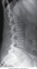
Why is this a bad lateral ?
|

Upper thorax region rotated, no marker
|
|
Why is this a bad L5-S1
|
L5-S1 joint space is not open
waist support of caudal angle needed underexposed |
|
Label
|
|
|
|
An AP lumbar radiograph reveals that the spinous processes are not midline to the vertebral column and the vertebral bodies are distorted. Name the positioning error
|
rotation of the spine
|
|
|
If on a LPO lumbar spine the downside pedicles and zygapophyseal joints are projected over the anterior portion of the vertebral bodies
|
it's insufficient rotation because the pedicles should be to midverterbral bodies.
|
|
|
For lumbar obliques, the pedicle demonstrated posteriorly on the vertebral body indicates ____________, and the pedicle demonstrated anteriorly on the vertebral body indicates ___________
|
For lumbar obliques, the pedicle demonstrated posteriorly on the vertebral body indicates overrotation and the pedicle demonstrated anteriorly on the vertebral body indicates underrotation
|
|
|
An AP axial coccyx projection reveals that the distal tip of the coccyx is superimposed over the symphysis pubis? what can be done to correct this?
|
increase angle to separate
|
|
|
An OBL lumbar radiograph reveals the downside pedicle and zygapophyseal joint are posterior in relation to the vertebral body, what needs to be done to fix this?
|
patient is over rotated, decrease rotation.
|
|
|
If a pt is in too much pain from a coccyx injury to perform an exam AP, what other options are available?
|
PA instead of AP and reverse the CR angle from caudal to cephalad.
|
|
|
Which degree of rotation will best demonstrate the zygapophyseal joints at the L1-L2 vertebral level?
|
50 degrees
|
|
|
Which of the following projections will best demonstrate the left zygapophyseal joints of the lumbar spine?
RPO LAO RAO Lateral |
RAO
_____________________ Posterior = the same Posterior = downside Anterior = opposite Anterior = upside |
|
|
which position best demonstrates advanced spondylolysis?
|
posterior and anterior obliques best demonstrate advanced signes of spondylolysis.
|
|
|
A patient has just had spinal fusion surgery, the surgeon wants to asses mobility of the spine at the fusion site, which position is best?
|
hyperflexion and hyperextension laterals
|