![]()
![]()
![]()
Use LEFT and RIGHT arrow keys to navigate between flashcards;
Use UP and DOWN arrow keys to flip the card;
H to show hint;
A reads text to speech;
39 Cards in this Set
- Front
- Back
|
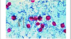
Cryptosporidum acid fast stain |
|
|
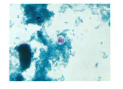
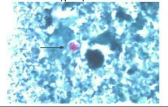
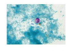
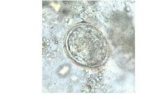
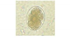
Cryptosporidium |
|
|
Cryptosporidium |
|
|
Cryptosporidium |
|
|
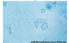
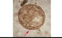
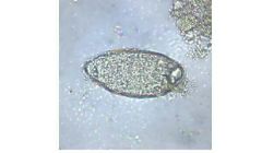
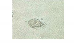
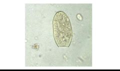
Balantidium coli |
|
|
Balantidium coli |
|
|
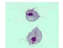
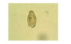
Trichomonas vaginalis |
|
|
Trichomonas vaginalis wet prep |
|
|
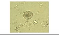
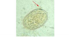
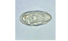
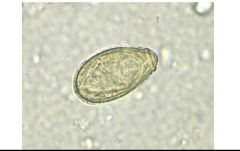
Hymenolepis nana |
|
|
H. nana |
|
|
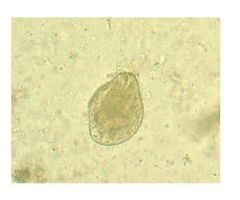
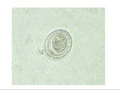
P. westermanni |
|
|
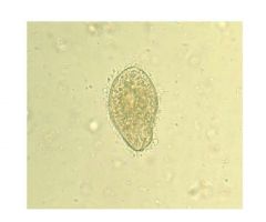
P. westermanni |
|
|
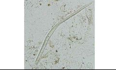
Plant hairs in a concentrated wet mount of stool. Plant hairs can be common in stool and may be confused for the larvae of hookworm or Strongyloides stercoralis. However, they are often broken at one end, have a refractile center and lack the strictures seem in helminth larvae (esophagus, genital primordium, etc). |
|
|
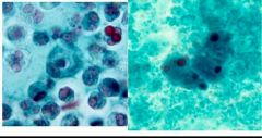
Epithelial and white blood cells are often seen in trichrome-stained |
|
|
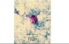
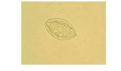
Cyclospora cayetanensis |
|
|
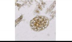
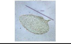
Plant cell in a concentrated wet mount of stool. Such material can be common in stool and may be confused for helminth eggs, although they are usually much larger than the eggs of most helminth species. |
|
|
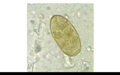
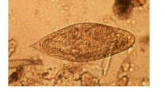
T. spiralis |
|
|
Trichinella spiralis |
|
|
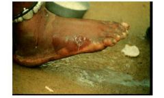
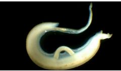
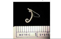
The female guinea worm induces a painful blister (A); after rupture of the blister, the worm emerges as a whitish filament (B) in the center of a painful ulcer which is often secondarily infected. |
|
|
Adults of S. mansoni. The thin female resides in the gynecophoral canal of the thicker male. |
|
|
Eggs of S. japonicum in unstained wet mounts |
|
|
Eggs of S. japonicum in unstained wet mounts |
|
|
Eggs of S. japonicum in unstained wet mounts |
|
|
Schistosoma intercalatum is related to S. haematobium, but restricted to east-central Africa. The eggs are similar to S. haematobium in general shape and in possessing a terminal spine, but are usually longer (140-240 µm), often have an equatorial (central) bulge and are shed in stool, not |
|

|
Schistosoma intercalatum |
|
|
Eggs of S. mansoni in unstained wet mounts |
|
|
Eggs of S. mansoni in unstained wet mounts- note side view |
|
|
Eggs of S. mansoni in unstained wet mounts |
|
|
Schistosoma haematobium |
|
|
Male small/ Fe large Schistosomia |
|
|
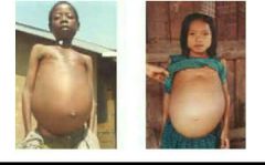
Schistosomiasis |
|
|
Schistosoma haematobium |
|
|
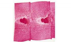
D. Latum showing uterus and eggs |
|
|
F. hepatica |
|
|
C. sinensis |
|
|
Trichuris trichiura (whipworm) |
|
|
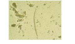
Strongyloides |
|
|
N. americanus |
|
|
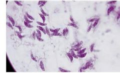
Toxoplasmosis |