Reading...
![]()
Play button
![]()
Play button
![]()
Use LEFT and RIGHT arrow keys to navigate between flashcards;
Use UP and DOWN arrow keys to flip the card;
H to show hint;
A reads text to speech;
94 Cards in this Set
- Front
- Back
|
What are the 5 divisions of the brain from top to bottom?
|
Telencephalon
Diencephalon Mesencephalon Metencephalon Myelencephalon |
|
|
What are the 3 layers of the meninges and what is different about each?
|
Dura mater ("hard mother") - outermost protective layer
Arachnoid membrane - spongy, spider-like web of tissue; Subarachnoid space - contains cerebrospinal fluid (CSF) and arteries Pia mater - delicate, innermost membrane |
|
|
What is the difference between grey matter and white matter?
|
Grey - nerve cell bodies (somas of neurons)
White - fiber bundles of neurons - white b/c of fatty myelin sheath |
|
|
What divides the 2 hemispheres?
|
Media longitudinal fissure
|
|
|
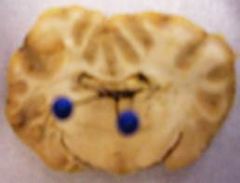
What do the superior colliculi do?
Where are they? |
Processes some simple aspects of visual stimuli.
Found in the tectum of the midbrain above the IC |
|
|
What do the inferior colliculi do?
Where are they? |
Processes some simple aspects of auditory stimuli.
Found in the tectum of the midbrain below the SC |
|
|
What does the cerebellum do?
Where is it? |
Critical for motor functioning, movements requiring aim and timing (e.g. finger to nose), coordinates w/vestibular system for balance (ex. when drunk can't walk heel-to-toe), some learning functions
|
|
|
Where is the Pons and what does it do?
|
Relays sensory information from cerebellum to cortex, contains reticular formation.
Found on the brainstem |
|
|
What does the reticular formation do?
|
Important for general arousal (respiration centers, muscle tone, heart rate, etc.)
|
|
|
What does the medulla do? Where is it?
|
Long, narrow structure (hence oblongata), controls autonomic functions such as breathing, heart rate, etc.
|
|
|
What structures make up the brain stem?
|
Together, the pons and medulla are sometimes called the brainstem.
|
|
|
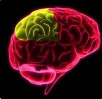
What does the frontal lobe do?
Where is it? |
Sensory and motor functions, higher-order cognitive processes in humans
Prefrontal cortex - receives input from all sensory modalities, is involved with planning, decision making, personality, impulse control, emotion Found at the rostral end of cortex |
|
|
What does the parietal lobe do?
Where is it? |
Somatic (body) sensations, spatial orientation, face recognition
Superior part of cortex |
|
|
What does the occipital lobe do?
Where is it? |
Vision
Most caudal part of cortex |
|
|
What does the temporal lobe do?
Where is it |
Learning, memory, hearing - contains primary auditory cortex, complex aspects of sight (e.g., face recognition in fusiform gyrus)
Most inferior part of cortex |
|
|
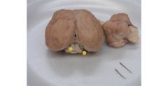
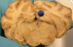
What does the pituitary gland do?
Where is it? (hint: dura matter) |
synthesizes and releases hormones
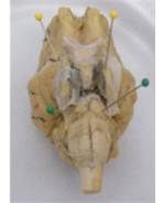
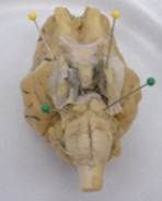
Found between the trigeminal nerves on the inferior part of brain |
|
|
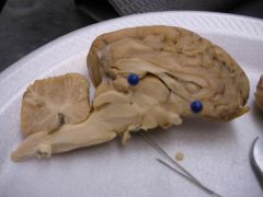
What do the cerebral peduncles do?
Where are they? |
motor system fibers from cerebral cortex that project to the brainstem areas and to spinal cord
Found inferior, anterior to pons |
|
|
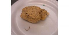
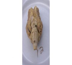
Point to the anterior/rostral and posterior/caudal directions of the brain?
|
Anterior/rostral - toward the nose end
Posterior/caudal - toward the tail end |
|
|
Point to the dorsal and ventral parts of the brain?
|
dorsal - toward the surface of the back and top of the head;
ventral - toward the surface of the chest or bottom of the head |
|
|
Explain medial vs lateral.
|
medial - toward the midline of the body;
lateral - away from the midline of the body |
|
|
Explain superior vs. inferior
|
superior - toward the top of the brain or above another structure;
inferior - toward the bottom of the brain or below another structure |
|
|
How many pairs of cranial nerves are there?
|
12
(On Old Olympus’ Towering Tops, A Friendly Viking Grew Vines And Hops) (Some Say Money Matters, But My Brother Says Big “Brains” Matter Most) |
|
|
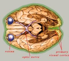
What is the optic chiasm?
Where is it? |
The place where the nerve becomes the tract; where 50% of the nerves cross over (in humans)
|
|
|
What is the difference between optic nerve and optic tract?
|
The optic nerve is before (anterior) the chiasm and the tract is after (posterior). In general nerves are outside the brain and part of the PNS, whereas tracts are inside the brain and part of the CNS.
|
|
|
Olfactory bulb:
What #? Motor, sensory, or both? What does it do? Where is it? |
# - 1
Sensory Smell Projects from the olfactory bulbs |
|
|
Optic nerve:
What #? Motor, sensory, or both? What does it do? Where is it? |
# - II
Sensory Vision projects from optic chiasm |
|
|
Oculomotor:
What #? M/S/B? What does it do? Where is it? |
# - III
Motor Control of eye movements, pupil constriction Projects from cerebral peduncles |
|
|
Trochlear:
What #? M/S/B? What does it do? Where is it? |
# - IV
Motor Control of eye movements, rolling outward and downward Projects in between pons and cerebellum laterally |
|
|
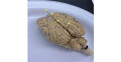
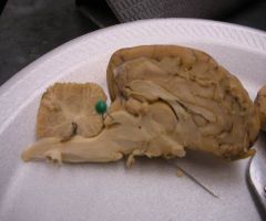
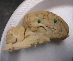
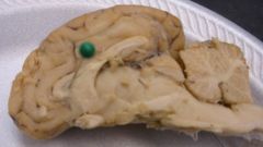
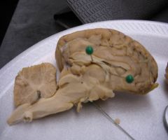
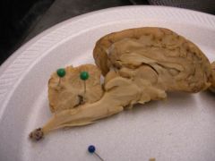
Trigeminal:
What #? M/S/B? What does it do? Where is it? |
# - V
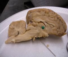
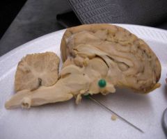
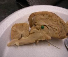
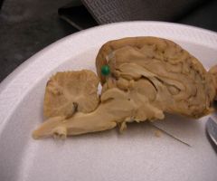
Sensory & Motor Touch and pain sensations from most of the face; Control of jaw muscles for chewing and swallowing, hot pepper taste Biggest nerves found lateral to pituitary gland (YELLOW) |
|
|
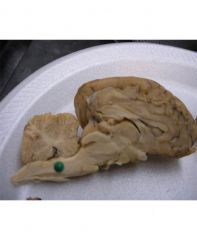
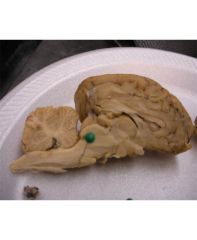
Abducens:
What #? S/M/B? What does it do? Where is it? |
# - VI.
Motor Control of eye movements, side to side Projects in between pons and medulla (GREEN) |
|
|
Facial:
What #? M/S/B? What does it do? Where is it? |
# - VII. (7)
Sensory & Motor Control of muscles of the face (facial expression, crying); Taste for anterior 2/3rd of tongue |
|
|
Stratoacoustic / Vesibulochochlear:
What #? M/S/B? What does it do? |
# - VIII (8)
Sensory Hearing and equilibrium |
|
|
Glossopharyngeal:
What #? M/S/B? What does it do? |
# - IX. (9)
Sensory & Motor Taste/sensation for throat and posterior 1/3rd of tongue; Control gag reflex and throat movements during speech |
|
|
Vagus:
What #? M/S/B? What does it do? |
# - 10
Sensory & Motor Sensations from neck & throat; sensations and motor control of many internal organs including heart, lungs and stomach. |
|
|
Spinal accessory:
What #? S/M/B? What does it do? Where is it? |
# - XI. (11)
Motor Control of neck and shoulder muscles for turning movements of the head Runs laterally to brain stem |
|
|
Hypoglossal:
What #? M/S/B? What does it do? |
# - XII (12)
Motor Control of the muscles of the tongue |
|
|
What is the difference between afferent and efferent?
|
Afferent - going toward the CNS - afferent nerves carry sensory info from skin, skeletal muscles, joints, eyes, ears, viscera, and so on to CNS; think of "a" as "approach", "arrive" - coming into CNS - sensory
Efferent - going away from the CNS - carry motor signals from the CNS to the skeletal muscles and viscera think of "e" as "exit" - going away from the CNS - motor |
|
|
Midsaggital View
Where is the Cingulate Gyrus? |
Above the corpus callosum.
|
|
|
What is the Thalamus?
Where is it? |
Sensory relay system of the brain, sending information primarily to the cortex
Circular structure superior to hypothalamus |
|
|
What is the Hypothalamus?
Where is it? |
Controls autonomic & endocrine (hormone) system; the 4 F's - feeding, fighting, fleeing, and fornicating; connects with pituitary for appetite
Inferior to thalamus, superior to optic chiasm |
|
|
What is the Pineal body?
Where is it? |
Aka - Pineal gland - responsible for secreation of melatonin - a hormone which, among other things, induces sleep
Rostral to tectum |
|
|
Where are the Superior Colliculi?
|
where
|
|
|
Where are the Inferior Colliculi?
|
where
|
|
|
Where is the Cerebellum?
|
where
|
|
|
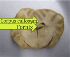
What is the Corpus Callosum?
Where is it? |
Band of white matter by which each cerebral hemisphere connects with the opposite cerebral hemisphere (allows for communication between the two)
Inferior to cingulate gyrus |
|
|
Midsagittal View
What is the Septum Pellucidum? Where is it? |
A thin layer of tissue that separates the two lateral ventricles from each other.
Medial portion of lateral ventricles |
|
|
Midsagittal View
What is the Fornix? Where is it? |
Band of white matter by which the hippocampus of each hemisphere connects to the hippocampus of the opposite hemisphere
Inferior to lateral ventricles |
|
|
Midsagittal View
What is the Arbor Vitae? Where is it? |
"Tree of life"; white nerve tissue of the cerebellum, has a tree-like outline.
|
|
|
Midsagittal View
Where is the Medulla? |
where
|
|
|
Midsagittal View
Where is the Pons? |
where
Pons bulges out at the bottom and medulla oblongates from it |
|
|
Midsagittal View
What is the Ventral Tegmental Area? |
Considered part of the pleasure system, or reward circuit. Activities which produce pleasure, such as psycho-stimulant drugs, activate this area
|
|
|
Midsagittal View
What is the Substantia Nigra? |
A layer of nerve cells in the midbrain that produce the neurotransmitter dopamine. One of the movement control centers of the brain. Also involved in addiction. Its destruction is seen in Parkinson's Disease.
|
|
|
Midsagittal View
Where are the Lateral ventricles? |

Two large empty spaces directly inferior to the corpus callosum
|
|
|
Midsagittal View
Where is the 3rd ventricle? |
Hollow space directly inferior to the fornix.
|
|
|
Midsagittal View
Where is the 4th Ventricle? |
Hollow space directly inferior to the cerebellum
|
|
|
Midsagittal View
What is the Cerebral Aqueduct? Where is it? |
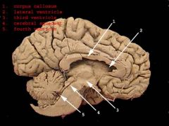
Short canal traveling through the midbrain; connects the 3rd and 4th ventricles
Found inferior to the IC |
|
|
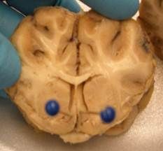
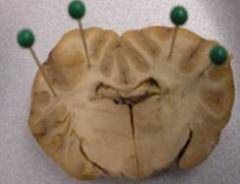
Coronal View
What is The Limbic System? |
A circuit of midline structures that circle the thalamus (limbic means "ring") - has several functions including motivating behaviors, learning, and memory
|
|
|
Coronal View
What is the function of the Thalamus? Where is it? |
located in the diencephalon, below the 3rd ventricles,
heart shaped; both process and relay sensory information selectively to various parts of the cerebral cortex regulating states of sleep and wakefulness; involved with consciousness; major role in regulating arousal, the level of awareness, and activity. Damage to the thalamus can lead to permanent coma; should see in the 3rd slice. |
|
|
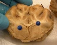
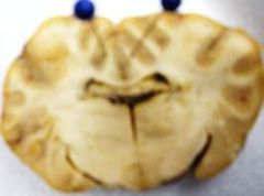
Coronal View
What is the function of the amygdala? |
Involved in visceral and autonomic responses, sexuality, food and water intake; also involved in emotions, emotional memories, fear, aggression, and pleasure
|
|
|
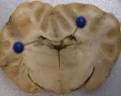
Coronal View
What is the function of the hippocampus? Where is it? |
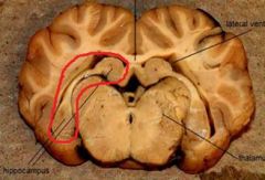
Involved in spatial memory (ex. put a rat into a maze, learns the mase, then lesion HC - after lesion can't remember how to get through maze); formation of new episodic memories; temporal information: dates, times, appointments, schedules, etc.; should see in the 3rd slice
Seahorse structure in 3rd slice |
|
|
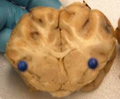
Coronal View
What is the septal nuclei responsible for? What happens if there is damage? Where is it? |
Connects with hippocampus, hypothalamus, amygdala, thalamus, and some structures in the midbrain; Role in reward and reinforcement along with nucleus accumbens; damage in humans can result in rage behavior and other strange behaviors; should see in the 1st slice
Lighter area inside septum; seen in 1st slice |
|
|
Coronal View
What are the mammillary bodies responsible for? What disease are they associated with? |
Play a role in memory; Korsakoff's syndrome - amnesia resulting from severe alcoholism
|
|
|
Coronal View
What does the Basil Ganglia do? |
Set of structures that play a major role in voluntary motor responses; sends motor messages thalamus and midbrain --> cortex --> medulla and spinal cord; also responsible for procedural memory
|
|
|
Coronal View
What is the caudate nucleus responsible for? Where is it? |
Responsible for learning and memory, specifically in receiving feedback; theorized that it is involved with OCD; found to function when falling in love; should see in 2nd and 3rd slices
Gray area medial to the internal capsule in slices 2 and 3 |
|
|
Coronal View
What is the putamen responsible for? Where is it? |
Involved in reinforcement learning and implicit learning; found to play a role in the perception of hate; should see in 2nd and 3rd slices
Gray area lateral to internal capsule in slices 2 and 3 |
|
|
Coronal View
What 2 structures make up the striatum? |
Caudate nucleus and Putamen
|
|
|
Coronal View
What does the globus pallidus do? |
plays an active part in pre-filtering external stimuli and may help reduce the amount of irrelevant information the brain can store.
|
|
|
Coronal View
Explain what Parkinson's disease is and what causes it. |
A disorder characterized by rigidity of muscles, resting tremors, and impairment of speech; results from deterioration of pathway from substantia nigra (midbrain structure) to the basal ganglia causing low levels of dopamine.
|
|
|
Coronal View
Where is the fornix? |
Should see in the 2nd and 3rd slices.
Corpus callosum is above the lateral ventricles which are above the fornix |
|
|
Coronal View
What is the corona radiate? Where is it? |
The radiating white matter of the neocortex carry nearly all of the neural traffic from and to the cerebral cortex. Should see in all 3 slices.
|
|
|
Coronal View
Where is the corpus callosum? |

Should see in all 3 slices
Corpus callosum is above the lateral ventricles which are above the fornix |
|
|
Coronal View
What is the internal capsule? Where is it? |
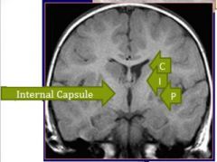
White matter surrounding subcortical structures; corona radiata --> internal capsule --> cerebral peduncle; runs between the caudate and putamen at some points; should see in 1st and 2nd slices.
runs between the caudate and putamen at some points; should see in slices 2 and 3 |
|
|
Coronal View
Where is the media longitudinal fissure? |

should see in all 3 slices.
superior medial space between hemispheres |
|
|
Coronal View
Where is the cingulate gyrus? |
Should see in all 3 slices.
“fox ears” above corpus callosum |
|
|
Coronal View
Where are the lateral ventricles? |
Should see in all 3 slices.
Lateral ventricles - all 3 slices; superior ventricles; inferior to corpus callosum |
|
|
Coronal View
Where is the 3rd ventricle? |
Should see in the 2nd and 3rd slices.
3rd ventricle – 2nd and 3rd slices; inferior to fornix |
|
|
Coronal View
Where is the pineal body (pineal gland)? |
Should see in the 3rd slice.
Center of 3rd cut Inferior to 3rd ventricle |
|
|
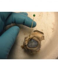
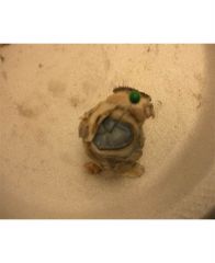
Eye Ball
What is the sclera and where is it? |
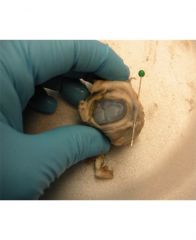
White protective covering of the eyeball (except cornea) - an extension of the dura mater.
|
|
|
What is the optic nerve and where is it (on the eye, not the brain)?
|
Carries visual information to the brain
carries visual information to the brain, extending from the ganglion cells of the retina |
|
|
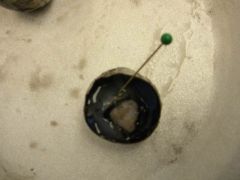
What is the nictitating membrane?
Where is it? |
Sheep and other animals it is a third eyelid; controlled by the abducens nerve
Eyelid on the side, has a dark edge |
|
|
Eye Ball
What is the conjunctiva? Swelling causes what? |
Delicate membrane that starts at the junction of the sclera and the cornea; lines inside of eyelid and provides lubrication; conjunctivitis = swelling of conjuctiva from infrection (aka pink eye)
|
|
|
Eye Ball
What is the cornea? Where is it? |
Area of the eye anterior to the iris; transparent protective covering; some accommodation occurs
|
|
|
Eye Ball
What is the iris and where is it? |
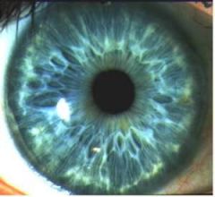
colored part of the eye; surrounds the pupil - orange on sheep
|
|
|
Eye Ball
What is the pupil? |
Hole in the iris controlled by muscles; it widens and narrows to allow the needed amount of light into the eye; controlled by the oculomotor nerve; failure to constrict to light indicates a concussion
|
|
|
What is the lens and where is it?
|
Transmits and refracts light to focus images near or far (accomodation) usually transparent
|
|
|
Eye Ball
What are the ciliary bodies? |
controls the suspensory ligaments which adjust the shape of the lens; there are also epithelium cells here that secrete the aqueous humor.
|
|
|
Eye Ball
What is the aqueous humor? |
liquid found in the anterior chamber of the eye; secreted and removed as waste.
|
|
|
Eye Ball
What is the vitreous body/humor and where is it? |
gel-like substance on the inside of the eyeball; gives support and shape.
|
|
|
Eye Ball
What is the retina and where is it? |
the neural tissue containing photoreceptive cells located on the inner surface of the posterior portion of the eye.
|
|
|
Eye Ball
What is the macula? |
The portion of the retina with the highest ability to resolve visual detail. The site of macular degeneration.
|
|
|
Eye Ball
What is the fovea? |
Center of the macula, made up only of cones; mediates find detail and color, the most acute vision
|
|
|
Eye Ball
What is the blind spot and where is it? |
location of the exit point from the retina of the fibers of the ganglion cells that form the optic nerve, blood vessels also exit the eye here; no photoreceptors
|
|
|
Eye Ball
What is the choroid and where is it? |
black substance of the back of the eyeball; behind the retina; absorbs any stray light; vascular layer
|
|
|
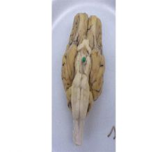
Eye Ball
What is the tapetum lucidum and where is it? |
blue-green iridescent membrane on back of sheep eye; reflective structure acts like a mirror, reflecting light back through the retina, allowing for better night vision.
|