Reading...
![]()
Play button
![]()
Play button
![]()
Use LEFT and RIGHT arrow keys to navigate between flashcards;
Use UP and DOWN arrow keys to flip the card;
H to show hint;
A reads text to speech;
52 Cards in this Set
- Front
- Back

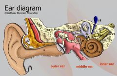
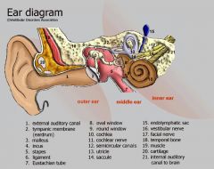
Identify #3. How is this structure used to divide the ear structures logically?
|

Eardrum. All structures external to this point are referred to as the external ear. Structures internal from this point to the ossicles are referred to as the middle ear.
|
|

Identify #1
|

Pinna (Auricle)
|
|

What is the formal name for earlobe. Where is it located on this diagram?
|

Lobule. Located in the very bottom right hand corner of the illustration, inferior and external to #2.
|
|

What is #2?
|

External acoustic meatus/canal/tube
|
|

What is #3?
|

Tympanic membrane
|
|

What space is located between #3 and #12?
|
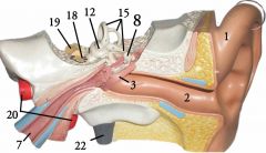
Tympanic cavity
|
|

What is #8?
|

The malleous or hammer.
|
|

What is #9?
|
The anvil or incus.
|
|

What is #11?
|

The stapes or stirrup.
|
|

What structure is underneath #11 (The stirrup) in this illustration?
|

The oval window
|
|
What is #14?
|

The round window
|
|

What "tube" does #7 lead to?
|

The nasopharynx
|
|

What is the reddish tube on this model between the hammer (#8) and the anvil (#9)?
|

The tensor tympani
|
|

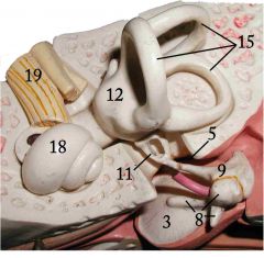
What is #18?
|

The cochlea
|
|

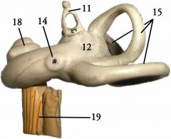
What is #15?
|

The semi-circular canals
|
|

Looking at the semi-circular canals on this model (#15), name each of the three canals
|

The canal oriented along the transverse plane (bottom right in this illustration) is the lateral canal.
The canal oriented along the sagittal plane (top left in this illustration) is the anterior canal. Finally, the canal oriented along the coronal plane (top right in this illustration) is the posterior canal. |
|

What is #12?
|

The vestibule
|
|

What structure is indicated by #15 in this illustration?
|

The endolymphatic sac
|
|
What nerve is indicated by #16?
|

The vestibulocochlear nerve
|
|

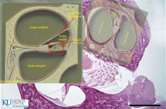
What is #1?
|

Scala vestibuli
|
|

What is #2?
|

Scala tympani
|
|

What is #3?
|

Vestibular membrane
|
|

What is #4?
|

Scala media
|
|

What is #5?
|

Organ of corti
|
|
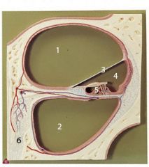
What is #6?
|
Spiral ganglion
|
|

What is #1?
|
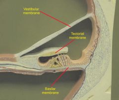
Vestibular membrane
|
|

What is #2?
|

Tectorial membrane
|
|
What is #3?
|

Basilar membrane
|
|
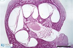
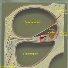
Identify the structures visible on this slide
|
Utilizing the top right section, from left to right, we see the scala vestibuli, then the vestibular membrane, the the scala media - containing the organ of corti, the tympanic membrane, and the scala tympani
|
|

What fluid is contained in #1 and #2?
|

Perilymph
|
|

What fluid is in #4 and #5?
|

Endolymph
|
|

What bone moves the fluid in your ear? What window does it hit? What cavity does that window push on? What cavity does it return through? And what window absorbs the shock?
|

The stapes. The oval window. The scala vestibuli. The scala tympani. The round window.
|
|

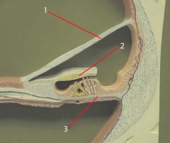
Identify 1
|

Scala vestibuli
|
|

Identify 2
|

Scala media
|
|

Identify 3
|

Scala tympani
|
|

Identify 4
|

Vestibular membrane
|
|

Identify 5
|

Tympanic membrane
|
|

Identify 7
|
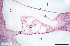
Organ of corti
|
|

Identify 6
|

Tectorial membrane
|
|

What is #13?
|

Utricle
|
|

What is #14
|
Saccule
|
|
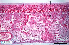
What is this a slide of? Identify the areas indicated by number
|

Olfactory epithelium (technically only the top edge, 1&2).
1. Hair cells 2. Supporting cell 3. Mucus glands |
|

Identify 1
|

Circumvillate papillae
|
|

Identify 2
|

Fungiform papillae
|
|
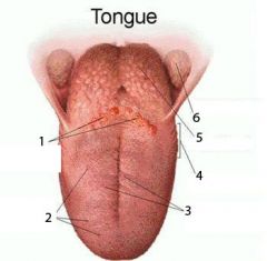
Identify 3
|

Filliform papillae
|
|
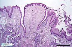
What is depicted here? How do you know?
|

Fungiform papillae on the tongue. Wider at the top than the bottom, like a mushroom.
|
|

What is depicted here? How do you know?
|
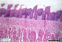
Filliform papillae on the tongue. Long and narrow structures.
|
|
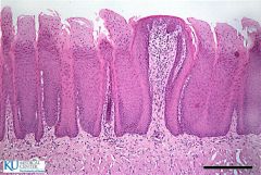
What is depicted here? How do you know?
|

Circumvillate papillae on the tongue. Large central structure with valleys on either side, taste buds visible along the edges of the valleys. (3 bumps in a row)
|
|
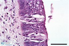
What is depicted here?
|

A taste pores with gustatory hairs
|
|
|
What is a Weber test?
|
A tuning fork is struck and placed medially on the subject's head. Sound should be heard equally in both ears. If so, the subject has equal hearing or equal hearing loss in both ears.
|
|
|
What is a Rinne test?
|
A tuning fork is struck and placed against the subject's mastoid process. Once the sound fades from perception this way, the fork is held next to the auditory canal; if the subject can hear it again via air conduction, hearing is not impaired.
|
|
|
What is a Barany test?
|
The subject is placed on a rotating chair or stool and spun. Spinning is stopped abruptly; the subject's eyes should continue scrolling back and forth and the subject should experience nystagmus. If not, injury to the semi-circular canals has occured.
|