![]()
![]()
![]()
Use LEFT and RIGHT arrow keys to navigate between flashcards;
Use UP and DOWN arrow keys to flip the card;
H to show hint;
A reads text to speech;
82 Cards in this Set
- Front
- Back
|
Sarcocystis neuronas life cycle |
DH: opossums IH: armadillo, felids, skunk, raccoon, sea otters, fishers dead end host: horses
1. DH sheds sporulated oocysts in feces 2. IH ingests and sarcocysts develop in muscles causing disease 3. DH ingests infeced carrion and gametogony occurs in GI tract
DEH ingests oocysts and merozoite crosses blood brain barrier; sarcocysts do not develop in muscles so it cannot transmit disease
|
|
|
Sarcocystis neuronas pathology and clinical signs |
pathology - causes focal hemorrhages and discoloration in brain and spinal cord - multifocal, non-suppurative myeloencephalitis
Clinical signs - non-specific neurological signs |
|
|
Sarcocystis neuronas diagnosis |
- clinical signs can be suggestive - run titers on both serum and CSF- CSF should be higher (serum alone is not sufficient because 75% of seropositive horses are not currently infected) |
|
|
Sarcocystis neuronas treatment goals and methods |
- reduce inflammation and brain and spinal cord edema-- banamine - get rid of agent (only effective if treated before too much inflammatory damage is done) -- ponazuril |
|
|
Sarcocystis neurona control |
prevent opossums from contaminating hay and/or pastures with feces |
|
|
Hepatozoon americanum life cycle |
DH: dogs and wild canids in US vectored by ambylomma maculatum (gulf coast tick)
1. dogs ingest tick with sporozoites 2. schizogony occurs in spleen, lymph nodes, liver, and muscles causing clinical signs 3. PMNs with gamonts are ingested by ticks 4. gamotogony occurs in tick 5. zygote enters tick GI (hemocoel) 6. oocysts form and undergo sporulation with production of sporozoites within hemocoel |
|
|
Hepatozoon americanum transmission |
- ingestion of infected tick - ingestion of vertebrate paratenic host with muscle cysts is suggested to be possible |
|
|
Hepatozoon americanum pathology and clinical signs |
pathology - widespread myositis - periostitis with new bone growth - large, onion like cystic schizonts in skeletal muscle
clinical signs - intermittent fever - anorexia - neutrophilic leukocytiosis - severe joint and muscle pain - stiffness - "master's voice stance" |
|
|
Hepatozoon americanum diagnosis |
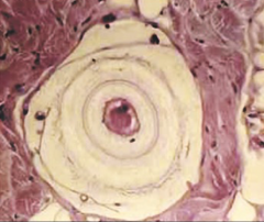
-see gamonts in neutrophils -schizonts in muscle biopsies (onion layer look and 250-500 um) |
|
|
Hepatozoon americanum treatment |
acute: sulfonamides and ponazuril chronic: decoquinate |
|
|
Hepatozoon americanum control |
tick control minimize predation |
|
|
Babesia spp. life cycle |
DH: dogs vector: Rhipicephalus sanguineus (brown dog tick)
1. vermicules injected into dog from salivary glands of ticks and they enter RBCs 2. in RBCs vermicules form piroplasms which reproduce by binary schizogony 3. merozoites enter other RBCs and repeat schizogony 4. ticks ingest merozoites 5. may or may not undergo gametogeny in tick GI (unknown) 6. undergo schizogony in tick GI and produce vermicules 7. Schizonts move to other tissues and repeat process, in ovary they enter developing tick larvae 8. vermicules eventually enter salivary glands from which they can infect new host |
|
|
Babesia canis vogeli vs. Babesia gibsoni transmission |
B. canis vogeli- through vector B. gibsoni transmission- through vector or blood exchange (transfusion, fighting) |
|
|
Babesia canis clinical signs |
highly variable: asymptomatic to peracute to chronic
peracute: - severe anemia - ictuerus - anorexia - bilirubinuria - hemoglobinuria
chronic - anemia (autoimmune) - ascites - SQ edema - jaundice
disease is regenerative so you will see immature RBCs |
|
|
Babesia canis diagnosis |
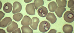
- piroplasms in RBCs on smears (most likely to see in acute- febrile stage) - ID based on size and shape (vogeli- 5 um, gibsoni- 1-3 um) - brain smears at necropsy |
|
|
Babesia canis treatment |
- aromatic diamidines and carbanilides - imozol won't work on B. gibsoni |
|
|
Babesia canis control |
vector control - keep R. sanguinus out of homes and kennels (can survive for years indoors) no dog fighting screen blood donors |
|
|
Cytauxzoon felis life cycle |
DH: felids (bobcats are natural host - no disease in them) vector: Ixodid ticks- Amblyomma americanum (lone star tick)
1. sporozoites from ticks infect cats and start schizogony in mononuclear phagocytes in vessels of lungs, spleen, and liver causing clinical signs - schizonts are large with syncytial cytomeres 2. merozoites (1-2 um) are produced and enter RBCs and form piroplasms 3. piroplasms are ingested by ticks during blood meal 4. in tick protozoa undergoes gametogeny, sporogony, and form sporozoites |
|
|
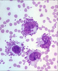
Cytauxzoon felis pathology and clinical signs |
pathology - pallor and icterus - engorged and distended abdominal veins - lumens of veins and venules occluded by large infected macrophages (worst in lungs) --> DIC - dark enlarged spleen - enlarged lymph nodes
clinical signs - rapidly fatal in domestic cats (100% mortality) 1. anorexia, depression, lethargy 2. fever (103-104), dehydration, icterus 3. marked dyspnea 4. fever abates 5. death |
|
|
Cytauxzoon felis diagnosis |
- piroplasms in RBCs on blood smear (only in late disease) - schizonts in aspirates or smears of spleen, lymph nodes, or bone marrow - PCR (important in early infection) |
|
|
Cytauxzoon felis treatment and control |
treatment - supportive care - tx often ineffective: atovaquone + azithormycin (60% recovery rate)
control - keep ticks away from cats (keep indoors or use preventative) |
|
|
Characteristics of flagellates |
- live in intimate association with mucous membrane - pyriform shape - possess 1 or more flagella (typically 4-6) - may or may not have nonmotile cyst stage - reproduce by longitudinal binary fission - have single axostyle |
|
|
intestinal flagellates |
trichomonas - no cyst stage - named based on number of flagella - may be commensal - herky jerky motion
giardia - parasitic - cyst and trophozoite stage - cyst is infective stage - tumbling (falling leaf) motion |
|
|
hemoflagellate characteristics |
- blood and tissue flagellates - vectored by blood sucking arthropods - mostly found outside US |
|
|
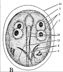
Giardia trophozoite characteristics |
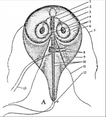
- found in small intestine and diarrhea - 12x8 um - pyriform shape (rounded anterior, pointed posterior) - 8 flagella (4 pairs); can have 3 or 5 pairs and would be called tri- or pentagiardia respectively - 2 nuclei - ventral sucking disk used for attachment to intestinal cells
|
|
|
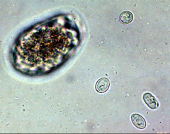
Giardia cyst morphology |
- found in formed feces - environmental dormant form that can survive wide range of temperatures and chlorine - 10x 8 um - elliptical - quadrinucleate - infective form |
|
|
Giardia life cycle |
direct 1. cyst is ingested and excystation occurs in small intestine 2. trophozoites develop and live in small intestine (cause clinical signs) 3. trophozoites multiply via longitudinal binary fission 4. cysts are formed and both trophs and cysts are shed in feces |
|
|
Sources of giardia transmission |
ingestion of cysts: - animal/human fecal contamination of water, food, and fomites - self grooming
highly infective so can be easily reinfected |
|
|
giardia pathology and clinical signs |
pathology - nutrient malabsorption due to blockage of intestinal epithelium and destruction of microvilli - lipid and carb maldigestion - inflammatory response leading to hypermotitiy and increased mucous production
clinical signs - malodorous, gray, greasy, and voluminous diarrhea (not hemorrhagic- parasite only on surface of cell) - flatulence |
|
|
Giardia diagnosis |
- trophs in direct smear of diarrhea (can't do ff for trophs because osmotic pressure will rupture them) - cysts on zinc sulfate ff of formed feces (sugar's osmolarity is higher and will crush cysts) - need at least 3 negative exams (done every other day) before negative diagnosis can be confirmed - ELISA (use with caution- many false negatives) |
|
|
Giardia treatment |
first choice is fenbendazole - if tx is unsuccessful and you are sure it isn't because of reinfection then: --metronidozole + fenbendazole for dogs -- metronidozole for cats
severe cases may require fluid therapy |
|
|
giardia control |
when treating you must implement control measures or reinfection WILL occur
- remove feces daily and prevent fecal contamination of food and water - treat all animals in household - bathe all animals - disinfect environment (drying is important) |
|
|
Trichomonas gallinae life cycle |
DH: columibs (pigeons and doves), chickens, turkeys, raptors
direct life cycle 1. trophozoites are transferred to new host 2. attach to mucous membranes of mouth and digestive tract where they cause clinical signs |
|
|
Trichomonas gallinae transmission |
ingestion in water regurgitative feeding (columibs) carnivorism by raptors |
|
|
Trichomonas gallinae morphology |
trophozoites only - found in upper digestive tract - 10 x 5 um - pyriform shaped - 4 anterior flagella and undulating membrane (no axial flagella) |
|
|
Trichomonas gallinae pathology and clinical signs |
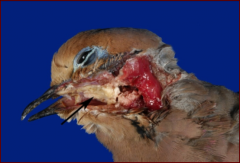
pathology - caseous necrotic masses in upper digestive tract - acute lesions in mouth, pharynx, esophagus, and crop - oral and liver lesions in raptors - lesions and clinical signs mainly caused by immune response
clinical signs - depression - excessive salivation and emaciation- can't swallow because of lesions - listless with ruffled, dull feathers - respiratory distress |
|
|
Trichomonas gallinae diagnosis |
- clinical signs and characteristic lesions - trophs in lesions, direct smear of caseous material - trophs in saliva - cultivation of organisms |
|
|
Tritrichomonas foetus characteristics |
DH: cattle trophozoite only - 17 x 10 um - pyriform - axostyle present - 3 anterior flagella - undulating membrane - single nucleus
causes bovine genital trichomoniasis |
|
|
Tritrichomonas foetus clinical signs |
cows - infertility - abortion - pyometra
bulls - no signs - maintained in preputial crypts
main complaint is not getting cows bread back (usually after getting a new bull or lending out bull) |
|
|
Tritrichomonas foetus diagnosis |
culture followed by PCR - 3 negative cultures are required to confirm a negative - sample is taken from preputial pouch and inoculated into test pouch that supports growth but prevents growth of other microorganisms |
|
|
Tritrichomonas foetus life cycle |
- reproduces by longitudinal binary fission in prepuce of bulls and genital tract of cows - maintained in preputial pouch of bull - transmitted to cow through coitus |
|
|
Tritrichomonas foetus treatment and control |
treatment - cull bulls - rest cows
control - buy only confirmed virgin bulls - use AI breeding program - negative culture non- virgin bulls - vaccine- efficacy is questionable |
|
|
Tritrichomonas blagburni disease
|
emerging feline large bowel disease
- associated with chronic diarrhea and bacterial overgrowth in cats - dribbling diarrhea - can lead to rectal prolapse |
|
|
Tritrichomonas blagburni diagnosis
|
direct smear looking for trophozoites
culture like in cattle PCR - often mistaken for giardia in microscopic exams |
|
|
Histomaonas meleagridis life cycle
|
DH: Galliforms
no cyst form (can't survive in environment) 1. eggs enter Heterakis gallinarium (nematodes) 2. nematode is inside earthworm 3. turkey eats earthworm (or can be transmitted directly from turkey to turkey in high density situations) 4. flagellated form (1 flagella) develops in cecum and replicates via binary fission 5. moves to liver where it looses flagella and becomes amoeboid form- causes liver abscesses |
|
|
Histomonas meleagridis host and disease
|
severe: turkeys and peafowl
moderate: quail, guinea fowl, chickens inapparent: pheasants and jungle fowl |
|
|
Histomonas melagridis pathology and clinical signs
|
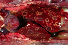
Pathology - pathoneumonic liver abscesses (target lesions) - caseous necrosis in cecum (this happens before liver abscesses) Clinical signs - sulfur yellow diarrhea -depression -fever -death within four days of |
|
|
Trypanosome general characteristics
|
- parasites of blood and tissues of vertebrates
- grouped based on type of transmission (fecal, saliva, or mechanical/venereal - 2 life stages: tryptomastigote (blood) and amastigote (tissue) - 1 central nuclei - 1 flagella on back |
|
|
Trypanosomea cruzi disease, host, and distribution
|
-disease: trypanosomiasis (Chagas' disease): considered most important cause of myocarditis in the world
-host: dogs, humans, cats, raccoons, rodents, etc -vector: reduviidae bugs (blood sucker) aka kissing or assassin bugs - distribution: south america, central america, southern US |
|
|
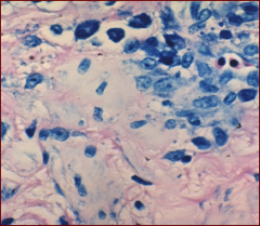
Trypanosomea cruzi morphology
|
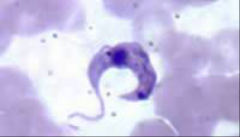
trypomastigoe
- low numbers in the blood -free flagellum and long, slender undulating membrane - 16-20 um amastigote - in cardiac, smooth and skeletal muscle cells, neuroglial cells, reticuloendothelial cells - small round cell structure |
|
|
Trypanosomea cruzi life cycle
|
1. kissing bug ingests trypomastagotes when it feeds on infected host
2. parasite moves to midgut of vector and multiplies by longitudinal fission 3. infective trypomastigote is either defecated by vector and ingested by new host (SA) or entire vector is ingested (NA) 4. trypomastigote moves through blood to tissues where it develops into amastigote and causes disease 5. amastigotes move to blood and become trypomastigotes which can be picked up by vector |
|
|
Trypanosomea cruzi transmission
|
- ingestion of vector or vector feces
- infected vector feces rubbed into open wound - transplacental - transmammary - transfer (via blood) from infected tissues of other animals |
|
|
Trypanosomea cruzi pathology and clinical signs
|
pathology
- infects macrophages followed by tissue cells (myocardial and neuralgia preferentially) - severe nectrotizing myocarditis - megaesophagus and megacolon - death from heart dysfunctions clinical signs (human) - swelling at sight of bite (chagoma) - lymphadenopathy -hepato and splenomegaly clinical signs (dog) - cardiac dysfunctions - pale mucous membranes - ascites - lymphadenopathy - hepato and splenomegaly - death within 28 days of clinical signs |
|
|
Trypanosomea cruzi diagnosis
|
- trypomastigoes in blood and fluids (smears)
- amastigotes in tissue samples from aspirates or biopsy (or necropsy) - blood culture - ELISA, PCR, IFA |
|
|
Trypanosomea cruzi treatment
|
benznidozole works best but it is not available in the US and causes severe nausea as a side effect
- most dogs are euthanized |
|
|
Trypanosomea cruzi control
|
- eliminate bugs
- improve living conditions (no dirt floors or thatched roofs - prevent bugs from biting |
|
|
Leishmania donovani disease, distribution, and hosts
|
host: dogs, fox, humans, rodents (foxhounds in US)
vector: sandflies disease: leishmaniasis distribution: all over but mostly in middle east and northern south america (killing off ISIS) |
|
|
Leishmania donovani life cycle
|
1. amastigotes ingested by fly during blood meal
2. transform and multiply in fly 3. migrate to fly salivary gland 4. enter mammal during blood meal 5. amastigotes develop in various tissues and cause clinical disease (NO TRYPOMASTIGOTE IN VERTEBRATE HOST) |
|
|
Leishmania donovani clinical signs
|
can be visceral or cutaneous
-progressive anemia - hepatosplenomegaly - lympadenopathy - chronic wasting - skin hyper pigmentation - ocular signs: (keratoconjunctivitis, uveitis, retinitis) - alopecia (dogs) - long brittle toenails - nose bleeds - mucocutaneous ulcers |
|
|
Leishmania donovani diagnosis |
CAN BE VERY DIFFICULT - amastigotes in tissue biopsies and cytologic preps (look identical to histoplasmosis (fungus) and trypomaniasis but leishmania amastigotes are only found in macrophages) - ELISA - IFA -PCR |
|
|
leishmania donovani treatment
|
no approved treatment
- pentavalent antimonials may help but must contact CDC to get |
|
|
Leishmania donovani control
|
humans- handle known infected pets with care and avoid contact with sores
- eliminate sandflies - control commensal rodents - treat/euthanize infected dogs |
|
|
Leishmania siamensis
|
DH: horses
- emerging in SE US - causes cutaneous nodules - Dx by biopsy and PCR |
|
|
Amebae general characteristics
|
- indefinite in shape
- move and phagocytize with pseudopodia - facultative parasites (mostly free living unless they get into a host) - may or may not produce cysts - trophozoite found within host |
|
|
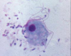
Entamoeba morphology
|
trophozoites
- found in large intestine - 20-30 um - blunt pseudopodia - non- foamy cytoplasm - uninucleate cysts - found in formed feces - 10- 20 um - spherical - quadrinucleate - resistant to environmental insults |
|
|
Entamoeba invadens host and disease
|
amebic dysentery in reptiles
- snakes, lizards, and some species of tortices |
|
|
Entamoeba histolytica life cycle
|
direct
1. host ingests quadranucleate cyst 2. excystation occurs in lower ileum 3. replication by binary fission 4. trophozoite is formed and lives in lumen and mucosa of colon 5. encystation and maturation in colon 6. host defecates mature cyst |
|
|
entamoeba histolytica host and disease
|
host: primates, dogs, and cats (humans serve as reservoir and pass it to other hosts
disease: amebic dysentery |
|
|
Entamoeba histolytica pathology
|
intestinal
- production of proteolytic enzymes - invasion of colonic mucosa (in monkeys it invades stomach) - hydrolysis of host tissue - ulceration - can erode submucosa and become extra-intestinal through blood extra-intestinal - liver infection- abscesses - lytic invasion into chest cavity, lungs, and brain |
|
|
Entamoeba histolytica clinical signs
|
vary depending on strain
- diarrhea - abdominal discomfort - bloody mucus - pulmonary and hepatic disease (if it gets this bad they are going to die |
|
|
Amebiasis diagnosis
|
throphs in diarrhea (smear)
cysts in formed feces (zinc sulfate float) - need 3 negative tests (every other day) before negative can be confirmed culture |
|
|
Amebiasis treatment
|
metronidazole
tetracyclines |
|
|
amebiasis control
|
prevent fecal contamination of food and water
|
|
|
General characteristics of Ciliates
|
- possess hair like cilia used for locomotion
- most species are commensal in large intestine or rumen of mammals - large- up to 100 um in diameter - oval to pyriform - 2 types of nuclei (micro and macro[dumbbell]) - can reproduce asexually (binary fission) or sexually (conjugation) |
|
|
What is the genus of parasitic ciliates of mammals?
|
Balantidium
|
|
|
balantidium coli host and disease
|
DH: swine, primates, and dogs (zoonotic mainly from swine to humans)
large intestine ulceration--> dysentery |
|
|
Balantidium coli morphology
|
trophozoites
- found in large intestine and diarrhetic feces - 30-150 um long - ciliated - micro and macro nuclei cysts - found in formed feces - 40-60 um - round to oval in shape - micro and macro nuclei - no cilia |
|
|
Balantidium coli lifecycle
|
1. cyst is ingested by host
2. excystation occurs in the small intestine 3. trophozoite multiples by transverse binary fission and moves to colon where it causes disease 4. encystation occurs and cyst is shed in feces |
|
|
Balantidium coli pathology and clinical signs
|
pathology
- deeply penetrating ulcers in large intestine clinical signs (more common in humans and primates) - diarrhea and dysentery with blood and straining |
|
|
Balantidium coli diagnosis
|
cysts on znso4 float of formed feces
throphs on direct smear of diarrhea PCR to determine species |
|
|
Balantidium coli treatment
|
usually only necessary in humans and primates
- tetracyclines - supportive care |
|
|
Balantidium coli control
|
prevent fecal contamination
- don't sleep and eat with pigs |