![]()
![]()
![]()
Use LEFT and RIGHT arrow keys to navigate between flashcards;
Use UP and DOWN arrow keys to flip the card;
H to show hint;
A reads text to speech;
28 Cards in this Set
- Front
- Back
|
Inferior Turbinate |
largest, most important director of air flow in nasal cavity |
|
|
Coronoid Process |
#3 triangular radiopacity; thin prominence of bone found on ramus |
|
|
Sigmoid Notch |
#2 aka mandibular notch; radiopaque curved depression between condyle and coronoid process on superior border of ramus |
|
|
Mandibular Foramen |

#4 ovoid radiolucency centered within ramus; actually a hole on lingual aspect |
|
|
Mandibular Canal |
#1 radiolucent band outlined by two thin radiopaque lines representing cortical walls of canal; travels length of mandible |
|
|
Mental Foramen |

#5 ovoid radiolucency apical to premolars |
|
|
Hyoid Bone |

#2 "floating" curved radiopacity at or below the inferior border of the body of the mandible; horseshoe-shaped bone that lies below the mandible, between the chin and thyroid cartilage |
|
|
Mental Fossa |
#3 radiolucent area above the mental ridge; scooped-out depressed area located on mandible *mental ridge: radiopaque prominence from premolar to incisor |
|
|
Lingual Foramen |
small radiolucent dot inferior to the apices of the mandibular incisors; tiny opening or hole on internal surface of mandible near midline |
|
|
Genial Tubercles |

#6 ring-shaped radiopacity surrounding lingual foramen; tiny bumps of bone on lingual aspect |
|
|
Mastoid Process |
large, rounded radiopacity posterior and inferior to the TMJ; prominence part of temporal bone |
|
|
Orbit |
#4 radioluncent compartment with radiopaque borders superior to maxillary sinuses; generally, only radiopaque inferior border of orbit is visible |
|
|
External Auditory |

#1 ovoid radiolucency anterior and superior to the mastoid process; hole in temporal bone |
|
|
Nasal Spine |

#5 V-shaped radiopaque area located at the intersection of the floor of the nasal cavity and nasal septum; sharp bony projection of maxilla |
|
|
Zygomatic Arch |

#9 appears as a "J" or "U" shaped radiopacity located superior to the maxillary first molar region; articulates with the zygoma (cheekbone) |
|
|
Hard Palate |

#8 horizontal radiopaque band superior to apices of maxillary teeth |
|
|
Maxillary Sinus |
#4 sinuses appear as paired radiolucent areas superior to the apices of the maxillary premolars and molars; floor is composed of dense cortical bone so it appears radiopaque; cavities of bone located within maxilla |
|
|
Nasal Cavity |
#14 large radiolucent area superior to the maxillary incisors; pear-shaped compartment of bone |
|
|
Vertebrae |
paired radiolucent; located bilaterally on radiograph |
|
|
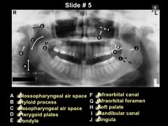
Nasopharyngeal air space |
#2 diagonal radiolucent band superior to soft palate and uvula (radiopaque); refers to space in pharynx posterior to nasal cavity |
|
|
Maxillary Tuberosity |
radiopaque bulge distal to third molar region; rounded prominence of bone |
|
|
Cancellous Bone |
soft, spongy bone located between two layers of dense cortical bone; appears predominately radiolucent (alveolar bone) |
|
|
Inferior Border of Mandible |

#1 radiopaque band that outlines lower border of mandible; composed of cortical bone |
|
|
Articular Eminence |
rounded radiopaque projection; projection of the temporal bone anterior to the glenoid fossa |
|
|
Lingula |
indistinct radiopacity anterior to the mandibular foramen; small, tongue-shaped projection of bone |
|
|
Pterygomaxillary Fissure |
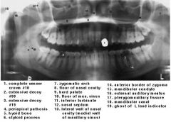
radiolucent area between the lateral pterygoid plate and maxilla; elongated and inverted tear drop outlined anteriorly by maxillary sinus; narrow space that separates these structures |
|
|
External Oblique Ridge |
#3 dense radiopaque band that extends downward and forward from the ramus to the molar region; located on external surface of mandible |
|
|
Floor of Maxillary Sinus |
radiopaque line; composed of dense cortical bone |