Reading...
![]()
Play button
![]()
Play button
![]()
Use LEFT and RIGHT arrow keys to navigate between flashcards;
Use UP and DOWN arrow keys to flip the card;
H to show hint;
A reads text to speech;
165 Cards in this Set
- Front
- Back
|
Define crural.
|
means leg (lower leg)
|
|
|
What is a tuberosity?
What is the opposite of a tuberosity? |
a projection on bone that serves as a point of attachment for muscles
opposite is a groove or a fossa |
|
|
What is a fibula named after?
|
clasp of a safety pin
|
|
|
What kind of angle does the thigh have?
Mnemonic? |
Varum/varus angle pointing medially
imagine varus pointing down toward his naked crotch |
|
|
What is the opposite of varum?
|
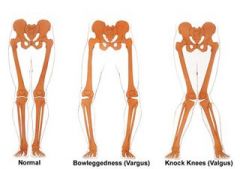
valgus
(pic up top is of the lower legs being deformed) |
|
|
mnemonic for valgum and varum?
|
L for pointing Lateral (the bottom is more lateral)
|
|
|
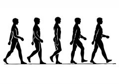
How is our lower limb arranged so that we can smoothly do bipedalism?
|
Our hips are far apart that our LE's were not in midline, but our legs come back toether so we don't waste energy with lateral motion.
Our lumbar spine is free from the hip for rotation. Our iliac bone is spread out so our hip extensors have good angle to pull back our leg. |
|
|
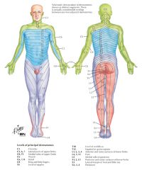
How can you use embryology to explain dermatomes of the arms and legs?
|
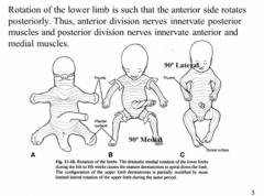
UE- lateral rotation
-why the higher up cervicals are at the back of the arms LE- medial rotation - why the femoral nerve, which is at a higher level wraps around the side to go down medially. |
|
|
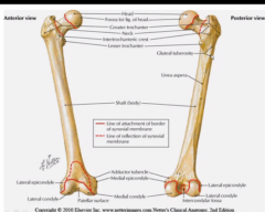
what does trochanter mean?
|
"to run"
makes sense cause all the running muscles attach to the trochanters |
|
|
difference between cndyle and epicondyle?
|
condyle is the bottom part and epicondyle is the lateral part above that
|
|

|
|
|
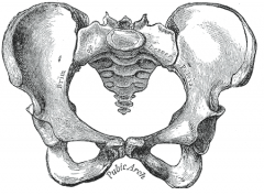
What does the pelvis look like?
|

a cromagnum koala bear
|
|
|
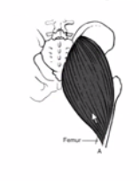
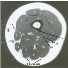
Via what fascia do muscles attach along the back of the thigh?
|
the fascia lata
|
|
|
What does lata mean?
|
thigh
|
|
|
What attaches to the adductor tubercle? What is it adduction?
What side is it on? |
the hamstring portion of the adductor magnus
adducting the thigh on the medial side of the condyles |
|
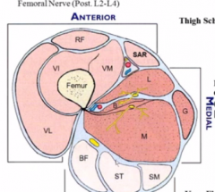
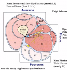
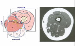
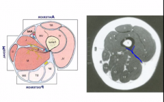
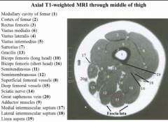
What is the MAIN action of each of these compartments?
|
anterior- knee extension (minor hip flexion)
medial- thigh adduction posterior- hip extension and knee flexion |
|
|
do the knee flexion muscles in the posterior compartment of the thigh ALWAYS work in knee flexion?
|
no, it depends on which part of the body is fixed. If the thigh is fixed, then yes!
If we are sitting down though, not really so much |
|
What do you notice about the swirl of this thigh section?
|
it's the same direction as they embryo twisted it's lower limb!
|
|
|
Which nerves control the anterior compartment? What muscles are in it?
|
L2-L4, but mainly L3
These are your quadripceps. |
|
|
What would happen if you had weak quads when you are standing?
|
you wouldn't be able to do knee extension and your knees would collapse while you are standing
|
|

What nerves innervate the medial comparentment? major nerve?
|
the same ones, from femoral
L2-L4 with most being in L3 |
|
|
How is this consistent with the dermatomes?
|
the anterior and medial portions of the thigh share the same dermatomes and L3 is the largest one
|
|

What nerves innervate the posterior compartment? major nerve?
|

L4-S3, but mostly S1
|
|
|
How can you use dermatomes to remember it is mostly S1?
|
S1 is the dermatome that runs exactly in the midline of the back thigh
|
|
|
how come the sciatic nerve stops at S3?
|
because S4 and 5 are just the rings around the butt
|
|
|
Describe and show the leg bones.
How are they connected in the shafts? Do they end at the same level? |
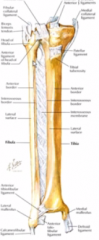
connected by interosseous membrane
fibula ends lower down to tarsal |
|
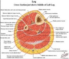
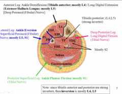
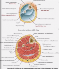
divide this leg into compartments
|

anterior, lateral, and deep and superficial posterior
|
|
|
What 3 muscles are in the anterior compartment? Which side of the tibia is this on?
|
lateral to the tibia in the anterior compartment:
tibialis anterior (duh) extensor haalicus and digitorum longus (the two major toe extensors, obviously they are the long ones) |
|
|
Where is digitorum vs hallicus longus compared to one another?
|
think according to their position on the foot
digitorum is more lateral so it must also be on top because otherwise they would be trying to cross one another hallicus is deeper and more medial |
|
|
What is the tibialis anterior mostly innervated by?
|
L4
|
|
|
What is the extensor hallicus longus mostly innervated by?
|
L5
|
|
|
Why does this make sense?
|
L4 and L5 split the shin right in half and L4 is more medial just like tibialis anterior
|
|
|
What are you testing if you plantarflex the pt's foot and ask them to flex?
(nerve and muscle) |
L4 aka tibialis anterior
|
|
|
What are you testing if you flex the pt's big toe and ask them to extend?
|
L5 aka extensor hallicus longus
|
|
|
How reliable are the tests?
|
very!
|
|
|
according to the line of your body's gravity, which way (front to back) would you fall on your ankles?
|
You're pretty front loaded so weak ankles would have you fall forward
|
|
|
How has the body managed to counter this flaw?
|
by have much larger muscles in the posterior compartment of the leg doing plantarflexion.
muscles involves are gastrocnemius and soleus |
|
|
What nerve serves the superficial posterior leg? how do you know?
|
S1 (**** still goes straight on down!)
|
|
|
What 3 muscles are in the deep posterior leg compartment?
Whys is this so easy to remember? |
flexor digitorum/hallicus longus
tibialis posterior they are just analogs to the anterior compartment! |
|
|
What nerve supplies the deep posterior leg compartment?
(say for each of the 3) |
S2- for the toe ones
L4/5 for TP |
|
|
What are the main actions of the deep posterior leg muscles?
|
also plantar flexion although they specialize in inversion and toe flexion especially
|
|
|
What is TA and TP's relation to one another in terms of their actions?
|
oppose each other in f/e
work together to invert |
|
|
So what nerves are responsible for inversion?
|
L4- TA
L4/5- TP |
|
|
Why does our skeleton design require strong inverters?
|
since we are somewhat valgus on our ankles, they have a natural tendency to pronate/evert.
the inverter counter this |
|
|
what muscle is responsible for the arch of our foot mainly?
|
tibialis posterior
|
|
|
What 2 muscle are responsible for ankle eversion?
|
peronela longus and brevis
|
|
|
What nerves supply peroneal muscles? Think about dermatomes!
|
L5/S1
|
|
|
What is the risk that comes along with strong inverters while running or doing sports?
|
if we pull the foot up and invert (like when we walk) and then come down too hard, we will have an inversion injury.
|
|
|
Why do we need everters?
|
when we are running, we don't want our feet to be inverted in the air so the everters will pull them stright so we don't twist our ankles when landing.
|
|
|
When we are walking and one foot is off the ground, what is the tendency for our hips to do?
|
adduct and collapse in on themselves
|
|
|
So what hold them in place?
|
very strong abductors
|
|
|
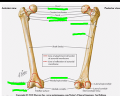
Is the intertrochanteric crest or line in the front?
Menmonic? |
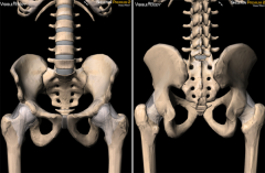
line is in front
crest is in back since the acetabulum is more in the front of the pelvis, the femur crest needs to bee in the back to scoop out with the shape of the pelvis more |
|
|
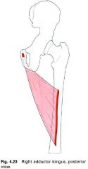
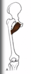
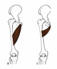
Where does the adductor magnus attach on the pelvis?
|
on the pubic/ischial rami
|
|
|
Where does it attach on the femur?
|
One part ges to the adductor tubercle on the medial epicondyle
Another part attaches to fascia from the linea aspera |
|
|
What does "aspera" mean?
|
rough
"rough line" |
|
|
So what are the actions of the adductor magnus?
|
1. adduction of the hip
2. lateral rotation of the hip 3. internal rotation using adductor tubercle only if leg is laterally rotated already |
|
|
Why are gluteal muscles so large?
|

they are abductors and prevent you from collapsing into hip adduction when you walk
|
|
|
What are the major abduction muscles? that stabilize the hip?
|
gluteus muscles (mostly medius and minimus though because they have a better angle)
plus the deep stabilizer muscles |
|
|
What action do the hip abductors pair with? Why?
|
internal rotation
medius and minimus have anterior fibers on the iliac crest and pull the greater trochanter forward |
|
|
What action do hip extensors pair with?
|
external rotation
|
|
|
Where are the hip abductors relative to the hip extensors? Why does this make sense?
|
more medial so they can get leverage for their motion.
|
|
|
Which nerve serves the hip abductors/internal rotators? Why?
|
Mostly L5
They attach on the lateral side of the leg just like L5 |
|
|
Which is the main nerve serving the hip extensors/external rotators?
|
S1
They attach at the linea aspera just like S1 |
|
|
What force does gravity place at the hip?
|
hip flexion
|
|
|
What does this mean for muscles to counter this?
|
we need good hip extensors (like gluteus maximus!)
|
|
|
What part of getting up from your chair is your gluteal muscle responsible for?
What does it pull on? |
the part where you straighten out your back after getting your legs up.
It pulls on the back of the sacral ilial area |
|
|
What innervation is the rest of the hamstrings?
|
Also S1!
|
|
|
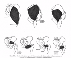
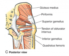
What are the "stabilizers of the hip joint"?
Show it. |
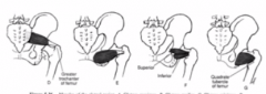
they are those small muscles that make sure the femur is nicely attached to the pelvis
|
|
|
What action do they all perform? Why?
|
a tiney bit of external rotation. No leverage for f/e and they all come out the back of the pelvis.
|
|
What is this? How do you know?
|
superior and inferior gemellus (they are TWINS!)'
|
|
What is this? How do you know?
|
obturator internus because it spans the medial obturator foramen
|
|
|
What does obturator mean?
Why are the muscles named this way? |
something that OBSTRUCTS
ironic that an opening is named this, but the muscles block the obturator foramen |
|
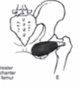
What is this? How do you know?
|
quadratus femorus
It is quad shaped covering just the span of the lateral pelvis to THE FEMUR. |
|
|
Where does the gluteus maximus originate and insert?
show it |
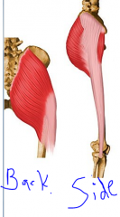
originate from the medial side of the pelvis
inserts into the iliotibial band |
|
|
How does the iliotibial band help the knee?
|
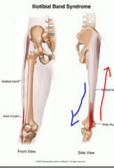
knee tends to want to become varus and the IT band pulls them back the other direction and help to suspend it.
(I probably have weak hip abductors like tensor fascia lata then) |
|
|
What are the connections of gluteus medius vs minimus?
|
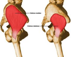
medius- all along the iliac crest
minimus- only in the more inside part of the back of the pelvis They both attach to the greater trochanter |
|
|
Now we talk layers, what do you see when you first peel off the butt skin?
|
gluteus maximus
|
|
|
What do you see next?
|
gluteus medius
piriformis all the deep hip stabilizers |
|
|
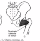
What is underneath gluteus medius? Show.
|

gluteus minimus
|
|
|
What muscle does the sciatic nerve pass underneath and which does it pass above?
Show |
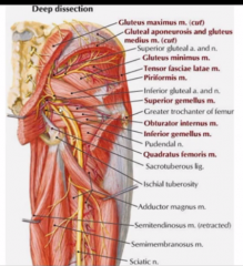
underneath piriformis
but above all the inferior lying deep hip stabilizers |
|
|
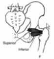
Which is most superior and inferior of the deep hip muscles?
show |

superior- gemellus/obturator/gemellus (think about where they originate in the pelvis)
inferior- quadratus femorus |
|
|
So which is the only muscle in the buttock that can compress the sciatic nerve?
why is this especially bad? |
the piriformis (made worse by them both coming out of the sciatic notch togeher.)
|
|
|
What nerve supplies gluteus maximus?
|
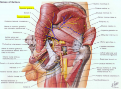
inferior gluteal nerve
coming off the sciatic |
|
|
What nerve supplies gluteus medius? What else does it supply?
|

superior gluteal nerve
coming off the sciatic also supplies the tensor fascia lata |
|
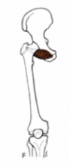
Where is the linea aspera? How do you know?
How can you tell what direction you are look at it from? |
It is tapered off due to it's attachments.
also the posterior muscles will be bulkier so look for that |
|
Get a general sense of what's going. Don't get bogged down.
|
|
|
|
What two muscles do both hip flexion and knee extension?
|
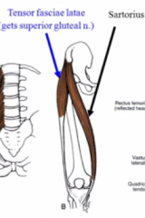
sartorius and tensor fasciae latae
|
|
|
What common evil action is sartorius responsible for? Mnemonic?
|
picking up your legs and crossing them
sartorius=sari girls in sari's will cross their legs |
|
|
Where do these two multifunctional muscles attach up top and on the bottom?
|

up top- both ASIS
bottom- on the two V type ridges on the tibia on opposite sides |
|

What nerve innervates each one?
|
tfl- superior gluteal
sartorius- femoral |
|
|
Why are the quads not really responsible for hip flexion? Which one does it slightly?
|
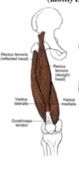
they don't attach onto the pelvis. except for recus femoris, but only a little even then.
|
|

What innervates the quadriceps?
|
femoral nerve aka L3
|
|
|
show what vastus intermedius looks like under the rest of the quads.
|
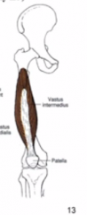
splits into 2
|
|
|
Mnemonic for which nerve extends the knee/
|
L THREE extends the KNEE
|
|
|
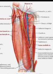
show how much breadth the fascia liga covers in the anterior thigh
|
|
|
|
What nerve passes through the inguinal ligament fairly laterally and superficially?
|
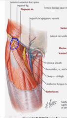
lateral cutaneous femoral nerve
|
|
|
What is this nerve prone to and what are the sx?
|
being squeeze by being trapped or inflamed during exercise resulting in pain and stinging down the anterolatero surface of the thigh
|
|
|
What artery likes to wrap around the inguinal ligament to go back up?
|
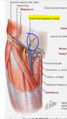
superficial epigastric vessels
|
|
|
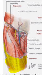
Show where the femoral nerve begins to branch out and to what
|
|
|
|
What nerve is most responsible for hip extension and knee flexion?
|
tibial nerve (S1)
|
|
|
Does the fibular nerve arise from the spine before or after the tibial nerve?
work it out by embryology. |
before (L4-5)
the embryo twists around |
|
|
What are the two main groups of hamstrings?
|
semimembranosis/tendonosis and biceps femoris
|
|
|
What other action are they all rsponsible for besides hip extension?
how do you know? |
knee flexion
they are all attached to the lower leg |
|
|
Why is it advantageous for them to manage both?
|
When we walk, we flex the knee, then bring the hip into extension in one smooth, hamstring controlled motion
|
|
|
What is similar and different about the attachments of the hamstrings? (show)
|
similar- both originate at the ischial tuberosity (sitting bone)
different- biceps femorus attaches to fibula semis attach to medial tibia |
|
|
Why are the semitendonosis and membranosis named what they are?
|
tendonosis- most of the muscle is tendon
membranosis- most of length is muscle belly |
|
|
What nerve innervates these hamstrings?
|
since they are the back of the thigh, S1 aka tibial
(except short head of biceps femoris) |
|
|
Is the tendonosis or membranosis more superficial?
|
tendonosis
|
|
|
Where does the sciatic nerve run in the layers of the hamstrings?
|
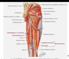
well protected underneath biceps femoris long head
|
|
|
Was that REALLY the sciatic nerve? What was it?
|
No, that was the common peroneal and tibial nerves running together
' |
|
|
Where does the sciatic nerve end?
|
it splits into those two underneath the piriformis
|
|
|
What nerve is most resposible for hip adduction?
mnemonic? |
obturator nerve (L3)'
hip adduction OBSCURES the genitals |
|
|
obturator externus
|
|
|
pectinius (attaches to the pelvic pectin- same as thoracis abdominus)
|
|

|
adductor magnus
|
|
|
abductor longus and brevis
|
|
|
Which hip adductors get femoral vs tibial innervation on top of obturator?
|
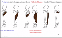
the ones that are higher up and shorter get femoral
the ones lower down get tibial |
|
|
where does the obturator branch off the femoral?
|
It doesn't lol! it get's it's own branches from the get go!!!
|
|

What is the adductor hiatus hole in the adductor magnus good for?
|
the femoral VAN pass through here to get to the popliteal fossa and become the popliteal VAN
|
|
|
Where does the tibialis anterior originate and attach to?
|
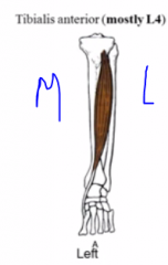
origin- more lateral part of tibia
insertion- arch of the foor |
|
|
What is TA innervated by?
|
deep fibular nerve L4
|
|
|
Where does extensor digitorum originate and insert?
|
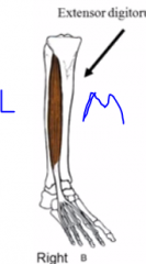
origin- between fibula and tibia
insert- all toe except big one |
|
|
What is EDL innervated by?
|
deep fibular nerve- L5
|
|
|
Where does extensor hallicus originate and insert?
|
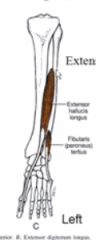
origin- fibulaish area
insertion- big toe |
|
|
Describe the general journey of the tibial and common fibular nerve.
Compare this to dermatomes |
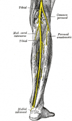
they branch right in the pectinius, but travel down the back of the thigh together.
At the knee, the common peroneal wraps around the front to innervate those muscles Tibial= S1 Peroneal= L4 and L5 |
|
|
What does the superficial vs deep fibular nerve innervate? Why does this make sense?
|
superficial- peroneal muscles (lateral compartment)
deep- anterior compartment the peroneal muscles don't run as deep as the anterior compartment ones |
|
|
Where to the peroneal longus and brevis originate and insert?
similar and different in what ways? |
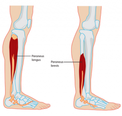
origin- different. longus is on the very top of the fibula and brevis is midway down
insertion- both are tucked behind behind the lateral malleolus at the lateral foot except longus goes all around the bottom as well |
|
|
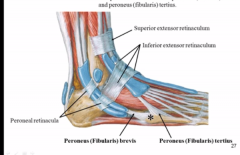
What is a retinaculum?
|
band around tendons that holds them in place
|
|
|
What is the name of the tendons that hold all the tendons from the anterior compartment down?
|
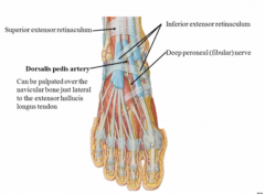
superior and inferior extensor retinaculum
RETINACULATING THE EXTENSORS! |
|
|
Why do all of the anterior compartment muscles do inversion?
|
they are all coming from more lateral and attaching more medial (they come from the other side of the tibia for goodness sakes!)
|
|
|
What are the muscles on the top of your foot that bulge when you dorsiflex?
|
those are the extensor hallicus and digitorum brevis muscle
|
|
|
What tendon is the dorsalis pedis next to?
|
just lateral to the extensor hallicus longus (large crest of the foot)
|
|
|
What is compartment syndrome?
What is the main warning sign of it? |
When the compartments gather up a very high pressure (inflammation/bleeding)
pain disproportionate to the injury |
|
|
What are the 4 P's of compartment syndrome?
Think about what is compressed |
Blood vessels
Pulselessness (not reliable) Pallor (have exceeded BP) Nerves (sensory and motor) Parenthesias (conpressing sensory nerves) Paralysis (compress LMN's) |
|
|
How do you know what compartment it is in?
|
they usually can't do the movement of that compartment because of the paralysis
|
|
|
What is the tx for compartment syndrome?
|
fasciotomy to relieve the pressure
|
|
|
What is peroneus tertius?
god ******* damn he's mentioned it like 3 times already |

a small everting muscle that comes off EDL and attaches to the same tarsal bone as fibularis brevis
|
|
|
What two retinaculum hold the peroneal tendons in place?
|

Pernoneal retinaculum- calcaneus to lateral malleolus
Inferior extensor retinaculum-starts at calcaneous and wraps over the top of the foot |
|
|
Why are fractures of the 5th metatarsal tricky to heal?
|
your two small peroneal muscles attach here and pull it out of place as you try to HEEL lol
|
|
|
What muscle gives off the achilles tendon?
alternate name? |
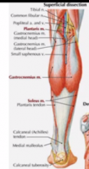
the gastrocnemius muscle
aka the calcaneal tendon |
|
|
Name what muscles border the popliteal fossa and what nerve creeps out of here.
|
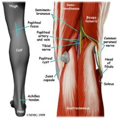
top middle- semi's
top lateral- biceps femorus bottom- two heads of gastrocnemius common fibular sneaks out of here and wraps laterally around to the front while tibial dives down between the gastrocs |
|
|
what major vessel follows the tibial?
|
the politeal vein and artery
|
|
|
Does the soleus attach both medially and laterally like the gastroc?
|
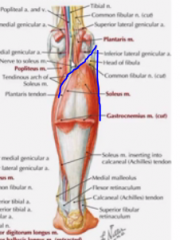
no it attaches laterally though it ends in the same achilles tendon
|
|
|
What passes behind the medial vs lateral malleolus?
|
medial- posterior compartment
lateral- lateral compartment So nice and NEAT! |
|
|
What retinaculum connects the medial malleolus and the calcaneous?
|
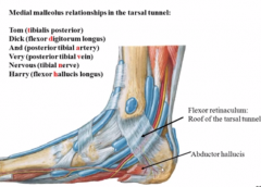
the FLEXOR retinaculum (cause it holds all the flexor tendons back!
|
|
|
What is the pathophysiology behind shin splints?
|
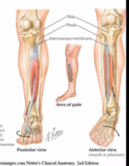
The tibialis muscles start to pull away from the interosseous membrane from overuse and this leads to inflammation.
|
|
|
What are 2 things that shin splints can lead to?
|
periostitis and bone remodeling
and compartment syndrome |
|
|
Now let's talk about heel fat.
How do we lose out "calcaneal fat pads"? (2 ways) |
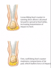
1. old age
2. loose fitting shoes that allow it to spread when we strike down. |
|
|
Where does the impact go if we have low calcaneal cushioning?
|
to the calcaneous
|
|
|
What happens to bones with repeated irritation?
|
bone spurs!
|
|
|
What syndrome do calcaneal bones spurs tend to create?
|
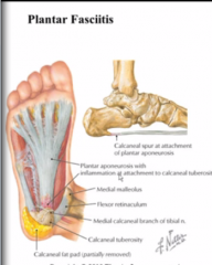
plantar fascitis because this whole aponeurosis is attached to the calcaneus
|
|
|
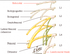
What are the two main nerves coming from L2-L4?
|
femoral and obturator?
|
|
|
How can you have two nerves from all these levels?
|
one comes from all the anterior divisions and the other comes from all the posterior
|
|
|
What nerves come from L4-S3?
|
the sciatic nerve although it is well divided into the common peroneal and the tibial nerves anteriorly and posteriorly
|
|
|
How do you remember which nerves come from which division?
|
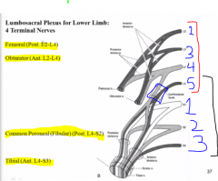
think about which ones innervate the front...
femoral and peroneal then remember that the leg twisted as an embryo so reverse it |
|
|
What is the lumbosacral trunk?
|

L4 overlaps between the 2 nerves because it also gives off a superior and inferior trunk and gives it's inferior to the sciatic.
Where it joins with L5 before splitting into anterior and posterior is the lumbosacral trunk, |
|
|
What happens in conus medullaris syndrome?
|
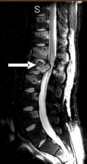
growth cutting off the front of the conus medullaris
|
|
|
What types of sx will present with this?
|
UMN type deficits of sacral levels
|
|
|
How quick will the onset be? Why?
compare with cauda equina syndrome. |
sudden. It has nowhere to hide.
the roots in cauda equina have a good amount of space to move to it is more gradual |
|
|
unilaterally affected or bilaterally?
compare with cauda equina syndrome. |
bilaterally cause everything is right there
CE is usually unilateral because a disc will slip toward one side or something. |
|
|
What area is affected in sensation in cauda equina vs conus medullaris? Mnemonic?
|
Cauda equine- saddle anesthesia (equina and saddle)
CM- perianal area |
|
|
Which one has more severe radicular pain? Why?
|
cauda equina because it is all root (radicular=root)
|
|
|
What pathology common to those with spina bifida can cause conus medullaris syndrome?
|
tethered spinal cord (filum terminale)
|
|
|
Why do we have such strong hip adductors even though we say we want o keep the knees from varusing too much?
|
they are the reason we varus in the first place, which is necessary for more streamlined walking with 2 leg.
|
|
|
Where do the hamstring originate?
|
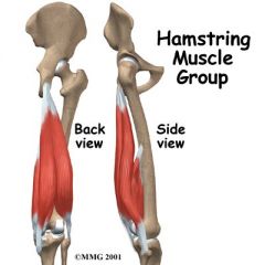
ischium
|