Reading...
![]()
Play button
![]()
Play button
![]()
Use LEFT and RIGHT arrow keys to navigate between flashcards;
Use UP and DOWN arrow keys to flip the card;
H to show hint;
A reads text to speech;
35 Cards in this Set
- Front
- Back
|
1. sphenoid
2. zygomatic 3. maxillary 4. palatine 5. lacrimal 6. ethmoid 7. frontal |
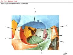
label the bones
|
|
|
what is the orbital linig that is continuous with the dura mater?
|
the orbital lining
|
|
|
what enters the orbit through the superior orbital fissure?
|
CN III, IV, VI, and V1
and opthalmic veins |
|
|
what enters the orbit through the optic canal?
|
CN II, opthalmic artery, and sympathetics
|
|
|
what enters through the inferior orbital fissure?
|
CN V2 (zygomatic nerve and infraorbital nerve) and artery and veins to pterygoid plexus
|
|
|
what is the relationship between the conjunctival epi and the corneal epi? What kind of epi are they?
|
they are continuous, stratified squamous epi
|
|
|
1. plica semilunaris
2. lacrimal caruncle 3. lacrimal puncta 4. lacrimal papilla |
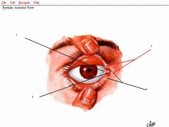
label
|
|
|
describe the flow of tears out of the eye.
|
lacrimal gland, lacrimal ducts, conjunctival sac, puncta, lacrimal canaliculi, lacrimal sac, lacrimal duct, inferior meatus of nasal cavity
|
|
|
1. anterior ethmoidal
2. posterior ethmoidal 3. opthalmic artery 4. central retinal 5. posterior ciliary 6. lacrimal 7. supraorbital 8. supratrochlear |

label the arteries
|
|
|
starting superiorly and then moving infero-medially what are three arteries that travel through the orbit and end up traveling on the periosteum?
|
supraorbital, supratrochlear, dorsal nasal
|
|
|
what artery is found in the orbital septum and what artery supplies it?
|
the palpebral artery from the lacrimal artery
|
|
|
what artery can be seen on the eye itself and what does it supply?
|
the anterior ciliary artery whcih supplies the anterior eye and the conjunctivum
|
|
|
1. cavernous sinus
2. superior opthalmic vein 3. vorticose veins 4. facial vein 5. inferior opthalmic vein 6. pterygoid plexus |
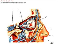
label
|
|
|
what type of pathology looks like a black eye but has no history of trauma? how does this occur?
|
a carotid fistula when the internal carotid has an aneurysm in the cavernous sinus, but luckily blood stays in the venous sinus system
|
|
|
1. sup. oblique
2. medial rectus 3. inferior rectus 4. lateral rectus 5. superior rectus 6. levator palpebrae superioris |
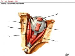
label
|
|
|
the occulomotor nerve innervates all the extrinsic muscles of the eye except what muscles (give the nerves that do innervate them)?
|
the sup. oblique from the trochlear nerve and the lateral rectus from the abducens nerve.
|
|
|
what fascia is on posterior eyeball and muscles? what ligs is it continuos with and what is the function of these ligs?
|
the bulbar fascia which is continuous with the check ligs which anchors the eyeball and muscles and restricts movement
|
|
|
all of the extrinsic eye muscles arise from the common tendinous ring except which ones? Where do all the muscles insert?
|
the levator palpebrae superioris, the inferior oblique and the superior oblique do not origniate from the ring. They all insert into the sclera
|
|
|
what are the individual actions of the extrinsic muscles?
|
SR: elevate
MR: adduct IR: depress SO: depress LR: abduct IO: elevate |
|
|
what combination of extrinsic eye muscles act together to adduct? abduct?
|
SR, MR, IR ->adduct
SO, LR, IO ->abduct |
|
|
paralysis of the extraoccular muscles will have pt's presenting with what?
|
opthalmoplegia and diplopia
|
|
|
the actions of the inferior and superior recti work best when the eye is in what postion? (used to test them)
|
when the eye is pulled laterally
|
|
|
the actions of the inferior and superior obliques work best when the eye is in what postion? (used to test them)
|
when the eye is pulled medially
|
|
|
what extraoccular muscles work to rotate the eye when laterally flexing the neck?
|
the IR and SR stabilize they eye while the IO and SO pull on the eye
|
|
|
what nerves innervate the intraocular eye muscles?
|
the ciliary body and the sphincter pupillae are innervated by parasympathetics from CN III while the dilator pupillae is innervated by sympathetics
|
|
|
what is anisocoria?
|
unequal pupil size
|
|
|
what are the muscles, and innervations for each, for the elevation of the upper eyelid?
|
the levator palpebrae superioris is innervated by GSE of CN III. The superior tarsal muscle is smooth muscle innervated by sympathetics (why poker players wear sunglasses bc eyelids open)
|
|
|
ptosis of which of the muscles for elevation of the upper eyelid is more prominant?
|
the levator palpebrae superioris
|
|
|
the extension of the optic nerve into the eye due to say a tumor or other force is called what?
|
papilledema or choked disc
|
|
|
1. opthalmic nerve (V1)
2. occulomotor 3. trochlear 4. abducens 5. semilunar ganglia 6. frontal 7. lacrimal 8. supraorbital 9. supratrochlear |
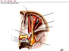
label
|
|
|
1. infratrochlear
2. ant. ethmoidal 3. post. ethmoidal 4. nasociliary 5. sensory from V1 to ciliary ganglion 6. sympathetics to ciliary 7. GSE to ciliary ganglion from oculomotor 8. long ciliary nerves (from V1) 9. short ciliary nerves 10. ciliary ganglion |
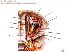
label
|
|
|
the sympathetic nerves follow what path from the cord to eventually innervate the intraocullar muscles?
|
up sympathetic chain, synapse in sup cervical ganglion, then jump on arteries (internal carotid in our case) and go through ciliary ganglion to get to eye
|
|
|
what path do the parasympathetics take to get to the intraocular muscles?
|
originate from the visceral motor Nu of CN III (edinger westphal) and then synapse at the ciliary ganglion and go to the short ciliary nerves to the eye
|
|
|
what is the path of the GVE parasympathetic message to the lacrimal gland to make it cry?
|
originates in superior salvatory nu of CN VII, then branches to the greater petrosal to the nerve of the pterygoid canal to the pterygopalatine ganglion where it synapses and travels to V2 (zygomatic nerve specefically) and then switches to the V1 (lacrimal nerve specefically) which goes to the lacrimal gland.
|
|
|
what is the result of occlusion of the opthalmic artery? central retinal artery?
|
opthalmic may not lead to complete vision loss bc of anastomotic connections with branches of the external carotid artery. If the central retinal is occluded, blindness will ensue.
|