![]()
![]()
![]()
Use LEFT and RIGHT arrow keys to navigate between flashcards;
Use UP and DOWN arrow keys to flip the card;
H to show hint;
A reads text to speech;
64 Cards in this Set
- Front
- Back

|
Retinal deatachment the separation of the neurosensory retina from the underlying |
|
|
a |
Corneal abscess, caused by a bacterial inflammation. Most bacteria are unable to |
|
|
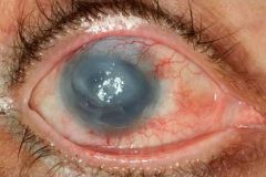
Acanthamoeba keratitis: is a rare type of keratitis (inflammation of the cornea) caused by the saprophytic protozoa acanthamoeba. It occurs usually in a patient who wears soft contact lenses but doesn't clean it well enough. Therapy: Since the corneal transparency is zero we have to do a keraroplasty - removing of the unclear corneal part and transplanting a new cornea from a donor. |
|
|
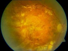
Exudative, age related macular degeneration: |
|
|
Anophtalmus. Patient is missing his right eye. This patient had retinoblastoma when |
|

|
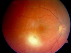
Central artery retinal occlusion: Retinal infarction are due to occlusion of an artery in the lamina cribrosa or a branch retinal artery occlusion. Paper-thin vessels and extensive retinal edema in which the retina loses its transparency are typical signs. In the middle a cherry red spot - the fovea centralis - can be seen because the other parts of the retina are swollen so compared to that it just appears cherry red (red of underlying choroid is showing under it). |
|
|
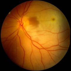
Retinal arterial occlusion (macular area). Remember the macula is always on the |
|
|
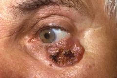
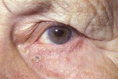
Basal cell carcinoma (basalioma) of the eyelid is a frequent, semi malignant tumor:Grows slowly but can cause severe tissue destruction.Rarely metastasizes.Can be removed surgically. |
|
|
Basal cell carcinoma, grows where the glasses contact the skin, it is and irritative |
|
|
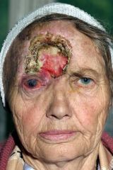
Basalioma recidivians, basal cell carcinoma, penetrates the frontal sinus in the |
|

|
Congenital glaucoma (end stage). Buphthalmus (abnormally large eye) is seen on both sides. This is caused because the iris inserts to far anteriorly in the trabecular meshwork and as a result an embryonic mesodermal tissue in the form of a thin transparent membrane (Barkanʼs membrane) covers the trabecular meshwork and impedes the flow of aqueous humor into the canal of Schlemm. Since this patient is at ends stage nothing can help this condition. In an earlier stage it could have been solved with a trabeculectomy (filtration surgery) which would |
|

|
Congenital glaucoma, patients left eye. Patient appears to have big eyes, visual acuity is still fine at this phase. Children with this disorder are irritable, poor eaters, and rub their eyes in an attempt to compress some fluid out of their eye. |
|

|
Traumatic cataract and rubeosis iridis - |
|

|
Nuclear Cataract: |
|

|
Mature cataract: diffuse, complete opacification of the lens. visual acuity under |
|
|
Focal Chorioretinitis: The chorion and the retina are always inflamated together. This is focal because we can see the focal white part in the middle lower part of the image. this Inflammation can be caused by toxocara, toxoplasma or leptospira for example. |
|

|
Corneal Cicatrix - cicatrix means scar. It is often caused by an accident, in this |
|

|
Congenital Iris Coloboma - A congenital anomaly that results from incomplete fusion of the embryonic optic cup which normally occurs in about the sixth week of pregnancy. Since this coloboma opens inferiorly it is safe to assume that it is congenital since in the development of the eye this is the last part closing. Surgical iris colobomas in cataract and glaucoma surgery are usually opened superiorly. |
|

|
Vernal conjuctivitis - vernal means seasonal, mosttly in springtime. The tarsal conjunctiva shows a cobblestone/paving stone appearance. The tool used to invert the upper eyelid is the Desmares spoon. |
|
|
Bacterial conjunctivitis - discharge inside the eyelids, its very common, painful, itchy and can cause blurry vision. Requires antibiotic eyedrops such as tobramycin or neomycin and it can be solved in a week. |
|

|
Acute bacterial conjunctivitis - same as the previous, bacterial discharge can be seen, appears to be more severe than the previous. |
|

|
Retinal hemorrage after ocular contousuion - can be caused by blunt trauma. It is dangerous because blood contains iron which is toxic. |
|

|
Conjuctival cyst: treatment depends, if its an old lady that had it for years than there is no need to treat. However, if it is a newly appearing cyst which is growing rapidly then you have to cut it out and send to histology. |
|
|
Iris tumor/cyst - the part on the right is rubeosis iridis ( a neo revascularization of the iris). |
|
|
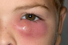
Acute dacrocystitis: inflammation of lacrimal sac. It is a painful condition which requires drainage. We also have to give local and oral systemic antibiotics. If it recurs in 4-6 weeks period we need to consider the possibility of a tumor. |
|

|
Allergic Dermatitis of the eyelids: severe allergy to eyedrops can be seen. |
|
|
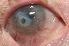
Descemetocele: The descement membrane of the cornea has a protrusion. in the middle (white). The black lines are 10.0 running suture because the patient underwent perforating keratoplasty. It is a serious condition because there is risk of perforation through which bacteria can go into the eye and cause endophtalmitis. |
|

|
Disinsertion of the levator superioris palpabrae muscle - |
|

|
Diabetic retinopathy-. We can see in this image is a severe neovascularization (small blood vessels on the top left of the image). Cotton white spots are debris. Retinal hemorrhages are also sen. how can you treat it? Laser treatment, a panretinal laser photocoagultive therapy. |
|

|
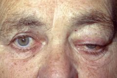
Cavernous hemangioma of the upper eyelid and dislocation of the bulb: This |
|
|
Dislocation of the bulb and ptosis of the upper eyelid : You have to ask the patient when did this begin because if it is relatively new than it is probably caused by a tumor, send this patient to do an MRI. |
|
|
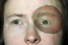
Ectoprotesis - full reconstruction of palpebral area. Patient probably underwent a very severe surgery in which the palpebral area and the full orbital cavity had to be cleared of tumor. This is the best they could do for him... |
|
|
Cicatriceal ectropion of the lower eyelid: |
|

|
Senile ectropion of the lower eyelid - Surgery includes tightening the lower eyelid |
|
|
check picture |
Endophtalmitis - The entire eye is involved, visual acuity is zero, maybe some light/dark perception. The patient needs IV vancomycin. If it is not solved then we will have to remove the whole eyeball in order to prevent the inflammation from going to the brain. |
|

|
Senile entropion of the lower eyelid: The eyelid is turned inside. The patient will feel itching, foreign body feeling . Kettesy operating method will solve the problem. It is caused by a scar. |
|

|
Congenital entropion of the lower eyelid - use glue stick until the age of 2, usually it helps. and if the condition persists then operate. |
|

|
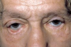
Exophtalmus - Graves disease. Protrusion of the eye. |
|

|
Exudative, age related, macular degeneration - |
|

|
Cooked Fish eye following alkali injury. What do you have to do in this case is first wash the eye for half an hour with any watery solution of neutral PH in order to remove all the alkalic material in the eye (Milk should be avoided as it increases the penetration of the burn by opening the epithelial barrier). Later this condition will require a special method for corneal transplantation - the sofort or ashor keratoplasty (a lamellar keratoplasty). |
|

|
Arcus Senillis (Gerontoxon): lipid containing circle. The Peripheral edge is sharp and separated from the limbus by a clear zone. Arcus senilis is the most common peripheral corneal opacity; it frequently occurs without predisposing systemic conditions in elderly individuals. Occasionally arcus may be associated with familial and non-familial dyslipoproteinaemias. |
|
|
a |
Protesis Bulbi: Prostesis located in left eye of the patient. Possible reasons for prosthesis are: very severe inflamation, severe accident. On the other eye the patient probably underwent cataract surgery and thats why this patient has aphakia (does not have a lens). |
|
|
a |
Hemangioma of the upper eyelid: Treatment for this would be waiting and seeing, this hemangioma is not in front of the pupil so it does not affect the vision. In most cases hemangiomas spontaneously resolves before age 7. |
|
|
a |
Herpes Zoster Ophtalmicus: a very severe reactivation of the virus, came from the |
|
|
a |
Hemangioma in Von-Hippel-Lindau disease (before and after treatment): |
|
|
a |
Foreign body in the lens: Some aluminum part from a hammer. The entire lens |
|
|
a |
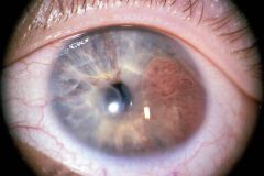
Kayser Fleischer ring: This golden-brown to yellowish-green corneal ring is |
|
|
a |
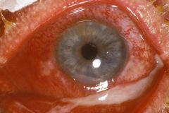
Esotropia: Left eye of the patient deviates inward. You can see that the light |
|
|
a |
Keratoprotesis: An artificial cornea. Just a plastic piece sutured to the conjuctiva |
|
|
a |
Dendritic Keratitis: Caused by HSV, called dendritic because it has the appearance of chinese letters or tree branches. It can be seen using fluorescein staining that stains the injured collagen on the cornea. Hypopyon (pus) can also be seen.Herpes simplex keratitis is usually very painful and associated with photophobia, lacrimation, and swelling of the eyelids. Vision may be impaired depending on the location of findings, for example in the presence of central epitheliitis. |
|
|
a |
Keratitis and Hypopyon: probably bacterial, you can see a hypopyon (pus in ant. chamber), an epithelial defect with an infiltrate. Chemosis is also present. |
|
|
a |
Marginal Keratitis: Marginal keratitis is probably caused by a hypersensitivity reaction against staphylococcal exotoxins and cell wall proteins with deposition of antigen-antibody complexes in the peripheral cornea with a secondary lymphocytic infiltration. The lesions are culture negative but S. aureus can frequently be isolated from the lid margins.Symptoms are mild discomfort, redness and lacrimation that may be bilateral. |
|
|
a |
Acute Keratoconus: Keratoconus is the most frequently encountered deformation of the cornea. In rare cases keratoconus can cause tears of Descemetʼs membrane due to the continuous stretching, then ant. chamber fluid will enter the stroma totally decreasing the transparency of the cornea. Symptoms of acute keratoconus include sudden loss of visual acuity accompanied by intense pain, photophobia, and increased tearing. |
|
|
a |
Deposits on the surface of contact lens. Jelly like deposits on the surface of the contact lens probably due to hygenic problems of the patient. |
|
|
a |
Penetrating Keratoplasty With 10.0 running sutures. |
|
|
a |
Penetrating Keratoplasty (three times): Smaller and smaller circles are seen. |
|
|
a |
Leucoma Cornea - Totaly white scar on the cornea. |
|
|
a |
Leucoma Cornea - This state is called Leucoma Vascularisatia Cornea. |
|
|
a |
Epibulbar Dermoid on the Limbus of the cornea: Epibulbar dermoid is a round dome-shaped grayish yellow or whitish congenital tumor. It is generally located on the limbus of the cornea, extending into the corneal stroma to a varying depth. It has to be removed because there can be hair, nails, inside... |
|
|
a |
Luxation of the lens into the anterior chamber and iris coloboma. This coloboma is acquired because it openes superiorly. A sublaxated lens can also be seen. |
|
|
a |
Retinal tear and retinal tamponade with elastic silicone sponge. The retinal tear is the whitish membrane seen. This retinal detachments was treated with a retinal tamponade with an elastic silicone sponge that was sutured to the outer surface of the sclera, a so-called budding procedure.This indents the wall of the globe at the retinal break and brings the portion of the retina in which the break is located back into contact with the retinal pigment epithelium. |
|
|
a |
Macula Cornea: A special scar of the cornea. If the entire cornea is |
|
|
a |
Exophtalmus Maligna: This patient has Graves disease and exhibits exophtahlamus. |
|
|
a |
Foreign body in the eye: A nail stuck in the eye. Visual acuity of the eye is |