![]()
![]()
![]()
Use LEFT and RIGHT arrow keys to navigate between flashcards;
Use UP and DOWN arrow keys to flip the card;
H to show hint;
A reads text to speech;
129 Cards in this Set
- Front
- Back
|
Zygomycota |
–Aseptate hyphae –Zygosporangium, sexual stage |
|
|
Deuteromycota |
–No known sexual stage |
|
|
Ascomycota |
Ascospores, sexual stage |
|
|
Basidiomycota |
Basidiospores sexual stage |
|
|
Asexual |
Anamorph Fungi Imperfecti Conidia Macroconidia Microconidia |
|
|
Sexual |
Teleomorph Perfect fungi |
|
|
Gram stain |
fungus appear gram positive Yeast: two to three times larger than g+c Hyphae: two to three times wider than g+b, stain irregularly |
|
|
for skin, hair or nails |
KOH prep |
|
|
India Ink |
For capsules around yeast |
|
|
Acid fast |
fungus - like bacteria (Nocardia) |
|
|
Media- Initial |
– Sabouraud Dextrose Agar – Sab with Brain Heart Infusion – Brain Heart Infusion with Blood |
|
|
Media- Subculture |
– Neutral Sabouraud (Emmon’s) – Potato Dextrose Agar |
|
|
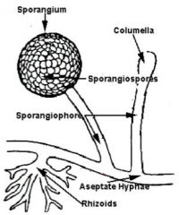
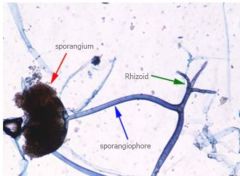
Zygomycetes: The hyphae are irregular in diameter, broad, ribbon - like and devoid of septations. •Root -like structures, called rhizoids, may or may not be present. • The sporangiophores terminate in a swollen columella • Each columella is surrounded by a bag like sporangium, in which spherical, pigmented sporangiospores are produced |
|
|
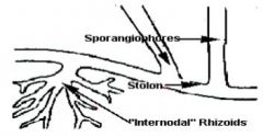
sporangiophores, stolon, internodal rhizoids |
|
|
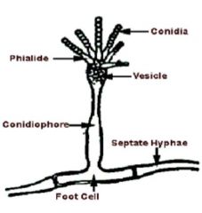
conidia, phialide, vesicle, conidiophore, septate hyphae, foot cell |
|
|
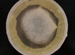
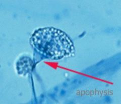
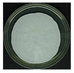
Colony Morphology: –Surface: Wooly gray colony, Fills plate with fluff –Reverse: Colorless • Microscopic– Aseptate Hyphae –Branching Sporangiophores between Rhizoids • Collarette remains after sporangial wall dissolves –Pear shaped Sporangia |
Absidia |
|
|
• Rapid growth; 4- 5 days to mature • Infections :Zygomycosis, keratomycosis |
Absidia |
|
|
Absidia |
|

|
Absidia |
|
|
Absidia |
|
|
Absidia apophysis |
|
|
Rapidly growing •Infections - recovered from sinuses or from organs when disseminated disease is present |
Cunninghamella |
|
|
Microscopic Morphology
– Sporangiophores are erect, branching into several vesicles that bear sporangioles and can be covered in long, fine spines •Macroscopic Morphology – Cottony colony that is initially white but becomes gray |
Cunninghamella
|
|
|
Cunninghamella |
|
|
Cunninghamella |
|
|
• Rapid growth, 4 days to mature •Infections: – Occasionally, zygomycosis; usually contaminant |
Mucor sp. |
|
|
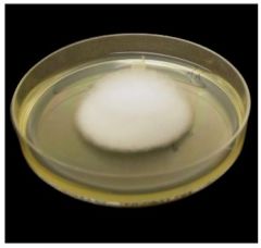
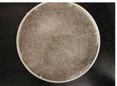
•Colony Morphology –Surface: Quickly fills plate with cotton candy like white fluff, becomes gray –Reverse: White |
Mucor sp. |
|
|
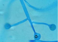
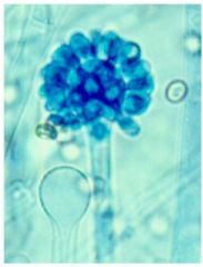
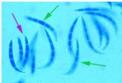
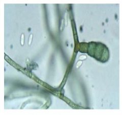
Microscopic: – Wide aseptate hyphae No Rhizoids – Long, often branched sporangiophores • Terminal round sporangia (50 - 300 um) • Round or slightly oblong spores • Columella left after sporangial wall dissolves, sometime with a collarette |
Mucor sp. |
|
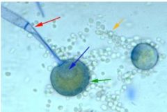
Red: Sporangiophore Blue arrow: Columella Green: Sporangium Orn: Sporangiospores |
Mucor sp. |
|
|
Mucor sp. |
|
|
Mucor sp. |
|
|
Rapid growth, 4 days to mature Infections: – Most common cause of zygomycosis |
Rhizopus sp. |
|
|
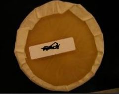
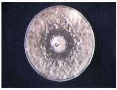
Colony Morphology – Surface: Quickly fills plate with cotton candy like white fluff, becomes gray – Reverse: White |
Rhizopus sp. |
|
|
Rapid growth, 4 days to mature Infections: – Most common cause of zygomycosis |
Rhizopus sp. |
|
|
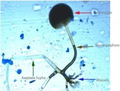
Microscopic: Wide aseptate hyphae • Rhizoids under sporangiophores – Long sporangiophores (up to 4 mm) • Terminal, dark, round sporangia (40 - 350 um) • Oval spores • Columella left after sporangial wall dissolves |
Rhizopus sp. |
|
|
Rhizopus sp. |
|
|
Rhizopus sp. |
|
|
Rapid growth • Infection - Rarely implicated in human disease – May cause cutaneous opportunistic infections |
Syncephalastrum |
|
|
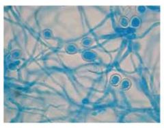
Microscopic – Erect sporangiophores – each has a large columella on which merosporangia containing stacks of sprangiospores are formed – This isolate is commonly confused with Aspergillus • Macroscopic – Colonies initially white and become gray with age |
Syncephalastrum |
|
|
Syncephalastrum |
|
|
Syncephalastrum |
|
|
Syncephalastrum |
|
|
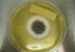
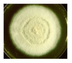
Rapid growth; 3 - 5 days to mature. • Infections: – Pulmonary, systemic, sinus, ear and other infections; produces aflatoxins |
Aspergillus flavus |
|
|
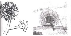
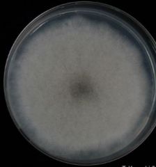
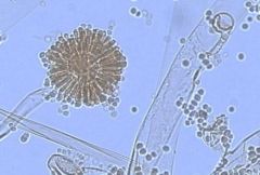
Colony Morphology – Velvety, yellow to green or brown. – Reverse goldish to red - brown. •Microscopic: – Conidiophores: variable length, rough, pitted, spiny – Uniseriate or Biseriate phialides, cover the entire vesicle, pointing out in all directions – Conidia in chains, round |
Aspergillus flavus |
|
|
Rapid growth; 3 - 5 days to mature. • Infections: – Most common cause of disseminated aspergillosis – Sinusitis |
Aspergillus fumigatus |
|
|
Colony Morphology – Velvety, white, then dark greenish to gray. – Reverse white to tan. • Microscopic: – Conidiophores: short (<300uM), smooth – Uniseriate phialides, on upper two thirds of vesicle, parallel to axis of conidiophore – Conidia in chains, round |
Aspergillus fumigatus |
|
|
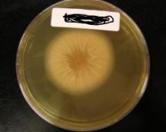
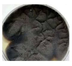
2 – 6 days to mature •Infections: – Otomycosis, Aspergilloma, nasal sinus infections Colony Morphology – Surface: cottony, flat and white, covered with Black Conidia (granular texture); Reverse: Colorless to ivory or pale yellow |
Aspergillus niger |
|
|
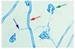
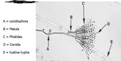
Microscopic: – Hyaline, closely septate, smooth parallel walls, 45o branching – Foot cell at junction of conidiophore with vegetative hyphae – Conidiophore – Wide (15- 20μm) up to 3 mm long – Spherical vesicles ~ 60μm– Biseriateconidial head composed of a series of secondary phialides topped with flask- shaped phialides bearing abundant conidia in columes. |
Aspergillus niger |
|
|
Aspergillus niger |
|
|
Aspergillus niger |
|
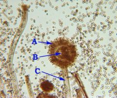
Arrow A: Phialide; Arrow B: Vesicle; A C: Conidiophore |
Aspergillus niger |
|
|
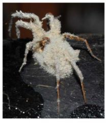
Moderately rapid growing •Infection – rare human isolate • A known insect pathogen |
Beauveria bassiana |
|
|
Microscopic – Abundant, single -celled, tear - shaped sympoduloconidia are formed on sympodulae which taper from a swollen base. Conidiophores may cluster to form radial tufts. • Macrosopic – Fluffy colonies, sometimes developing a powdery surface that may look similar to T. mentagrophytes |
Beauveria bassiana |
|

|
Beauveria bassiana |
|
|
Beauveria bassiana |
|
|
Moderate growth rate • Infection – rare cause of disease. – Have been recovered from opportunistic nail and skin lesions |
Chrysosporium |
|
|
Microscopic: – simple, wide - based, single - celled conidia. The conidiogenous cell disintegrates to release conidia • Arthroconidia and aleurioconidia may be seen • Macroscopic: – Hyaline that can develop light shades of pink, gray or tan pigment |
Chrysosporium |
|
|
Chrysosporium |
|
|
Chrysosporium |
|
|
Rapid growth; 4 days to mature • Infections: – Most common cause of keratomycosis – Burn lesions, onychomycosis, otomycosis, varicose ulcers, mycetoma, osteomyelitis following trauma, disseminated infections |
Fusarium |
|
|
Colony Morphology – Surface: White cottony colony becomes lavender, sometimes yellow or orange – Reverse: Light |
Fusarium |
|
|
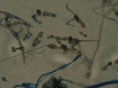
Microscopic: – Septate hyphae – Conidiophores single or branching, tapering phialides • Macroconidia: 2 – 5 celled, banana or cylinder shaped, foot cell at attachment point • Microconidia: single celled, occur in balls • If macroconidia are not present, may be confused with Acremonium • Chlamydoconidia are common |
Fusarium |
|
|
Fusarium |
|
|
Fusarium |
|
|
Fusarium |
|
|
Fusarium |
|
|
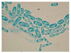
• Pulmonary disease • Microscopic – Abundant arthroconidia fromed from the vegetative hypha that occur singly or branched |
Geotrichum
|
|
|
Macroscopic: – Colonies are white to cream, yeast - like and can be confused with Trichosporon spp. Aerial mycelium can sometimes form and resemble C. immitis |
Geotrichum |
|
|
Geotrichum |
|
|
Geotrichum |
|
|
Microscopic – Similar to the structure of Penicillium spp. – Longer phialides – Verticillate pattern– Spindle shaped (a little cylindrical) conida form Macroscopic – Rapid growth – Form granular to velvety colonies in shades of tan, brownish gold, or mauve. |
Paecilomyces |
|
|
No green or blue- green colors are seen Extremely difficult to treat and manage |
Paecilomyces |
|
|
Paecilomyces |
|
|
Paecilomyces |
|
|
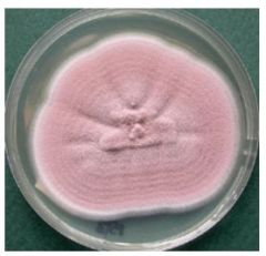
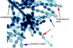
Rapid growth, 4 days to mature • Infections: – Found in a variety of diseases, etiology is uncertain |
Penicillium sp. |
|
|
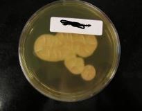
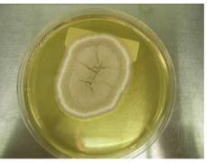
Colony Morphology – Surface: White turning to a powdery bluish green center with white border – Reverse: Usually white (If red reverse with pigment in the agar, consider P marneffei) |
Penicillium sp. |
|
|
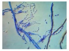
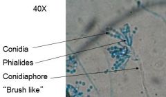
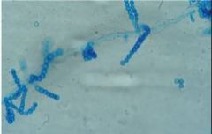
Microscopic: – Septate hyphae, with branched or unbranched conidiophores – Conidiophores have secondary branches (metulae) which hold flask shaped philides that hold unbranched chains of round conidia. Characteristic brush appearance. |
Penicillium sp. |
|
|
Penicillium sp. |
|
|
Penicillium sp. |
|
|
Penicillium sp. |
|
|
Penicillium sp. |
|
|
Penicillium sp. |
|
|
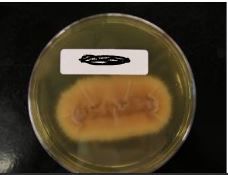
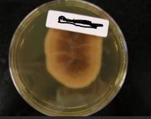
Rapid growth, 5 days to mature Infections: Known to infect nails (especially toenails) Colony Morphology – Surface: Initially white, becoming powdery light brown (granular) with tan periphery –Reverse: tan with darker center |
Scopulariopsis |
|
|
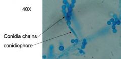
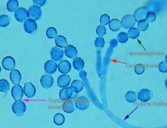
Microscopic:–Septate hyphae, with short often branched conidiophores – Conidiophores bear cylindrical or tenpin shaped annellides •Annellides are in brush - like groups •Conidia are in chains, rough, thick walled and spiny when mature •Conidia are cut off at the base, forming a “neck” |
Scopulariopsis |
|
|
Scopulariopsis |
|
|
Scopulariopsis |
|
|
Scopulariopsis |
|
|
Scopulariopsis |
|
|
Scopulariopsis |
|
|
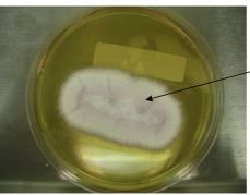
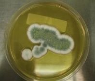
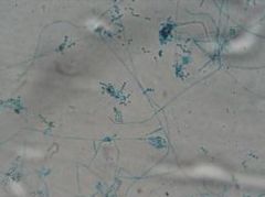
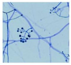
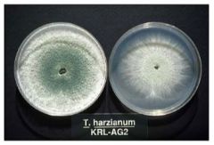
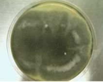
Rapidly growing • An emerging pathogen • Infections – pulmonary and skin Microscopic – Hyaline hyphae that give rise to yellow - green to green patches of conidia formed on clusters of tapering phialides. Conidia may remain clustered in balls at the phialide tips •Macroscopic – Intensely green and granular with an abundance of conidia |
Trichoderma |
|
|
Trichoderma |
|
|
Trichoderma |
|
|
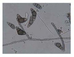
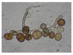
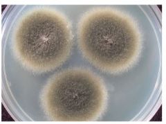
Rapid growth 5 days to mature •Infections: – Keratomycosis, skin infections, osteomyelitis, pulmonary disease, and nasal septum infection |
Alternaria sp. |
|
|
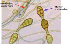
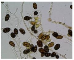
Colony Morphology – Surface: Young colony is gray and wooly. Rapidly matures to dark greenish black or brown – Reverse: black reverse • Microscopic: – Septate hyphae, may be dark – Conidia are club shaped, containing vertical and horizontal septations, tips are narrower than base |
Alternaria sp. |
|
|
Alternaria sp. |
|
|
Alternaria sp. |
|
|
Alternaria sp. |
|
|
Alternaria sp. |
|
|
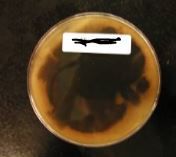
Can be caused by contaminated dialysis lines and other similar devices • Usually contracted in wet conditions |
Aureobasidium |
|
|
Microscopic – Hyaline hyphae give rise to hyaline conidia directly from the vegetative hyphae – As it ages, dematiaceous hyphae develops and it breaks up into anthroconidia which do not have hyaline conidia |
Aureobasidium |
|
|
Macroscopic – The anthroconidia that develop microscopically are responsible for the darkening colony morphology |
Aureobasidium |
|
|
Aureobasidium |
|
|
Aureobasidium |
|
|
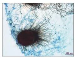
has been reported in the brains of patients with central nervous system diseases – Several patients have been reported as being intravenous drug abusers –These fungi can be found in the environment –Can destroy printed literature and library holdings–Can also cause indoor air quality problems |
Chaetomium |
|
|
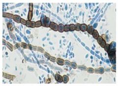
Microscopic – Numerous perithecia can be seen – Pineapple shaped – Have curled “hairs” or setae
|
Chaetomium |
|
|
• Macroscopic – The asci contained within the perithecia are evanescent so at maturity, the pigmented, lemon shaped ascospores are released with in the perithecium. Colonies are moderately to rapidly growing and begin dirty gray, becoming dematiceous with age –Some species produce a diffusible pigment that turns the agar completely red |
Chaetomium |
|
|
Chaetomium |
|
|
Chaetomium |
|
|
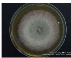
Moderate growth; 7 days to mature • Infections: – Keratomycosis and allergies |
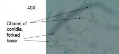
Cladosporium sp |
|
|
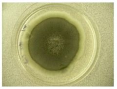
Colony Morphology Surface: Powdery or Velvety, heaped or folded, dark gray green to black Reverse: black Microscopic: Dark, septate hyphae – Short chains of 1 - 4 blastoconidia arise from forked conidiophores |
Cladosporium sp |
|
|
Cladosporium sp (Deuteromycota) |
|
|
Cladosporium sp (Deuteromycota) |
|
|
Cladosporium sp (Deuteromycota) |
|
|
Cladosporium sp (Deuteromycota) |
|
|
Infection – Mostly found in chronic sinusitis in immunocompetent patients – Usually found in grass, leaves, and decaying vegetation |
Curvularia |
|
|
Microscopic – Multicelled conidia are produced on sympodial conidiophores – Easy to identify because of the frequently crescent shaped conidia with three to five cells of unequal size and enlarged central cell |
Curvularia |
|
|
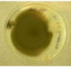
Macroscopic – Form rapidly growing dematiaceous colony that is cottony and dirty gray to black |
Curvularia |
|
|
Curvularia |
|
|
Curvularia |
|
|
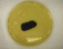
Opportunistic Infections: Secondary to Trauma |
• Phoma • Pithomyces • Ulocladium |
|
|
Rapid growth • Microscopic – Produce pycnidia, which appear as black fruiting bodies that are globose and lined inside with short conidiophores – Large numbers of hyaline conidia are generated in the pycnidium and flow out a small apical pore • Macroscopic – Gray to brown colony |
Phoma |
|
|
Phoma |
|
|
Phoma |
|
|
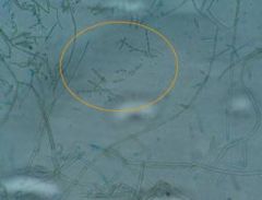
Rapid growing • Microscopic: – Conidia are barrel shaped, formed singly on simple, short conidiophores – Conidia have transverse and longitudinal cross - walls – Often echinulate • Macroscopic – Dematiaceous colonies (i.e., dark colored) |
Pithomyces |
|
|
Pithomyces |
|
|
Pithomyces |
|
|
Rapid growing • Microscopic – Conidophores bear dark, multicelled conidia on sumpodial conidiophores – Conidia have angular cross walls • Some species have echinulate surfaces
• Macroscopic – Range from olive to brown |
Ulocladium |
|
|
Ulocladium |
|
|
Ulocladium |