Reading...
![]()
Play button
![]()
Play button
![]()
Use LEFT and RIGHT arrow keys to navigate between flashcards;
Use UP and DOWN arrow keys to flip the card;
H to show hint;
A reads text to speech;
135 Cards in this Set
- Front
- Back
- 3rd side (hint)
|
What type of system is the nervous system?
|
The nervous system is a cellular, electrochemical communication
system. |
|
|
|
What are the 4 main functions of the nervous system?
|
1. It receives sensory messages from the environment (internal and external)
2. It integrates new inputs with information that is already stored 3. It uses integrated information to send out messages to muscles and glands 4. It provides the basis for conscious experiences and cognitive abilities |
|
|
|
Where do AFFERENT pathways go?
|
Go AWAY
A is also superior so these pathways go up toward our brains afferent neurons (otherwise known as sensory or receptor neurons), carry nerve impulses from receptors or sense organs towards the central nervous system. |
|
|
|
What are EFFERENT pathways?
|
Opposite the Afferent
efferent nerves, otherwise known as motor or effector neurons, carry nerve impulses away from the central nervous system to effectors such as muscles or glands (and also the ciliated cells of the inner ear). |
|
|
|
What can the peripheral nervous system be broken down into?
|
Nothing
tricked ya hahahha :) |
|
|
|
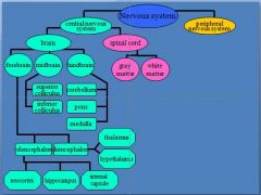
What is the central nervous system broken down into?
|
Brain and Spinal cord
|
|
|
|
What is the spinal cord divided into?
|
Gray and White Matter
|
|
|
|
divisions of the NS
Picture |
Know this and be able to draw it out!
|
|
|
|
What is Gray Matter?
|
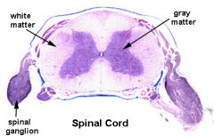
Cell Bodies****
Grey matter contains neural cell bodies*** In contrast to white matter, which does not and mostly contains myelinated axon tracts |
|
|
|
What is White Matter?
|

Axons and Myelin Layers
In cross-section, the peripheral region of the cord contains neuronal white matter tracts containing sensory and motor neurons. |
|
|
|
What are the 3 divisions of the brain?
|
1. Forebrain
2. Midbrain 3. Hindbrain |
|
|
|
What can the forebrain be divided into?
|
Telencephalon and Diencephalon
|
|
|
|
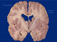
What can the Telencephalon be divided into?
|
Neocortex
Hippocampus Internal Capsule |
|
|
|
What can the Diencephalon be divided into?
|
Talamus
Hypothalamus |
|
|
|
What can the midbrain be divided into?
|
Superior colliculus
Inferior colliculus |
|
|
|
What can the hindbrain be divided into?
|
cerebellum
pons medulla |
|
|
|
What is the difference between the inside layers and outside layers of the brain?
|
Inside Layer: core physiological functions
Outside Layer: More higher order functions |
|
|
|
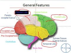
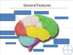
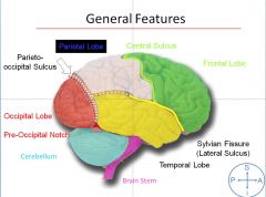
What are your 3 sulcuses of the brain?
|
Central
Lateral aka Sylvian Parietal |
|
|
|
3 sulcuses
picture form |
|
|
|
|
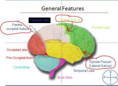
Are the lobes of the brain real anatomical structures?
|
NO
We made them up |
|
|
|
What are the 4 lobes of the brain?
|
Frontal
Temporal Parietal Occipital |
|
|
|
lobes pic
|
|
|
|
|
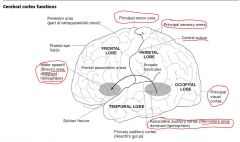
What does the temporal lobe function in?
|
Hearing****
Heschl's gyrus: Primary auditory cortex Wernike's Area: Associated Auditory cortex |
|
|
|
Just in case she asks:
What is the difference between Broca's and Wernicke's? |
Broca's: Higher order inability to speak. Think Broca's Brocken Boca
Wernicke's: Fluent aphasia with impaired conprehension. Wernick's area = Superior Temporal Gyrus Wernicke's = What? |
|
|
|
What does the occipital lobe function in?
|
vision
|
|
|
|
What does the parietal lobe function in?
|
Sensation
Tacile Tempo / Pressure Sense of Touch |
|
|
|
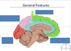
What are brain sulci?
|
Treches or ditches in brain
|
|
|
|
What are brain gyri?
|
Hills or top of mounds on the brain
|
|
|
|
picture of cerebral cortex functions
first Aid |
|
|
|
|
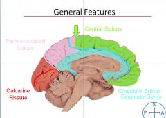
What sulcus forms the Cingulate Sulcus?
|
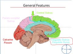
The Cingulate Gyri
|
|
|
|
What do the pre and post central gyrus do?
|
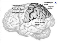
Pre -central gyrus – motor function
Post-central gyrus – sensory strip |
|
|
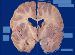
Label this picture
|
|
|
|
|
|
|
|
|
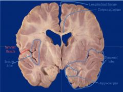
What is the Insular Cortex?
|
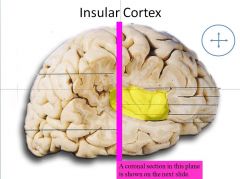
In each hemisphere of the mammalian brain the insular cortex (often called insula, insulary cortex or insular lobe) is a portion of the cerebral cortex folded deep within the lateral sulcus between the temporal lobe and the frontal lobe.
The insulae play a role in diverse functions usually linked to emotion or the regulation of the body's homeostasis. These functions include perception, motor control, self-awareness, cognitive functioning, and interpersonal experience. In relation to these it is involved in psychopathology. |
|
|
|
|
|
|
|
|
|
|
|
|
|
|
|
|
|
|
|
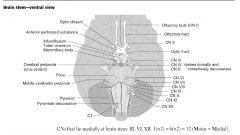
What is the optic tract?
|
Beyond the optic chiasm
NOT the optic nerve |
|
|
|
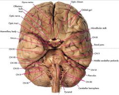
What nerves operate the eye?
|
2,4,6
|
|
|
|
What is the function of cranial nerve:
1 |
Olfactory
smell |
|
|
|
What is the function of cranial nerve:
2 |
Optic
vision |
|
|
|
What is the function of cranial nerve:
3 |
Oculomotor
Move eyeball move eye lid pupil |
|
|
|
What is the function of cranial nerve:
4 |
Trochlear
move eyeball |
|
|
|
What is the function of cranial nerve:
5 |
Trigeminal
movement & sensation of head and face |
|
|
|
What is the function of cranial nerve:
6 |
Abducens
move eyeball |
|
|
|
What is the function of cranial nerve:
7 |
facial
move face move scalp taste |
|
|
|
What is the function of cranial nerve:
8 |
Vestibulocochlear
Audition and Position Movement of Head |
|
|
|
What is the function of cranial nerve:
9 |
Glossopharyneal
Swallowing and Taste |
|
|
|
What is the function of cranial nerve:
10 |
Vagus
Heart Larynx Trachea Visceral |
|
|
|
What is the function of cranial nerve:
11 |
Spinal Accessory
palate Larynx Pharynx Shoulder Neck |
|
|
|
What is the function of cranial nerve:
12 |
Hypoglossal
Tongue movement speech |
|
|
|
Which Cranial Nerves function in sensory, motor, or both?
|
Some Say Marry Money But My Brother Says Big Breasts Matter Most
S= sensory, M=motor, B=both |
|
|
|
The spinal nerves
first aid |
|
|
|
|
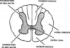
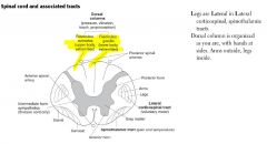
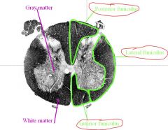
What does the spinal cord look like?
|
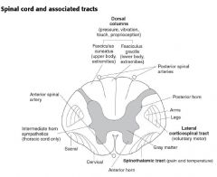
Butterfly-shaped central gray composed of collections of cell bodies. Surrounding mantle of white matter composed of bundles of axons that are either ascending or descending
|
|
|
|
Where are the neurons located in the spinal cord?
|
Gray Matter
Internal Structure of Spinal Cord -Each half of spinal cord has posterior (dorsal) gray horn (extends posterolaterally almost to surface) and anterior (ventral) gray horn which extends ventrally but does not reach surface. A gray commissure, connecting the gray matter of the two sides, encompasses the central canal. -In thoracic segments, a small pointed lateral horn is evident near base of anterior horn (associated with autonomic nervous system). Arranged in 10 laminae (layers). Lamina I is dorsal, lamina IX is ventral, and lamina X surrounds the central canal. -Dorsal gray horn contains neuronal somata concerned with sensory information, ventral gray horn contains neuronal somata concerned with motor information. |
|
|
|
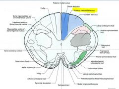
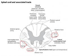
At what spinal Level do the Gracilus and Fasiculus appear?
EXAM**** |
T7
|
|
|
|
What about above and below T7?
EXAM**** |
Posterior intermediate sulcus – only above T7
below do not have that**** |
|
|
|
Picture of:
Posterior Intermediate Sulcus |
|
|
|
|
What are the Fasciculus cuneatus and Fasiculus gracilis?
|
|
|
|
|
What do the fasciculus cuneatus and gracilis do?
|
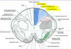
Fasciculus cuneatus:
Upper Body Extremities More Lateral Fasciculus gracilis Lower Body Extremities Located More Medial |
|
|
What is the circled area?
|
Anterolateral System
functions in PAIN and THERMAL sense |
|
|
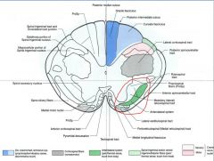
What is the circled area?
|
Dorsal Column Lemnisus System
Proprioception Vibratory sense Descriminative Touch |
|
|
|
What is the circled gray area?
|
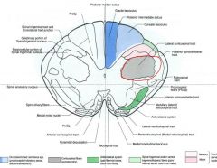
Corticospinal fibers
somatomotor |
|
|
|
There are 2 main pathways for sensory and motor:
|
1. one set for the entire body except for the orofacial complex
2. one set for the orofacial complex |
|
|
|
What is another name for the "entire body except for the orofacial complex"
|
Ascending Spinal Pathways
|
|
|
|
What are the 2 ascending spinal cord pathways
|
1) Dorsal column/ medial lemniscus pathway
2) Anterior Lateral System (ALS) |
|
|
|
What does the Dorsal column/ medial lemniscus pathway function in?
|
Carries information concerned with fine touch, proprioception, vibration.
Uses these following receptors: Pacinian corpuscles Joint receptors Ruffini corpuscles Meissner’s corpuscles Golgi tendon organs Muscle spindles Peripheral processes of dorsal root ganglion cells provide axons to these receptors. Axons pick up information from the receptors and project to spinal cord. |
|
|
|
Central processes of dorsal root ganglion (DRG) cells enter the spinal cord in the posterior horn and enter the posterior funiculus.
Central processes of dorsal root ganglion (DRG) cells enter the spinal cord in the posterior horn and enter the posterior funiculus. |
The posterior funiculus on each side is divided by the posterior intermediate septum above T7 into two separate fasciculi - fasciculus gracilis (medial) and fasciculus cuneatus (lateral).
|
|
|
|
|
|
|
|
At what levels is the Fasciculus gracilis found?
|
present at all spinal levels - contains long ascending branches of fibers from sacral, lumbar and lower 6 thoracic dorsal roots.
|
|
|
|
At what levels is the Fasciculus cuneatus found?
|
first appears around T7 - contains long ascending branches of fibers from upper 6 thoracic and all cervical dorsal roots.
|
|
|
|
Where are the 1st order neurons for the Dorsal Column Tract?
|
Neurons in dorsal root ganglion which send their central processes ascending in fasciculus gracilis and cuneatus are considered to constitute the 1st order neuron for this pathway.
Fibers in the fasciculus gracilis and cuneatus ascend ipsilaterally and terminate (synapse) on nuclei in the medulla - the nucleus gracilis and nucleus cuneatus, respectively. |
|
|
|
Where are the 2nd order neurons for the Dorsal Column Tract?
|
Nucleus gracilis and cuneatus give rise to 2nd order fibers which project ventromedially as internal arcuate fibers
Internal arcuate fibers decussate (cross the midline) and form a compact fiber bundle located on each side of the brain stem – the medial lemniscus. The medial lemniscus ascends through the contralateral half of the brain stem, and its fibers terminate in the ventral posterolateral (VPL) nucleus of the thalamus |
|
|
|
Where do 3rd order neurons for Dorsal Column Tract appear?
|
From VPL of thalamus, 3rd order neurons send fibers to postcentral gyrus of cerebral cortex (somatosensory cortex).
|
|
|
|
Neumonics to help:
|
Very pretty line drive – VPL - body
VPM – very pretty music - head |
|
|
|
What should you think of the thalamus as?
|
you mommy
tells you what to do........that inner voice lol |
|
|
|
Mnemonic for remembering that Fasciculus gracilis caries lower extremity information
|
Feet In the Grass
|
|
|
|
Picture of Dorsal Column Pathway
|
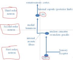
2nd order is in the medulla
VPL is in the thalamus |
|
|
|
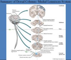
Summary of Dorsal Column
Medial Lemniscus System |
|
|
|
|
Which are fast fibers?
Pain or Fine touch? |
Fine touch****
- Fine touch are heavy myelinated and fast - pain is slow fibers - second order neuron |
|
|
|
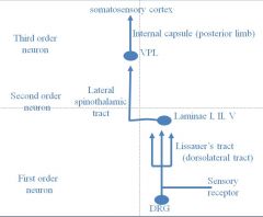
What are the 3 neurons systems for the Anterior Lateral System?
|
First order neuron- DRG neurons (A-sigma and C fibers)
Second order neuron- neuron in dorsal horn to VPL Third order neuron- VPL to somatosensory cortex |
|
|
|
What is the function of the ALS?
|
Conducts pain, temperature, and crude touch information
|
|
|
|
What is Lissauer's Tract?
|
First order neuron receives input from sensory receptors and projects to the dorsal horn where the neuron bifurcates (splits). These branches travel 1 to 3 segments up and down the cord in the dorsolateral tract, also known as Lissauer’s tract. The axons then enter the dorsal horn, where they synapse (mostly in laminae I and II, but also in V).
|
|
|
|
Where do the axons of Lissauer's tract enter?
|
First order neuron receives input from sensory receptors and projects to the dorsal horn where the neuron bifurcates (splits). These branches travel 1 to 3 segments up and down the cord in the dorsolateral tract, also known as Lissauer’s tract. The axons then enter the dorsal horn, where they synapse (mostly in laminae I and II, but also in V).******
|
|
|
|
where are 2nd order neurons formed for the ALS tract?
|
The second order neurons from the dorsal horn send axons that immediately cross the cord in the ventral white commissure. They ascend the spinal cord in the anterior lateral system (sometimes called the lateral spinothalamic tract) and synapse in the VPL. VPL neurons project to the somatosensory cortex.
|
|
|
|
What is Somatotropic organization?
|
In the lateral spinothalamic tract new neurons are added on the medial side, thus creating a somatotopic organization.
|
|
|
|
Where are other places that the ALS 2nd prder neurons make synapses?
|
On their way to the brainstem
1. Periaquaductal Gray matter 2. Reticular Formation 3. Intralaminar thalamic nucleus |
|
|
|
What does the periaquaductal gray matter in the midbrain do?
|
activates descending pain control mechanims
|
|
|
|
What does intralaminar thalamic nucleus do?
|
provides emotional and arousal aspects of pain
|
|
|
|
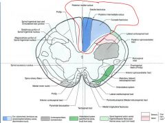
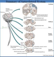
Diagram of the ALS
|
|
|
|
|
Summary of ALS
|
|
|
|
|
What pathways function to convey information regarding specific sensory modalities ultimately to cerebral cortex for conscious perception
|
somatosensory pathways
|
|
|
|
What are the somatosensory pathways?
|
There are 2 sets:
1. one set for the entire body except for the orofacial complex 2. one set for the orofacial complex |
|
|
|
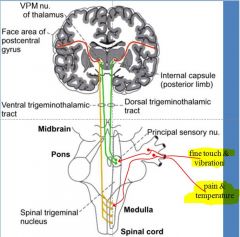
What are the trigeminal nerve cranial nerve 5 pathways?
|
1. a trigeminal pathway for fine touch and vibration
2. a trigeminal pathway for pain and temperature |
|
|
|
How does the trigeminal nerve (5) function in touch?
|
The primary sensory neuron has its soma in the trigeminal ganglion, and projects into the pons where it synapses in the chief (principle) sensory nucleus on the ipsilateral side
|
|
|
|
The second order neuron (with its soma in the chief sensory nucleus) projects through the brainstem and synapses bilaterally in the ventral posterior medial thalamic nucleus (VPM) of the thalamus
|
The third order neuron, with its soma in the VPM, projects to the primary somatosensory cortex. More details to follow…
|
|
|
|
Picture of trigeminal nerve pathways
|
|
|
|
|
now let's review pain with the trigeminal nerve....cranial nerve 5
|
The primary sensory neuron has its soma in the trigeminal ganglion, and projects into the brainstem where it synapses in the spinal trigeminal nucleus on the ipsilateral side
|
|
|
|
The second order neuron (with its soma in the spinal trigeminal nucleus) projects to the contralateral side of the brainstem and ascends in the ventral trigeminal tract to synapse in the VPM.
|
The third order neuron (with its soma in the VPM) projects an axon to synapse in the primary somatosensory cortex.
|
|
|
|
wow get ready
|
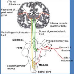
The primary sensory neuron has its soma in the trigeminal ganglion, and projects into the brainstem where it synapses in the spinal trigeminal nucleus on the ipsilateral side. The second order neuron (with its soma in the spinal trigeminal nucleus) projects to the contralateral side of the brainstem and ascends in the ventral trigeminal tract to synapse in the VPM. The third order neuron (with its soma in the VPM) projects an axon to synapse in the primary somatosensory cortex.
|
|
|
|
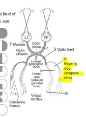
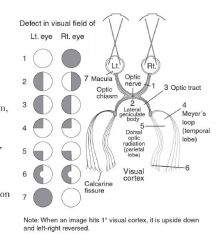
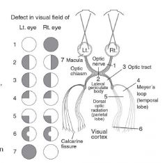
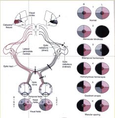
What does each OPTIC TRACT Carry?
|
Each optic tract carries information from the lateral ipsilateral retina and the medial contralateral retina.
Stated another way, each optic tract carries information from the contralateral visual field. |
|
|
|
Visual Field Pathways
Make sure you understand these |
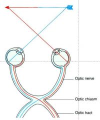
Each optic tract carries information from the lateral ipsilateral retina and the medial contralateral retina.
|
|
|
|
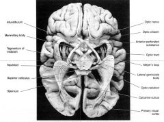
Where do all visual fibers eventually go?
|
Lateral geniculate bodies
Right and Left |
|
|
|
|
|
|
|
Why is Meyer's Loop Important?
|
Meyer’s loop very important – stroke or vascular system if parts of visual field get knocked out and can lose blood supply
|
|
|
|
What artery supplies the Meyer's Loop?
|
Middle cerebral artery territory! Classic question on exam!
|
|
|
|
Just know that the things you see in the periphery are deeper in the brain then things that are seen in the middle of your vision
|
|
|
|
|
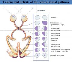
You have to know these lesions!
|
|
|
|
|
What would happen if I cut the optic nerve?
Cut left eye |

Blindness in the ipsilateral eye
Left eye blindness |
|
|
|
What would happen if i cut the optic nerve?
|
Bilateral hemianopia
#2 |
|
|
|
What would happen if you cut the RIGHT optic tract?
|

Homonymous hemianiopia
#4 Could not see anything on LEFT side of either eye |
|
|
|
What if you cut the RIGHT Meyes's Loop?
|

LEFT Quadrant anopia
|
|
|
|
this is important.......
what either diagram is not showing you is: 1. Meyer's Loop 2. Dorsal Optic Radiation |
Meyer's Loop:
Inferior retina Loops around inferior horn of lateral ventricle Dorsal Optic Radiation: Superior retina goes through internal capsule |
|
|
|
What if you cut both Meyer's Loop and Dorsal Optic Radiation on the LEFT side?
|
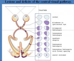
LEFT Homonymous Hemianpoia with Foveal Sparing
|
|
|
|
What happens if you hit in the back of the head with a baseball bat?
|

Central Scotomas BOTH eyes
|
|
|
|
know these cold lol
|
|
|
|
|
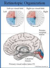
What fissure is the visual cortex located in?
|
Calcarine Fissure
|
|
|
|
What is the CORTICOBULBOSPINAL TRACT
|
Cell bodies are in the cerebral cortex (mostly pre-central gyrus -Brodmann’s area 4 but also in 6)
They descend through the CNS and make synapses in the brainstem (corticobulbar axons) and in the ventral spinal cord (corticospinal axons). Corticobulbal synapses are made on red nucleus and cranial nerve nuclei III, IV, V, VI, VII, X, XI, XII. |
|
|
|
From cortex, fibers converge in the corona radiata and travel in the posterior limb (corticospinal) and genu (corticobulbar) of the internal capsule. At midbrain levels, the corticospinal fibers form the middle 1/3 of the crus cerebri (cerebral peduncles), while the corticobulbar fibers are located just medial to the corticospinal fibers.
|
In the pons, the fibers of the corticobulbospinal system are broken up into scattered fiber bundles in the basilar (ventral) portion of the pons.
|
|
|
|
In the medulla, the fibers of this system coalesce again into the medullary pyramids. Nearly all of the axons cross the midline in the medulla at the pyramidal decussation. The crossed fibers travel through the spine in the lateral corticospinal tract and the uncrossed fibers (<10%) travel in the anterior corticospinal tract.
|
The fibers of the lateral corticospinal tract enter into the ventral horn at all spinal levels and synapse on motor neurons and interneurons. The fibers in the anterior corticospinal tract cross the midline in the anterior white commissure at the same level they enter the ventral horn and make similar synapses as the lateral corticospinal tract.
|
|
|
|
What does the Lateral Corticospinal tract do?
|
movements of the extremities
****EXAM This is the MOST clinically important descending motor pathway |
|
|
|
What does the Anterior corticospinal tract do?
|
movement of axial muscles
|
|
|
|
Diagram of the corticospinal tract
|
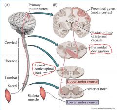
Motor – just a 2 neuron pathway, desicates in the medulla
Most important is the lateral Lower motor neuron lesion – lose of reflexes Upper motor neuron lesion – very spastic and very ridgid |
|
|
|
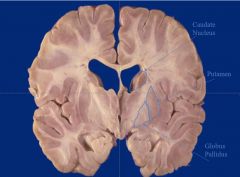
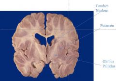
What is the basal ganglia?
|
The basal ganglia are a set of nuclei that function as a “consultant” to the cerebral cortex.
They provide the crucial physiological link between the idea of movement and the motor expression of that idea.**** EXAM |
|
|
|
Neuronal activity in the descending motor systems is closely correlated in time with the expression of a particular motor act. In contrast, most neuronal activity of the basal ganglia occurs before a particular movement begins.
|
Disturbances in the function of the descending pathways result in paralysis or paresis, but lesions to the basal ganglia causes disturbances in the initiation or cessation of a motor event
|
|
|
|
Basal Ganglia Picture
|
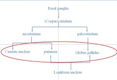
Know the 3 in middle – help cortex need do what it wants to do
|
|
|
|
Direct Pathway
|
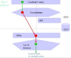
This pathway involves a circuit beginning in the cerebral cortex with connections to the caudate and putamen (together: neostriatum), which then project to the medial segment of the globus pallidus (GPm), which projects on to the thalamus, and then back to the cerebral cortex. Without input from other areas, GPm neurons are tonically active and inhibit thalamic neurons with GABA, preventing them from exerting an excitatory influence on the cerebral cortex. Activation of the direct pathway causes excitation of the neostriatum by the cerebral cortex. Neostriatal neurons secrete GABA and are inhibitory to neurons of the GPm. Therefore, when activated, neostriatal neurons will inhibit GPm cells, thus preventing them from inhibiting thalamic neurons (disinhibition). Thus, the result of activation of the direct pathway is increased output from the thalamus with a resultant increase in activation of the motor regions of the cerebral cortex.
Release of dopamine by substantia nigra compacta (SNc) cells onto neostriatal neurons with D1 receptors facilitates activity in the direct pathway. |
Direct allows you to do what you want lol
|
|
|
Indirect Pathway
|
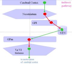
This pathway includes an additional loop through the lateral globus pallidus (GPl) and the subthalamic nucleus (ST). As with the direct pathway, the indirect pathway begins with excitatory projections from the cerebral cortex to the neostriatum. However, neostriatal output in this pathway is to the lateral, rather than medial, segment of the globus pallidus. These neostriatopallidal fibers are inhibitory (GABA is transmitter). Neurons of the GPl in turn send inhibitory connections (also GABAergic) to the subthalmic nucleus. These fibers to the subthalamic nucleus show high levels of spontaneous activity and tonically inhibit subthalamic neurons. Inhibition of GPl neurons by the neostriatum prevents GPl cells from inhibiting the subthalamic nucleus (disinhibition of ST). When active, subthalamic neurons are excitatory (glutamate is transmitter) to neurons of the GPm. Remember from the description of the direct basal ganglia pathway, that GPm neurons inhibit thalamic neurons. Thus, when the indirect basal ganglia pathway is activated, the result is decreased activity of the thalamus and therefore decreased activity of the motor regions of cerebral cortex.
|
|
|
|
Basal Ganglia mnemonics
|
1. Caudate Hotel
“book a date at the Caudate” 2. Putamen-Amen 3. Globus Palidus – two words, two parts |
|
|
|
|
|
|
|
We have to learn Blood Supply on our own so THANK YOU FIRST AID! :)
|
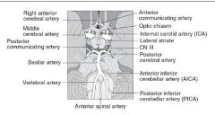
Know the circle of willis
|
|
|
|
Know what hemispheres supply these lobes of the brain for the CEREBRAL ARTERIES
|
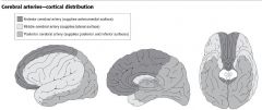
Anterior: anteromedial surface
Middle: Lateral suface Posterior: posterior and inferior surfaces |
|
|
|
What is an epidural hematoma?
|
usually an artery torn following skull fracture
high pressure arterial blood forms a rapidly expanding intracranial mass displaces brain – increased intracranial pressure Presentation of epidural hematoma: initial unconsiousness (KO) return to consciousness secondary unconsiousness death |
|
|
|
What is a subarachnoid hematoma?
|
usually a vein
low pressure blood causes slowly increasing intracranial mass displaces brain Compromised mental function and clouding of consciousness may take days – weeks to develop Surgery required to alleviate bleeding and decompress brain |
|