Reading...
![]()
Play button
![]()
Play button
![]()
Use LEFT and RIGHT arrow keys to navigate between flashcards;
Use UP and DOWN arrow keys to flip the card;
H to show hint;
A reads text to speech;
84 Cards in this Set
- Front
- Back
- 3rd side (hint)
|
Gram +
Rod shaped Bacillus Cereus |
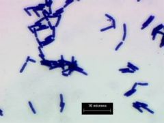
The following has what gram reaction and what morphology? give a possible species.
|
|
|
|
Gram -ve
Rod shaped E. Coli |
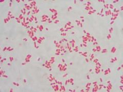
what are the gram reaction and morphology of the bacteria? Name a potential species.
|
|
|
|
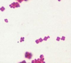
+ ve
Coccus StaphyloCOCCUS aureus |
what gram reaction and cell morphology. Name a potential species
|
|
|
|
why is nutrient broth undefined media?
|
Because we do not know its exact chemical formula
|
|
|
|
How to use spectrophotometer?
|
First set to 0 with left knob
Then put in blank set to 0 with right knob Put in the actual substance to test and read absorbance from right to left. |
|
|
|
TFTC vs. TNTC
|
Plate count must be between 30 and 300.
Less than 30 TFTC More than 30 TNTC |
|
|
|
Plate dilution-
say 36 counted on a 10-5 plate dilution. How many bacteria? |
36 X 10^5= 3,600,000
no need to add an extra fold. Plate dilution is the multiplication factor. |
|
|
|
Why is plate more accurate than spectrophotmetry?
|
In spec some of the dead colonies are counted as well as any debris.
|
|
|
|
What was the point of the Sabouraud plate?
|
it was used to demonstrate person-to-person contact spread of microorganisms.
|
|
|
|
Baker's yeast
|
Sacchaomyces cerevisiae
|
|
|
|
Crystal violet, nigrosin and methelene blue are examples of...
|
Simple stains
|
|
|
|
What is the purpose of a wet mount?
|
To check for motility in microorganisms.
|
|
|
|
What cellular structure is differentially stained using the Gram technique?
|
The cell wall
(peptidoglycan (+ve) vs. none (-ve)) |
|
|
|
What is the bacterial cellular structures that confers motilitiry
|
Flagellum
|
|
|
|
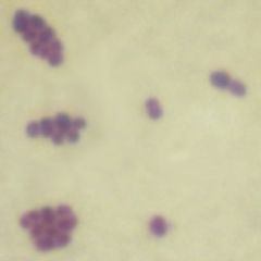
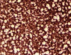
How can we disinguish between Micrococcus luteus and bacillus cereus?
|
Both are gram positive and have a cocci morphology BUT
Micrococcus luteus are often found in tetrads or pairs. |
|
|
|
Why doesnt nigrosin stain the cell?
|
Because nigrosin is a negative dye it does not stain the cell it stains the background and organisms appear White.
|
|
|
|
When studying wet mounts, which organism moved very quickly?
|
Pseudomonas aeruginosa
|
|
|
|
When studying wet mounts,What organism moved slowly?
|
Bacillus Cereus
|
|
|
|
What is brownian motion?
|
vibrating motion caused by bacteria being struck by H20 molecules.
|
|
|
|
When studying wet mounts, what organism did we observe and identify as engaging in brownian motion?
|
Staphylococcus Auerus
|
|
|
|
In the gram stain what was the:
Primary stain mordant decolorizer differential stain |
Crystal violet
Gram's iodine alcohol safranin |
|
|
|
The acid fast stain served what purpose?
|
used to differentiate the genus Mycobacterium which can cause TB and leprosy. CLUMPED RED RODS!
|
|
|
|
Why is the gram stain the most important staining procedure in microbiology?
|
Because it differentiates MANY bacteria. Basically cuts in half.
|
|
|
|
What is expected in a capsule stain?
|
The background is dark (crystal violet) and the capsule appears in white with the black bacteria enclosed in the white capsule
|
|
|
|
What is expected in an endospore stain?
|
round endospores are green because they opened due to heat and the bacterial rods (pink- safranin)should surround the endospores.
|
|
|
|
Selective/differential
for what? how? CRYSTAL VIOLET AGAR |
selective media for gram -ve
Does not allow gram +ve cell growth b/c hinders cell walls |
|
|
|
Selective/differential
for what? how? BLOOD AGAR |
differential media.
shows to what degree bacteria can hemolyse blood. |
|
|
|
Selective/differential
for what? how? Manitol Salt agar plate |
Differential and selective.
High salinity only allows Staphylococcus genus to grow. while the manitol sugar is broken at different rates to differentiate between species. |
|
|
|
Selective/differential
for what? how? MacConkey agar |
Maconkey is differential and selective.
It has crystal violet (selective) no gram positive)) and lactose w/ indicators (differential) |
|
|
|
Selective/differential
for what? how? EMB |
EMB- Eosin/Methylene blue is both differential and selctive
Selective- not perfect does not allow gram +ve again differential- obvious (E.coli metallic green) |
|
|
|
Which organism should not grow on a Crystal violet agar plate, why?
Staph Aureus or E. coli |
Staph aureus should not grow because crystal violet is a selective media for gram -ve bacteria. (e-coli should grow)
|
|
|
|
What are the classes of hemolysis in the blood agar plate and what does that mean? Identify potential organisms.
|
Beta- complete breakdown
Alpha- partial breakdown Gamma- no breakdown Beta- S. pyogenes Alpha-S. mitis Gamma- E. faecalis |
|
|
|
If given a MSA plate broken in 5 pieces and 2 show no growth. and of the three with growth 1 has made the plate yellow. what can you conclude?
|
The two that did not grow are not Stapylococcus.
The two that grew but no yellow are staphylococcus but not maniotol fermenters. and the one that grew and changed to yellow is both and probably Staph Aureus. |
|
|
|
If given an EMB plate broken in 5 and 2 did not grow, 1 grew but no change in color, 1 grew and changed ot pink, and a last grew and became green. what can you conclude?
|
The 2 that did not grow are probably gram +ve. the one that grew but no change in color is either gram positive or negative but CANNOT FERMENT LACTOSE. the one that converted to pink is a mediocre fermenter and the metallic green is an excellent fermenter of lactose (E. coli)
|
|
|
|
If given a MacConkey plate where 2 did not grow and 2 others are pinkish red instead of colorless.
|
MAC is selective:
Gram negative organisms will grow, Gram positive organisms will not. This is due to the addition of bile salts and crystal violet. MAC is differential: Lactose fermenters will appear pink, non-lactose fermenters will appear colorless. This is due to the addition of the indicator, neutral red. |
|
|
|
In MSA plate change from red to yellow indicates
|
the organism can ferment mannitol and the acid produced as byproduct lowers pH to YELLOW. (Phenol red)
|
|
|
|
MacConkey plate: organisms changing from colorless to pink.
|
Lactose fermenters will appear pink, non-lactose fermenters will appear colorless. This is due to the addition of the indicator, neutral red.
|
|
|
|
Which plate would you use if you were checking whether your patient's upset stomach was salmonellosis or traveler's diarrhea?
|
Use manitol salt agar plate. if the organism does not grow it is most likely Salmonelosis. if it does and turns yellow (Staph aureus= travelers diarrhea).
To be sure also innoculate Crystal violet and gram -ve salmonella typhimurium should grow and Staph not. |
|
|
|
Bacteria that thrive in cold climates
|
Psychrophiles
(P. Fragi) |
|
|
|
Bacteria that thrive in very hot climates
|
Thermophiles
(B. stearothermophilus) |
|
|
|
Bacteria that thrive in room temperature
|
Mesophiles
(E. coli, S. Marcescens) |
|
|
|
pH conditions: E coli
|
Grows in alkaline pH
(your intestines) |
|
|
|
pH conditions: A. faecalis
|
grows in alkaline pH
|
|
|
|
pH conditions: L.Lactis
|
grew in acidic conditions (pH 3-4)
|
|
|
|
Why use buffers in the testing of pH conditions?
|
Because the organisms themselves will begin to change the pH by fermentation and/or wastes.
|
|
|
|
Why are buffers NOT necessary in nutrient broths?
|
because they contain natural buffers in the form of zwitterions from amino acids which can donate or accept protons
(brownsted-lowry definition of acid/base). |
|
|
|
Obligate aerobes
|
Require Oxygen
|
|
|
|
Obligate anaerobes
|
Cannot have oxygen
|
|
|
|
Facultative Anaerobes
|
Dont need Oxygen but make use of it and grow more abundantly b/c oxygen is their final electron acceptor. (E.coli)
|
|
|
|
Aerotollerant Anaerobes
|
those that can live in the presence of Oxygen or not with no difference.
|
|
|
|
Microaerophiles
|
Species that grow if provided only with a little bit of oxygen.
|
|
|
|
When using the GasPak system, to determine oxygen requirement, the strip contains methylene blue is obviously blue until...
|
no oxygen is present in which case the strip will turn colorless.
|
|
|
|
Growth curve:
|
Lag phase- cells adapt to new surroundings no major growth
Log phase- where growth outnumbers death Stationary phase- where they are equal (carrying capacity) Death phase- where more organisms are dying than reproducing. |
|
|
|
extracellular enzymes
|
Produced within the cell but work outside the cell often to breakdown large substrates into smaller transportable units (amylase, lipase, caseinase, gelatinase)
|
|
|
|
Intracellular enzymes
|
are produced inside the cell and function there. (Catelase)
|
|
|
|
Catabolic reactions
|
breaking down substrates= generates energy or subunits to be used by the cell.
|
|
|
|
Anabolic reactions
|
Synthesizing reactions usually to build cellular constituents at the expense of ATP energy.
|
|
|
|
Amylase
|
Along with maltase breaks down starch.
|
|
|
|
Lipase
|
general term for enzymes which hydrolize lipids to glycerol and fatty acids; extracellular
|
|
|
|
Caseinase
|
The enzyme that breaks the protein CASEIN in milk into its constituent amino acids.
|
|
|
|
Gelatinase
|
reduced gelatin to its amino acids. (Psuedomonas Aeroginosa)
|
|
|
|
Phenol red carbohydrate broths (red to yellow)
|
Organism fermented the carbohydrate.
|
|
|
|
Phenol red carbohydrate broths (red outside yellow inside)
|
organism OXIDATIVELY break down the peptones in the broth~ PROTEOLYSIS (pH goes up)
|
|
|
|
IMVIC test is useful for
|
the genus Enterobacteriaceae
specifically to distinguish between e. coli and e. aerogenes. |
|
|
|
Indole test
|
If indole is produced from tryptophan (by the enzyme tryptophonase) a floating red ring will form.
|
|
|
|
Methyl red test
|
When glucose is oxidized it may produce a final acid product (RED) or another final product will not be acidic (Yellow).
|
in other words the methyl red test rules out...
|
|
|
Voges-Proskeaur Test
|
If the final of further glucose oxidation is ACETYLMETHYLCARBINOL (AMC) it will produce an orangey-red broth after 30 minutes of vortexing.
|
|
|
|
Citrate test
|
The ability to use citrate as the sole carbon source is detected by a change from green to deep blue. (bromethyl blue)
|
|
|
|
SIM agar deep test
|
In this test if the gas released is hydrogen sulfide it reacts with the iron in the medium to turn black.
|
|
|
|
Carbohydrate phenol red test (gas in durham tube)
|
This means that the carbohydrate was fermented and CO2 was most likely produced and released as a by-product.
|
|
|
|
SIM test stands for...
|
the three tests it can do:
Sulfide- turn black Indole- unreliable motility- unreliable |
|
|
|
Urease test
|
Many organisms convert urea to ammonia and water using urease.
In this case the medium will become hot pink from an orangey-yellow color. (phenol red indicator will show when alkaline ammonia is produced). |
|
|
|
2 major causes of UTI's. how to test for it.
|
Proteus Vulgaris and Proteus Mirabilis
Urease test |
|
|
|
Major cause of Stomach ulcer and how to test for it
|
Helicobacter pylori; urease test.
|
|
|
|
Nitrate reduction test
|
Possible products are: (red is always terminal)
Nitrogen- Air in Durham tube if nitrate is reduced to N2. Nitrite- if after addition of 2 reagents turns red. Nitrate- if Zinc is added and broth turns red, nitrate was never reduced Ammonia- no color change nitrate was never reduced. |
|
|
|
Production of Catelase test
|
Catelase- breaks H202 into water and oxygen. The reason most anaerobes cant live in oxygen.
Add H202 to the slant if it fizzes the organism has catelase to break it down. |
|
|
|
TSI agar test used to identify members of
|
Enterobacteriaceae
|
|
|
|
TSI- triple sugar iron medium contains:
|
three sugars:
glucose, lactose, and sucrose. ph indicator: phenol red iron- to detect production of H2S |
|
|
|
TSI possible results:
red slant, red butt= red slant yellow butt= yellow slant yellow butt= Black= |
red/red= nothing fermented no H2S produced.
red/yellow= only glucose fermented yellow/yellow= glucose and either both or one of the two sucrose/lactose was fermented black= H2S was produced and fermentation of sugars is obscured. |
|
|
|
Litmus milk test serves two purposes:
|
1.) redox indicator- showing whether the medium was reduced or oxidized by changing color.
2.) pH indicator- showing if the medium is acidic or basic following the activities. |
|
|
|
Litmus milk as a pH indicator
|
lactose fermentation yields acid:
Pink- lactose fermented Pink on top only and curd- excessive fermentation Deep blue- breakdown of milk proteins (casein) Brown and purple/blue at top-proteolysis of milk protein |
|
|
|
Both are gram positive and coccus shape, BUT M. luteus clump in duplets or quads.
|
The following organism has what gram reaction and what morphology and yet it is known not to be Staphylococcus Aureus, why?
|
|
|
|
Capsule, capsules enter the body without detection (NO PHAGOCYTOSIS)
|
What kind of stain is this? what medical importance does this serve?
|
|
|
|
Endospore
|
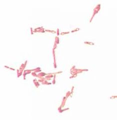
What kind of stain is this?
|
|