Reading...
![]()
Play button
![]()
Play button
![]()
Use LEFT and RIGHT arrow keys to navigate between flashcards;
Use UP and DOWN arrow keys to flip the card;
H to show hint;
A reads text to speech;
63 Cards in this Set
- Front
- Back
|
describe Skin 3 layers
|
epidermis: keratinized stratified squamous epithelial cells, repair by desquamation. dermis: thick, dense layer with connective tissue to support, give strength and thickness to skin. hypodermis: subcutaneous fat in lobules separated by connective tissue.
|
|
|
epidermis 5 layers
|
1. stratum basalis, 2. stratum spinosum, 3. stratum granulosum, 4. stratum lucidum, 5. stratum corneum
|
|
|
stratum basalis
|
cuboidal cells, germinativum, produce keratinocytes; some columnar of dermis
|
|
|
stratum spinosum
|
polyhedral cells with intercellular desmosomes
|
|
|
stratum granulosum
|
release keratohyaline granules, and hydrophobic membrane bounded lipid into aqueous barrier, and enzyme to digest organelles as immunogenic molecule
|
|
|
stratum lucidum
|
loss function of keratohyalin replaced by eleidin (only in thick skin: sole and palmar skin)
|
|
|
stratum corneum
|
two layers: (deep) intercellular desmosomal link and intracellular keratin filaments; (surface) keratinized, with soft keratin, desquamation, lose, surface
|
|
|
special features of skin list 7
|
hair, sebaceous gland, sweat gland, meissner's corpuscle, merkel cells, pacinian cells, free nerve ending exteroceptor
|
|
|
cell types in epidermis (origin and function) 5 pt
|
Keratinocytes from ectoderm produce ectoderm; Melanocytes from neural crest produce melanin in melanosomes at SB sent process to SS; Langerhans cells at SS antigen presenting cells with Birbeck granules; Merkel's cells at SB as exteroceptor (touch)
|
|
|
Release of keratohyalin granule to cytoplasm triggers the final aggregation of keratin filaments to form tonofilament bundles thus transforming the granular cell to cornified cells
|
Release of keratohyalin granule to cytoplasm triggers the final aggregation of keratin filaments to form tonofilament bundles thus transforming the granular cell to cornified cells
|
|
|
SG, SL, SC, keratinocytes are reinforced by ______
|
involucrin fibrous protein
|
|
|
dermis layer
|
papillary layer (loose) and reticular layer (dense and thick, coarse)
|
|

free nerve ending cells (nature, location, function)
|
unmyelinated, small diameter, slow conduction of nerve signal, for pain touch temperature; from dermo-epidermal junction extend to SG
|
|

meissner's corpuscle
|
elliptical, tactile cells with nerve ending, transversely located dermis under hairless skin (nipple, lip, eyelid, finger tip)
|
|
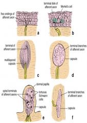
pacinian corpuscle
|
oval shape, nerve ending, skin of the hands and feet, pressure sensitive,
|
|
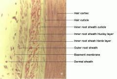
hair follicle layers 7pt
|

M, Cx, Cu, IRS, ERS, GM, CT
|
|
|
Sebaceous gland
|
holocrine; secrete sebum (oil) into neck of hair follicle, arrector pili
|
|
|
sweat glands 2pt
|
Merocrine: Secrete a watery fluid, hypotonic, directly to skin surface; Apocrine: adjacent hair follicles via duct areolae of breasts, genital regions, viscid, milky secretion
|
|
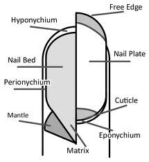
Structure of Nail
|

Eponychium, Nail, NM (germinating center) Nail bed, hyponychium
|
|
|
Ceruminous
|
cuboidal (inactive) or columnar (active); ear wax, waxy, yellowish, semisolid, prevent desiccation and irritation
|
|
|
4 layers of GI tract
|
mucosa (epithelial lining, lamina propria, muscularis mucosae , submucosa, Muscularis externa , external layer
|
|
|
oesophagus
|
submucosal cardiac glands (simple tubular, mucous), gastroesophageal and pharyngoesophageal sphincters. muscularis externa: upper striated, middle mix, lower smooth
|
|
|
stomach
|
three layers of muscle in muscularis externa
|
|
|
division of stomach
|
Cardia, Fundus and body of stomach, Pylorus
|
|
|
5 cell types in stomach
|
Mucous neck cells (mucus) , undifferentiated cell in neck and isthmus, Parietal cell (HCl, stomach intrinsic factor), chief cell (zymogen)
|
|
|
enteroendocrine glands
|
secrete peptide hormones, neuroendocrine system; involve in amine precursor uptake and decarboxylation.
|
|
|
example of enteroendocrine
|
Gastrin secrete more HCl , somatostatin inhibit release of gastrin, Urogastrone ( inhibit HCl secretion)
|
|
|
4 cell types in small intestine
|
columnar absorptive cell, paneth cell (zymogen and lysozyme), goblet cell (mucus) enteroendocrine, mucosal glands in crypts of Lieberkuhn; Brunner's glands submucosal gland.
|
|
|
folding of intestine 3pt
|
plicae circulares, villi, microvilli,
|
|
|
duodenum special external layer
|
adventitia
|
|
|
Peyer's patches
|
in ileum, Gut-associated lymphoid tissue (GALT): mucosal immunity; Antigen presenting epithelial cells (M cells) -> deliver them to macrophages -> IgA producing plasma cells
|
|
|
large intestine
|
goblet cells, and enteroendocrine, no villi, no paneth cell, haustra coli, teniae coli,
|
|

name the bones of nose 3
|
nasal bone, nasal part of frontal bone, frontal process of maxilla
|
|

name the bones of nasal septum
|
vomer, perpendicular plate of ethmoid, septal cartilage
|
|
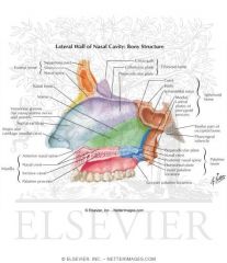
name the bones that make up the roof of nasal cavity
|
sphenoidal bone, frontonasal, ethmoid bone,
|
|
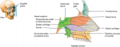
name the bones make up the floor
|
palatine process of maxilla, horizontal process of palatine
|
|

lateral wall is formed the three curvature plates known as _______ ?
|
conchae.
|
|
|
underneath each curvature plate, you will find a potential space called ________?
|
meatus
|
|
|
functions of nose
|
1. respiration, 2. Humidification, 3 filter of dust,4 Olfaction
|
|
|
function of meatus
|
drainage paranasal sinuses and nasolacrimal ducts
|
|
|
nerve supply to nose
|
cranial nerve I : olfactory nerve; cranial nerve V trigeminal nerve
|
|
|
3 Arteries to nose
|
branches of 1. maxillary artery (external carotid a.), 2 facial artery (external carotid a.), 3 ophthalmic artery (internal carotid a.)
|
|
|
4 veins drain the nose
|
pterygoid plexus, facial vein, infraorbital vein, ophthalmic vein
|
|
|
paranasal sinuses
|
frontal (frontal sinuses); ethmoid (ethmoid sinuses); sphenoid (sphenoid sinuses); and maxilla (maxillary sinuses)
|
|
|
nerve, artery to sinuses
|
trigeminal nerve, maxillary, ophthalmic arteries
|
|
|
3 layers of pharynx
|
1. mucous membrane (ciliated columnar epithelium at nasopharynx; stratified squamous in oropharynx and laryngopharynx), 2. fibrous tissue, 3. muscle tissue
|
|
|
oropharynx
|
behind the mouth to C3; collection of lymphoid gland palatine tonsils
|
|
|
laryngopharynx
|
C3 to C6 , continue by oesophagus at C6
|
|
|
larynx (lower respiratory)
|
C3 to C6; anterior: hyoid, supra- and infra muscles; posterior is laryngopharynx and oesophagus
|
|
|
trachea
|
C6-T4
|
|
|
artery to trachea
|
inferior thyroid artery (a branch of subclavian artery) and bronchial artery (from thoracic aorta)
|
|
|
vein to trachea
|
inferior thyroid vein and brachiocephalic vein
|
|
|
nerve supply to trachea
|
vagus nerve, recurrent laryngeal nerve, sympathetic cervical ganglion
|
|
|
respiratory bronchioles and alveoli involve in _____ list 4
|
external respiration, immune defense, warming and humidification of air
|
|
|
2 type of alveolar epithelial cells
|
1. single layer forms the alveolar wall; 2. The production of surfactant
|
|
|
respiratory zone components
|
Respiratory bronchioles, Alveolar ducts, Alveolar sacs
|
|
|
describe the cell wall structure of Gram-negative bacilli
|
pentapeptide bridges linking N-acetylmuramic and N-acetylglucosamine polymer called peptidoglycan. external is a lipopolysaccharide.
|
|
|
two ways to prevent candidal infection.
|
Fluconazole prophylaxis for transplant recipients; Avoid antibiotics overuse to prevent overgrowth
|
|
|
Helminths: 3pt
|
Nematodes, Trematodes, Trematodes
|
|
|
iceburg of disease
|
Iceberg of disease describes the phenomenon that what disease we see is a small proportion of the total, much of which is developing.
|
|
|
Upstream model of public health
|
It states that if we know how and why disease develops, we might be able to prevent its progression by surveillance and prevention.
|
|
|
List one essential criterion that makes a method of detection of cancer a benefit
|
It should lead to improved survival and quality of life.
|
|
|
what is phagocytosis
|
Phagocytosis is the ingestion and digestion of extracellular particulate material, which may include whole pathogenic microorganisms.
|