![]()
![]()
![]()
Use LEFT and RIGHT arrow keys to navigate between flashcards;
Use UP and DOWN arrow keys to flip the card;
H to show hint;
A reads text to speech;
18 Cards in this Set
- Front
- Back
|
What are the biochemimcal features of apoptosis? |
- activation of caspases - DNA and protein breakdown - membrane alteration and recognition by phagocytes |
|
|
Give evidence for DNA breakdown? |
- Gel electrophoresis of apoptotic cell DNA - show laddering - cleavage by endonucleasses into internucleosomal units of 180-200 bp |
|
|
What are the nuclear effects seen during apoptosis? |
- Hallmark cleavage of chromosomal DNA due to fragmentation by CAD action (conserved) - Inactivation of enzymes involved in DNA repair or replication (topoisomerase) - breakdown of structural proteins |
|
|
What is the key phagocytosis signal? |
- Phosphatidylserine (PS) - macrophages and neutrophils recognise it - PS normally found in inner layer of plasma membrane - during apoptosis some PS molecules move to outer layer (caspase mediated) |
|
|
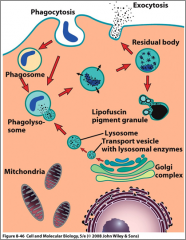
What are the steps of the phagocytic pathway? |
- material to be degraded is engulfed and internalised by the phagocyte in a phagosome -phagosome fuses with a lysosome to form a phagolysosome where degradative enzymes destroy the cellular contents - residual bodies form and contents of RBs can be eliminated by exocytosis or retaind formin lipofuscin granules |
|
|
What are the steps of the phagocytic pathway (image)? |
|
|
|
In C. elegans: 1090 somatic cells are generated during the hermaphrodite development. How many of them will die via apoptosis? |
- Exactly 131 - 113 somatic cells during embryonic development (mainly neurons) - 18 somatic cells during larval state (mostly neurons) - germ cell death - oocytes die during oogenesis in the adult hermaphrodite |
|
|
Morphological characteristics of C. elegans apoptotic cells in light microscope level: |
- increase in refractivity of the cytoplasm and nucleus - dying cells or corpses ingested (but by neighbouring cells not phagocytosis) |
|
|
Morphological characteristics of C. elegans apoptotic cells in electron microscope level: |
- reduction in cell volume - condensation of nuclear chromatin - organelles appear normal until late process - appearance of apoptotic bodies, autophagic vacuoles and membrane whorls |
|
|
Three classes of proteins mediate apoptosis: |
- Regulators -> Adaptors -> Initiator/Effector Caspases |
|
|
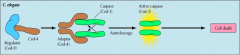
Regulators and effectors of apoptosis in C. elegans. |
- three ced genes - cell death abnormal: - Ced-9 (negative regulator, binds Ced-4 preventing Ced-3 activation) - Ced 4 (adaptor, allows 2 molecules of Ced-3 to bind together) - Ced-3 (caspase, undergoes autocleavage aided by ced-4) |
|
|
Regulators and effectors of apoptosis in C. elegans (image) |
|
|
|
What happens in step 1 in apoptotic cells? |
- Egl-1 (egg-laying defective), pro-apoptotic, contains one BH3-only-domain; binds to ced-9 causing a conformational change that releases ced-4 - Ced-9 - anti-apoptotic protein, contains 4 BH domains, bound to surface of mitochondria; interacts with ced-4 dimer |
|
|
What is characteristic of pro-apoptotic proteins? |
- has a single BH3 domain only |
|
|
What happens in step 2 in apoptotic cells? |
- Activated ced-4 interacts and facilitates ced-3 autoactivation - two ced-4 dimers join to form a tetramer - binds ced-3 = formation of apoptosome - ced-3 autocleavage is initiated and apoptosis is triggered - mitochondrial proteins CPS-6 and WAH-1 are also released and aid in DNA degradation of dying cell |
|
|
What is the apoptosome? |
- scructure formed by adaptor and initiator caspases (and other molecules) - necessary for the activation of the caspase |
|
|
What happens in C. elegans if Ced-3 is mutated? |
- survival of cells normally targeted to die - mutants still have normal life-span |
|
|
How are apoptotic cells engulfed? |
- ingested by neighbours - Ced genes play a role in two groups converging on ced-10 - ced-2, ced-5, ced-12 -> ced10 (induces cytoskeletal rearrangements for migration and engulfment) - ced-1 - receptor-like transmembrane protein; mutation results in persistant cell corpses |