![]()
![]()
![]()
Use LEFT and RIGHT arrow keys to navigate between flashcards;
Use UP and DOWN arrow keys to flip the card;
H to show hint;
A reads text to speech;
39 Cards in this Set
- Front
- Back
|
Pelvic Region |
Area of transition b/w trunk and lower limbs Enclosed by bones, ligaments and muscles. |
|
|
Pelvic Girdle |
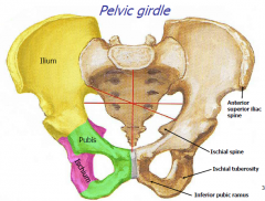
Ilium, Pubis, Ischium. These three bones are fused together. |
|
|
Joints of the Pelvic Girdle |
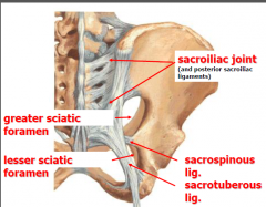
Sacroiliac joint (b/w ilium and sacrum) Sacrospinous ligament Sacrotuberous ligament Greater sciatic Foramen- formed b/w ligaments Lesser sciatic Foramen- below. Formed b/w sacrospinous lig and sacrotuberous lig |
|
|
Pelvic inlet |
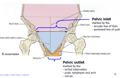
Marked by the arcuate line of ilium Pecteneal line of pubis Superior Open to abdominal cavity |
|
|
Pelvic outlet |
marked by the ischial tuberosities pubic symphysis and arch, coccyx Inferior Closed by pelvic diaphragm (levator ani and coccygeus) |
|
|
Sex differences in pelvis |
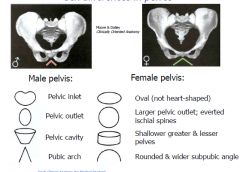
See picture |
|
|
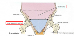
Pelvic cavity |

Funnel shaped space bound by bones of pelvis. -Greater pelvis: b/w ala of ilium and pelvic inlet -Lesser pelvs: b/w pelvic inlet and pelvic outlet Continous with abdominal cavity Conatins urinary bladder, terminal part of ureters, pelvic genital organs and rectum |
|
|
Perineum |
Triangular area of trunk b/w thighs and buttocks extending from the pubis to coccyx. Seen best in lithotomy position (diamond shaped) separated from pelvic cavity by the pelvic diaphragm |
|
|
Perineum contents |
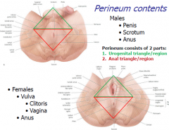
2 parts: 1. urogenital triangle/region 2. Anal triangle/region Males: Penis, Scrotom, Anus Females: Vulva (clitoris and Vagina), Anus |
|
|
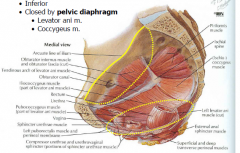
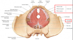
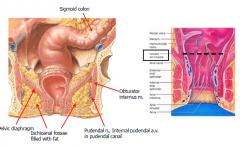
Muscles of Pelvic Wall and Floor |
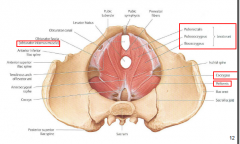
Obturator internus, Coccygeus, Piriformis and Levator Ani (puberctalis, pubococcygeus, and iliocyccygeus) |
|
|
Greater Sciatic Foramen |
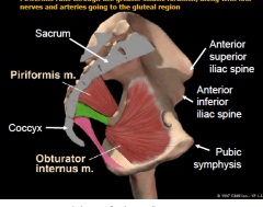
B/w ligaments, Piriformis runs throug this along with nerves and arteries going to the gluteal region. |
|
|
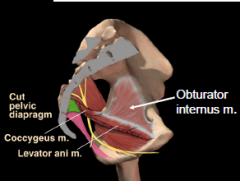
Pelvic Diaphragm |
Levator Ani + Coccygeus Forms floor of pelvic, supporting abdominiopelvic organs Aids in urinary and fecal continance |
|
|
Tendinous arch of levator ani |
Formed from the thicken fascia of obturator internus muscle. Origin of levator ani muscle. |
|
|
Function of pelvic diaphragm |
Supports abdominopelvic viscera during couging, sneezing etc. Also raises the pelvic floor during urination, vomiting, coughing and weight lifting increasing the intra-abdominal pressure. Aids in urinary and fecal continance During parturition, it supports the fecal head while the cervix dialates for delivering a baby |
|
|
Walls of pelvic cavity |
Posterior (sacrum, piriformis) anterior (pubic rami, pubic symphysis) lateral (obturator internus) Inferior (levator ani muscles, coccygeus) Superior (open to abdominal cavity) |
|
|
List of arteries in pelvis |
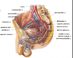
Aorta, common iliac, external iliac (becomes femoral in lower limb), internal iliac, iliolumbar, lateral sacral, superior gluteal, inferior gluteal, internal pudendal, middle rectal, superior vesical, inferior vesical, and umbilicial A. |
|
|
Blood supply to pelvic viscera |
Umbilical A---> superior vesical aa. Inferior vesical A (males)/ Uternine A (females) Middle Rectal A |
|
|
Blood supply to walls of pelvis |
Lateral sacral A Iliolumbar A |
|
|
How does blood exit pelvis? |
Exits to perineum: Internal pudendal A---> inferior rectal A Exits to lower limb: Obturator A Superior gluteal A Inferior gluteal A **Gonads (testies and ovaries receive blood from abdominal aorta, rectum receives blood from inferior mesenteric A (superior rectal) |
|
|
Peritoneal recesses |
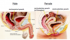
Deepest points of the peritoneal cavity, these are sites where infection and fluids typically collect. Males: rectovesicle pouch Females: rectouterine pouch (of douglass) and vesicouterine pouch |
|
|
Rectum Anal Canal and Anus |
Rectum: superior to levator ani Anal canal: inferior to levator ani Anus: Opening |
|
|
Urinary System |
Ureters: drain urine from kidneys to urinary bladder Urinary bladder: temporarily stores urine Urethra: conducts urine from urinary bladder to exterior ** in adults, urinary bladder is in the retropubic space w/in lesser pelvis. in children bladder is in abdomen, enters greater pelvis @ age 6 and lesser pelvis @ age 16. |
|
|
Urinary bladder muscles/sphincters and innervation |
Wall of bladder made of detrusor muscle. Internal (involuntary) and external (voluntary) urethral spincters help maintain continence. Sympat. innerv(T11-L2) maintain continence by contraction of internal sphincter Parasymp innerv (S2-S4) allows urination by relaxing internal sphincter and contracting detrusor muscle. |
|
|
Male urethra parts (superior to inferior) |
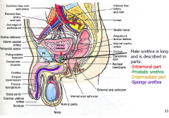
Intramural part Prostatic urethra Intermediate part Spongy urethra |
|
|
Ductus Deferens (Vas deferens) |
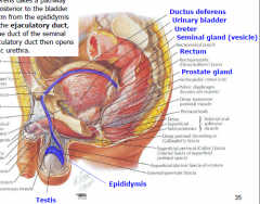
Takes a pathway superior, then posterior to the bladder to transmit sperm from epididymus (stores sperm prior to ejaculation) of testis to ejaculatory duct where is joins the duct of the seminal gland. The ejaculatory duct then opens into the prostatic urethra. |
|
|
% of semen made where |
65%-75% of semen is made of the fluid secreted by the seminal glands. 25%-30% of semen is made of secretions from the prostate gland. Sperm is only 2-5% of semen. Bulbourethral gland secrete a lubricating fluid during sexual arousal (>1% of semen) |
|
|
Corpus cavernosum |
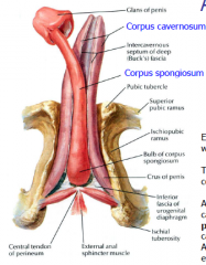
Erectile tissue- spongy tissue that fills with blood to become swollen. @ base of penis corpus cavernosum is called crus of penis |
|
|
Corpus spongiosum |

Erectile tissue-spongy tissue that fills with blood to become swollen. Spongy urethra runs thru this. At the base of the penis corpus spongiosum is called the bulb of the penis. @ tip of penis, it expands into glans of the penis. |
|
|
Muscles @ base of penis |
Help maintain erection Ischiocavernous m. covers the crus of the penis. bulbospongiosus m. covers the bulb of the penis These muscles are innervated by pudendal N (S2-S4) |
|
|
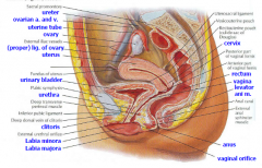
Female pelvic viscera |
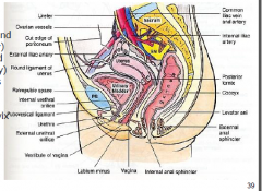
Female urethra is short. Vaginal canal lies b/w urethra and bladder (anteriorly) and anal canal and rectum (posteriorly) |
|
|
Orientation of uterus |
The uterus is normally bend anteriorly (anteverted). Turning backward of the uterus is called retroverted uterus; usually seen in older women. |
|
|
Broad ligament |
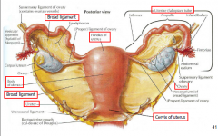
Secures Uterus, uterine tubes and ovaries to lateral pelvic walls. Double layer of peritoneum. |
|
|
Parts of uterus |
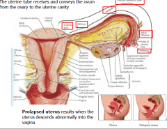
Uterine tube recieves and conveys the ovum from the ovary to the uterine cavity Prolapsed uterus results when uterus descends abnormally into vagina. |
|
|
Erectile tissues in female |
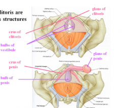
Homologous to erectile tissues of males. Penis and clitoris are homologous structures. |
|
|
Female genitalis |
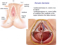
Ischiocavernosus muscle covers crus of clitoris. Blulbospongiosus muscle covers bulbs of vestibule (the vestibule is the region between the labia minora) |
|
|
Innervation of pelvis and perineum SEXUAL FUNCTION |
Parasympathetic: S2-S4, erection- relaxation of smooth muscles in the walls of arteries allowing blood to fill erectile tissue. Sympathetic T12-L2, ejaculation in males, contraction of smooth muscles in walls of vas deferens, seminal vesicles, prostate and urethra orgasm in females- rhythmic contractions of the smooth muscles in the walls of vagina Voluntary (somatic) control (S2-S4, pudendal n.) contract bulbospongiosus and ischiocavernosus to assist in erection Somatic Sensory (S2-S4 pudendal N) general sensation to external genitalia for arousal |
|
|
Innervation for pelvis and perineum DEFECATION and URINATION |
Parasympathetic (S2-S4): relaxation of smooth muscle internal sphicters of bladder and rectum, contraction of smooth muscles in walls of these organs Sympathetic (T12-L2): contraction of smooth muscle internal sphincter to maintain continence. Voluntary (somatic) control (S2-S4, pudendal N,): contraction of striated muscle external sphincters to maintain continence Visceral Sensory: stretch for defecation and urination reflexes and pain/cramping. |
|
|
Pelvic Pain Line |
Above Pelvic pain line: returns via sympathetic nerves to T12-L2 Below Pelvic pain line: returns via parasympathetic nerves to S2-S4 |
|
|
Hormones |
Functions of ovaries, uterus, and testies are controlled primarily by hormones- ovulation, spermatogenesis, uterine contractions during menstration and childbirth. |