Reading...
![]()
Play button
![]()
Play button
![]()
Use LEFT and RIGHT arrow keys to navigate between flashcards;
Use UP and DOWN arrow keys to flip the card;
H to show hint;
A reads text to speech;
7 Cards in this Set
- Front
- Back
|
What is the difference between methemoglobinemia and sulfhemoglobinemia?
|
Sulfhemoglobinemia is a rare condition that can
result from exposure to any substance containing a sulfur atom with the ability to bind to hemoglobin. Cases of sulfhemoglobinemia have been reported from ingestions of phenacetin, dapsone, and sulfonamides.1-3 Sulfhemoglobinemia should be considered in cases presenting with oxygen desaturation and cyanosis, especially if methemoglobinemia can be excluded. Unlike methemoglobinemia, which is reversible with a known antidote, methylene blue, sulfhemoglobinemia is irreversible with no known antidote. Exposure to exogenous oxidizing drugs and their metabolites (such as benzocaine, dapsone and nitrates) may accelerate the rate of formation of methemoglobin up to one-thousandfold, overwhelming the protective enzyme systems and acutely increasing methemoglobin levels. Other classical drug causes of methemoglobinemia include antibiotics (trimethoprim, sulfonamides and dapsone[2]), local anesthetics (especially articaine and prilocaine[3]), and others such as aniline dyes, metoclopramide, chlorates and bromates. Ingestion of compounds containing nitrates (such as the patina chemical bismuth nitrate) can also cause methemoglobinemia. |
|
|
What are Dohle bodies?
|
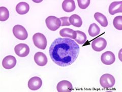
Döhle bodies appear as single or multiple light blue or gray staining areas in the cytoplasm of a neutrophil. They are rough endoplasmic reticulum containing ribonucleic acid (RNA) and may represent localized failure of the cytoplasm to mature. Döhle bodies are found in infections, poisoning, burns, and following chemotherapy.
|
|
|
What are Pappenheimer bodies?
|
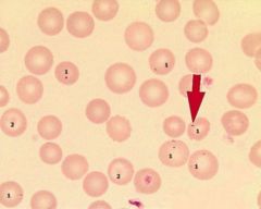
Pappenheimer bodies are abnormal granules of iron found inside red blood cells on routine blood stain.[1] They are a type of inclusion body formed by phagosomes that have engulfed excessive amounts of iron. They appear as dense, blue-purple granules within the red blood cell and there are usually only one or two, located in the cell periphery. They are seen in diseases such as sideroblastic anemia, hemolytic anemia, and sickle cell disease.
|
|
|
What are Howell-Jolly bodies?
|
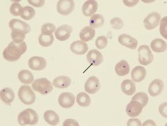
Nuclear remnants (DNA) seen in erythrocytes with asplenia, hemolytic anemia, megaloblastic anemia, hereditary spherocytosis, and myelodysplastic syndrome (MDS).
|
|
|
Buttock cells are associated with which hematologic malignancy?
|
Commonly follicular lymphoma, less so mantle cell
|
|
|
Which two cell surface markers can help differentiate CLL from follicular lymphoma?
|
CD5 and CD23 are negative in follicular lymphoma.
|
|
|
What is platelet satelittism?
|
Clumping of platelets to the Neutrophils in a rosette formation is known as ‘Platelet Satellitism’. In 1963 this in-vitro phenomenon was reported for the first time by Field & MacLoad in peripheral blood collected in EDTA 1. Since then around 100 cases have been reported in literature 2. The mechanism of this platelet adhesion to PMN is not clearly understood, but there is ample evidence that autoantibodies directed to the glycoprotein IIb/IIIa complex on the platelet membrane, as well against the neutrophil Fc receptor, are involved 3.
There is a possibility that the epitopes for those antibodies are usually hidden in leukocyte and platelet membranes and that membrane changes induced by EDTA expose them; which would explain why satellitism is observed exclusively with this specific anticoagulant. Platelet satellitism being a rare finding, as only about 100 cases have been described in the literature concerning platelet satellitism.6, may be seen in any age; although most of individuals revealing this phenomenon are asymptomatic. The finding has been reported in a variety of clinical conditions, such as pregnancy, autoimmune disorders, Behcet’s disease, thromboembolism and malignant disorders like mantle cell lymphoma |