Reading...
![]()
Play button
![]()
Play button
![]()
Use LEFT and RIGHT arrow keys to navigate between flashcards;
Use UP and DOWN arrow keys to flip the card;
H to show hint;
A reads text to speech;
45 Cards in this Set
- Front
- Back
- 3rd side (hint)
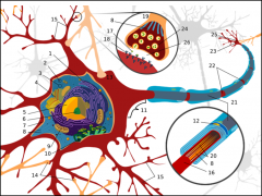
Name it, bitchees!
|
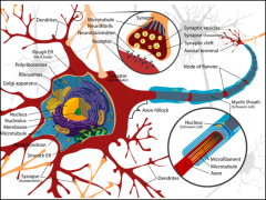
BOOM! Headshot
|
|
|
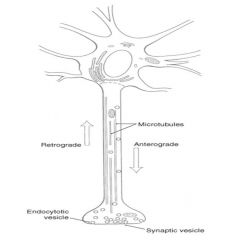
Which method of transport (antro or retrograde) can only move fast? Which one causes your brain to get messed up via RABIES?!
|

|
|
|
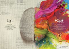
Low yield fun fact: Which side of the brain has more neurons?
|
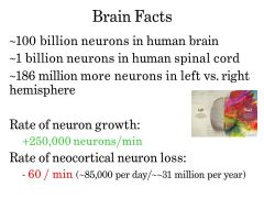
Left side
|
|
|

WTF... What the eff is the difference between a
o Nerve o Neuron o Nerve fiber o Neurites o Neuropil |
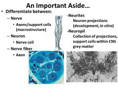
Nerve= Axons/support cells (macrostructure)
Neuron= Nerve cell Nerve fiber = Axon Neurites = Neuron projections (development, in vitro) Neuropil= Collection of projections, support cells within CNS grey matter |
|
|
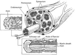
Look at the different layers of a peripheral nerve-
1.) Name them 2.) What are the different layers ? |
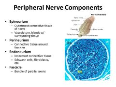
• Epineurium
– Outermost connective tissue of nerve – Vasculature, blends w/ surrounding tissue • Perineurium – Connective tissue around fascicles • Endoneurium – Innermost connective tissue – Schwann cells, fibroblasts, etc. • Fascicle – Bundle of parallel axons |
|
|
|
How Do Neurons “Know” Where to Go?
|
Adhesive gradients (Non-specific outgrowth cues, or specific pathway)
Contact guidance (Response to contact with substrate) Diffusible cues -Chemotaxis |
|
|
|
NETRIN signaling has been well established as a soluble molecule that ATTRACTS axons to the midline
On the other hand, ROBO and SLIT are important to midline crossing: 1. Robo is a receptor mediating REPULSION 2. Slit is the ligand expressed in the MIDLINE Commissural/Crossing axons don’t have high levels of Robo. Why does this make sense? What happens the robo levels after the axons cross the midline? |
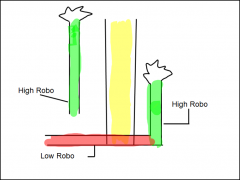
This is because they have a product (“comm”) that keeps expression of Robo low (comm may force Robo into a degradation pathway)
Once they cross the midline, Robo levels are increased and they will therefore not recross |
|
|
|
What are the intracellular pathways important to axon guidance?
|
Calcium
Rho signaling Cyclic nucleotides |
|
|
|
SUPER HIGH YIELD::: What are the (4) classes of guidance molecules??
|
1. Early Patterning
2. Neurogenesis and neuronal fate specification 3. Neuronal Migration 4. Neuronal Contact Formation -Pre-synaptic axon terminals and post synaptic contacts (ie dendritic spines) -The axonal growth cone- better studied than its dendritic counterpart Molecules important to axon guidance during development are likely important to post-injury repair and synaptic plasticity |
|
|
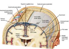
Which one of the meninges here is the only part that touches the brain?
|
PIA MATER
**ps Inferior and superior saggital sinuses are also important--> drains the blood in the brain |
|
|
|
What is it about a lumbar puncture that makes it so much safer than a thoracic or cervical punture?
|
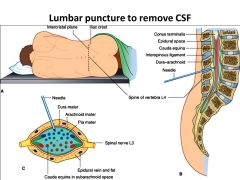
CNS DOES NOT REGENERATE, PNS does
|
|
|

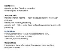
What is so special about each region? What are they usually associated with?
|
|
|
|
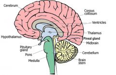
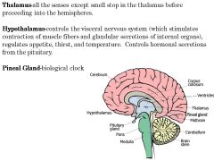
All of the senses (EXCEPT SMELL) stop in the _______ before proceeding into the hemispheres.
_______ controls the visceral nervous system & hormonal secretions. ________ is the biological clock |
|
|
|

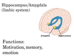
When you are really scared of something, what gets activated
|
|
|
|

If you are a Tony Montana, which part of his brain would be activated right now?
|

|
|
|
|
There are two different longitudinal axis of the brain. On is relative to the forebrain , the other _________
|
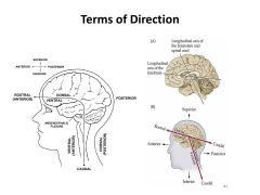
brain stem and spinal chord
|
|
|
|
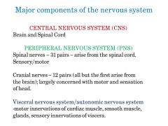
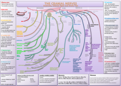
Which major component of the nervous system is largerly concerned with motor/ sensation of the head?
|
|
|
|
holy shjt
|
Just know there is 12
|
|
|
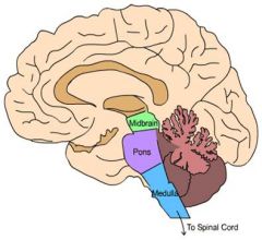
_____ connects with spinal chord... respiration & HR
___relays between cerebral hemisphreres & cerebellum _____ Relay between cerebral cortex and spinal chord. Also involved with visual & auditory reflex patterns. ________ involved in muscle coordination, balance; writing and walking. |

|
|
|
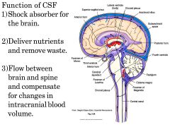
As you can see, cerebral spinal fluid has many SUPER important functions... (shock absorber, nutrient deliver, and homeostasis of blood volume). So what all makes up the VENTRICULAR SYSTEM that is filled with CSF
|
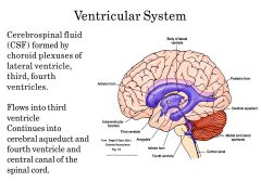
Choroid plexus: Lateral, third and fourth ventricles
flows via 3rd--> cerebral aqueduct--> 4th--> central canal of spinal chord |
|
|
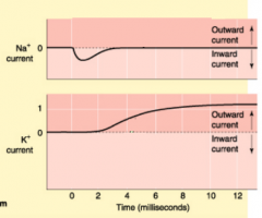
Which one is TTX poisoning and which one is TEA poisoning? How can you tell which is which, and why is this useful?
|
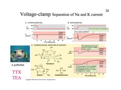
TEA blocks K+ current, revealing Na+ current
TTX(pufferfish) blocks Na+ current, revealing K+ current |
|
|
|
What are the two different driving forces determing flux and direction?
|
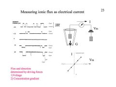
Remember current is the movement of positive charges...
|
|
|
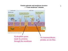
Look at this picture and think carefully about different membrane proteins....
DEFINE the terms “Active” and “Passive” transport mechanisms. DESCRIBE the difference between Na/K pump and ionic channels. |

|
|
|
|
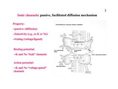
DEFINE the terms “gated” and “non-gated” channels and DESCRIBE the four types of channels contribute to resting and action potential.
|
|
|
|
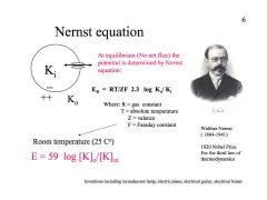
DEFINE electrochemical equilibrium, DESCRIBE the Nernst equation and USE it calculate trans-membrane potentials
|
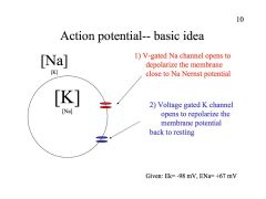
*Don't forget, at rest ALL NERVE CELLS ARE NEGATIVELY CHARGED
resting potential about -60 mV |
|
|
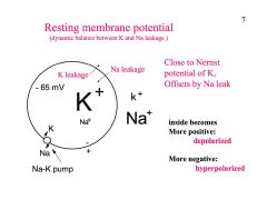
DEFINE membrane “resting” and “action” potential,
|
|
|
|
|
DEFINE conduction velocity and DESCRIBE the factors (space and time constant, myelination) that can change conduction velocity.
|
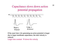
Faster:
* larger diameter (aka larger SPACE/ length constant) good! *smaller capacitance (myelination) (aka smaller TIME constant) * * |
|
|

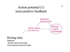
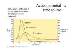
DESCRIBE different phases of action
potential-- initial phase, overshoot, repolarization |
|
|
|

Which ion channel SPECIFICALLY can make the difference between ABSOLUTE and RELATIVE refractory period?
|
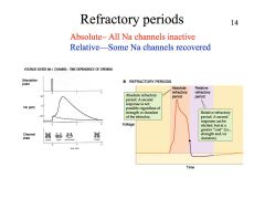
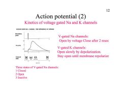
Don't forget this is made possible because there are 3 different states of voltage gated Na channels:
closed open and inactive |

No matter HOW MUCH your coach is yelling at you, if it still can't get the team to respond, this would be the ABSOLUTE refractory period...
All Na channels INACTIVE. But once some Na channels become ACTIVE again, and he yells at you loud/long enough, he might get you to do what he wants... (relative refractory period) |
|
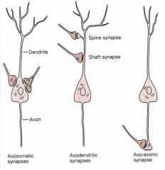
Which one of these synapses is INHIBITORY? Excitatory?
|
Axosomatic synapses
Axon to cell body Inhibitory Axodendritic synapses Axon to dendrite Excitatory Axo-axonic synapses Axon to terminal endings Presynaptic inhibition Reduced polarity |
|
|
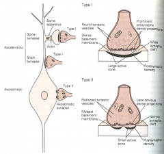
Which one of these is the GABA-ergic/ inhibitory synapse? Gray I or Gray II
|
GRAY type 2!!!
(contacts the cell body) |
1. Gray type I
Glutamatergic/excitatory Typically contact on spines & less commonly contact the shafts of dendrites 2. Gray Type II GABA-ergic/ inhibitory Contact the cell body |
|

Seriously... stare at this picture for a long ass time.... UNDERSTAND the contribution of the UPS (ubiquitin-proteosome) and AL (autophagy-lysosome) pathway. What types of dieseases occur when these pathways are no longer working??
|
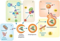
(1) reduced autophagy induction (alzheimers)
(2) enhanced autophagy repression (Huntington’s) (3) altered cargo recognition (Huntington’s, Parkinson’s) (4) inefficient autophagosome/lysosome fusion, (Huntington’s, spinal muscular atrophy) (5) inefficient degradation of the autophagic cargo in lysosomes. (Lysosomal storage disease) |
Examples of neurodegenerative disorders for which cross-talk has been shown to be relevant are indicated in the red boxes.
MVB, multivesicular bodies CMA, chaperone-mediated autophagy UPS, ubiquitin proteasome system AD, Alzheimer's disease HD, Huntington's disease PD, Parkinson's disease FTD, frontotemporal dementia ALS, amyotrophic lateral sclerosis SMA, spinal muscular atrophy |
|
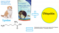
What role does lysine play during normal cell apoptosis/ cells death?
|
UBIQUITIN CONJUGATION PATHWAY
|

|
|
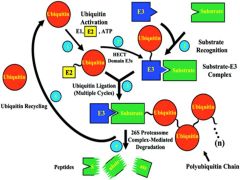
What the EFF is going on here?
|

|
|
|
|
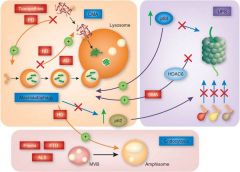
DONT CONFUSE YOURSELF WITH STUPID FLOWCHARTS-- Describe the relationship between neurodegenerative diseases and cell death mechanisms…
|
In neurodegeneration, cell death is associated with accumulation of proteins that become toxic and impair cellular quality control mechanisms, leading to apoptosis.
|
|
|
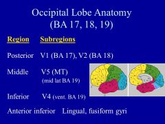
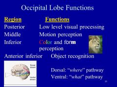
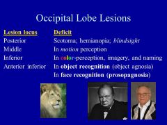
What happens in the OCCIPITAL lobe? What happens with a lesion in this area?
|
|
|
|
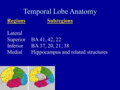
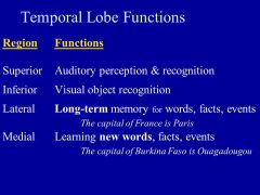
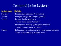
What happens in the TEMPORAL lobe? What happens with a lesion in this area?
|
|
|
|
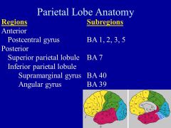
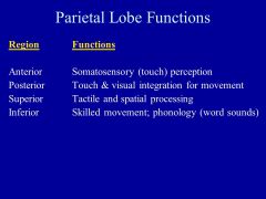
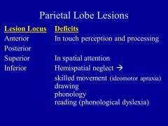
What happens in the PARIETAL lobe? What happens with a lesion in this area?
|
|
|
|
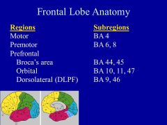
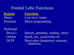
What happens in the FRONTAL lobe? What happens with a lesion in this area?
|
|

|
|
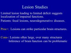
What are the pros/ cons of using LESION studies for figuring out cortical functions?
|
|
|
|
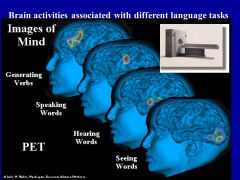
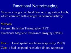
What are the pros/ cons of using FUNCTIONAL NEUROIMAGING studies (PET, fMRI) for figuring out cortical functions?
|
|
|
|
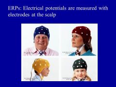
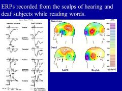
What are the pros/ cons of using EVENT RELATED POTENTIALS (ERPs) for figuring out cortical functions?
|
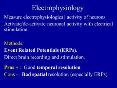
|
More
|
|
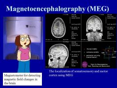
What are the pros/ cons of using MAGNETOENCEPHALOGRAPHY (MEG) for figuring out cortical functions?
|
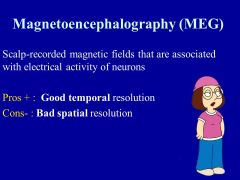
|
|
|
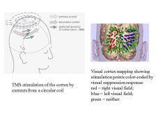
What are the pros/ cons of using TRANSCRANIAL MAGNETIC STIMULATION (TMS) for figuring out cortical functions?
|
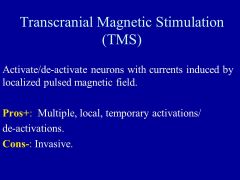
|
|
|
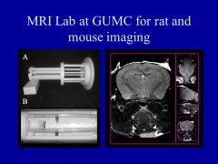
What are the pros/ cons of using ANIMAL STUDIES for figuring out cortical functions?
|
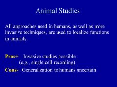
|
|