Reading...
![]()
Play button
![]()
Play button
![]()
Use LEFT and RIGHT arrow keys to navigate between flashcards;
Use UP and DOWN arrow keys to flip the card;
H to show hint;
A reads text to speech;
167 Cards in this Set
- Front
- Back
|
FIRST, WE TALK ABOUT MYELINATING GLIAL CELLS.
|
WOOT!
|
|
|
How many nerves do oligodendrocytes vs schwann cells myelinate?
|
oligodendrocyte- up to 50
schwann cell- only one |
|
|
Hopw can you deduce this function using the cell names?
|
oligodendrocyte- multiple dendrites reaching out and supplying myeline
schwann cell- named after discover |
|
|
Do both axons in the CNS and PNS have nodes of ranvier?
|
yes
|
|
|
What is concentrated at the membrane of nodes of ranvier?
|
sodium channels
|
|
|
NOW WE TALK ABOUT MS!
|
YAY!
|
|
|
BASIC DYSFUNCTION: What happens to myelin to damage it in the CNS?
|
It undergoes an inflammatory reaction if it is made by oligodendrocytes
|
|
|
BASIC DYSFUNCTION: What happens to nerve trasmission when myelin is inflamed?
|
It is impaired or blocked
|
|
|
BASIC DYSFUNCTION: What do we call areas that are demyelinated? Are the borders sharp or blurry?
|
plaques, which are sharply demarcated.
|
|
|
BASIC DYSFUNCTION: Where do plaques tend to show up?
|
in axons that run close to the the pia mater surfaces in the brain and brain stem. PERIVENTRICULAR
|
|
|
BASIC DYSFUNCTION: What happens to the oligodendrocytes in MS?
|
they scar over (called gliosis)
|
|
|
SYMPTOMS OF MS: What is the basic tenet about the symptoms of MS as far as when/where they show up?
|
The symptoms of MS are lost in time and space.
|
|
|
SYMPTOMS OF MS: Why are they lost in space?
|
they have no symmetry and can be sensory and motor
|
|
|
SYMPTOMS OF MS: Why are they lost in time?
|
the signs and sx may come and go.
|
|
|
DIAGNOSIS OF MS: What would a doctor look for as a time and space sign?
|
If 2 or more sensory or motor systems are affected in separate attacks.
|
|
|
BASIC DYSFUNCTION: Are any cranial nerves affected by MS?
|
Only CN I, the optic nerve because it is actually a tract.
|
|
|
MICROBIOLOGY OF MS: What would you expect to find in the CSF in MS?
|
elevated IgG, T-lymphocytes, and normal glucose
|
|
|
MICROBIOLOGY OF MS: wHY WOULD YOU FIND ELEVATED IGg AND LYMPHOCYTES IN THE csf?
|
Because this is a chronic attack that the immune system has seen before.
|
|
|
MICROBIOLOGY OF MS: What is the antigen being targeted in MS?
|
myelin so the antibody is anti-myelin
|
|
|
MICROBIOLOGY OF MS: What gene is associated with MS? (Mnemonic)
|
HLA-DR2 (Dr. needs to look for 2 or more systems affected)
|
|
|
PATHOLOGY OF MS: What is the triad of MS? (mnemonic)
|
Triad of MS is a SI(I)N
Scanning speech Intention tremor Internuclear opthalmoplegia/ Nystagmus |
|
|
PATHOLOGY OF MS: What structure is affected in Internuclear opthalmoplegia/ Nystagmus?
|
the MLF
|
|
|
EPIDEMIOLOGY OF MS: What demographic is most likely to get MS?
|
Caucasion women 20-40 living far from the equator
|
|
|
TREATMENT OF MS: What is it?
|
immunosuppressive therapy and IFN-b aimed at reducing severity and relapse
|
|
|
NOW WE TALK ABOUT GUILLAIN-BARRE SYNDROME
|
GI-AN BARAY!
|
|
|
PATHOLOGY OF GBS: What is attacked in GBS and by what?
|
myeline made from schwann cells by our own immune cells.
|
|
|
PATHOLOGY OF GBS: What event is GBS usually preceded by?
|
respiratory or gastrointestinal illness
|
|
|
PATHOLOGY OF GBS: Why does the immune system attack scwann cells in this case?
|
We don't fully know why, but it has to do with it being very amped up.
|
|
|
SYMPTOMS OF GBS: What is the main symptom produced by GBS?
|
motor weakness with deep, aching pain
|
|
|
SYMPTOMS OF GBS: What is the distribution and progression?
|
bilateral symmetric ascending muscle weakness
|
|
|
PATHOLOGY OF GBS: Which cranial nerves are affected in GBS? Why?
|
CN 5 and 7 because they control a lot of major muscles in the head. (5-chewing 7-smiling)
|
|
|
PATHOLOGY OF GBS: What percentage of people will get facial paralysis?
|
50%
|
|
|
PATHOLOGY OF GBS: What is the major risk of GBS? How many people die of this?
|
demyelination of the phrenic nerve and loss of breathing. Less than 5% of people.
|
|
|
PATHOLOGY OF GBS: Is there a cure?
|
yes, it will go away with time and the immune attack subsiding.
|
|
|
PATHOLOGY OF GBS: What regular recommended doctor thing can cause GBS (think Dhruv)?
|
vaccinations
|
|
|
PATHOLOGY OF GBS: What exact part of the PNS is affected? Why is it only motor?
|
The VENTRAL ROOTS!
|
|
|
PATHOLOGY OF GBS: Can this affect autonomic nerves?
|
Yes, this can cause cardiac dysfunction and decreased regulation of blood pressure
|
|
|
PATHOLOGY OF GBS: Does it present with fever?
|
No
|
|
|
EPIDEMIOLOGY OF GBS: Are males or females more likely to get GBS?
|
they are equally likely
|
|
|
NOW WE TALK ABOUT NERVE DEGNERATION
|
YEAAAH!
|
|
|
What happens to the distal neuron if you sever the axon?
|
it undergoes anterograde degeneration (wallerian degeneration)
|
|
|
What does anterograde mean again?
|
anything moving away from the cell body
|
|
|
What kinds of axons have the capacity to regenerate? (mnemonic with polio)
|
only nerves with scwann cell (why you can partially recover from polio)
|
|
|
How do scwann cells help regenerate?
|
They will grow out a sheath in which the axon can grow into
|
|
|
Can axons in the CNS regenerate?
|
NOOOO! you can get a nobel prize if you discover how.
|
|
|
Peyton manning underwent peripheral nerve degeneration from a herniated disc. Why did it take him out for a whole year?
|
peripheral nerve axons can only regenerate at the rate of 1-3mm/day
|
|
|
Contrast this with how fast can axons transport things in anterograde and retrograde direction?
|
anterograde- 400 mm/day
retrograde- 200 mm/day |
|
|
BACK TO THE BASICS OF THE SPINE and SPINAL CORD!
|
YESHHH!
|
|
|
How many pairs of cranial vs spinal nerves do we have?
|
12 pairs of CN's
31 pairs of spinal nerves |
|
|
What are the two types of GANGLIA we have?
|
sensory and autonomic
|
|
|
Embryologically, why are the sensory cell bodies outside of the CNS? What kind of neurons are these?
|
they are derived from the neural crest cells and are pseudounipolar neurons
|
|
|
Why don't we have somatic motor ganglia? Where are those cell bodies?
|
because those cell bodies are contained in the anterior horn and derived from neural tube cells
|
|
|
Are there cell bodies in the dorsal horn? What kind?
|
Yes, but not of the PNS sensory neurons because they have dorsal root ganglia. (the cell bodies in the dorsal horn are like clarke's nucleus and substantia gelatinosa)
|
|
|
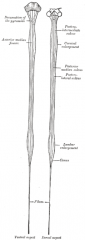
Where does the tapered end of the spinal cord end?
|
L2
|
|
|
What generalization can you make about branches of spinal nerves when you see them in dissection?
|
they are all mixed (sensory AND motor)
|
|
|
SPINAL CORD: What divides the anterior and lateral columns?
|
the lower motor neurons exiting out the anterior horn.
|
|
|
What is the intermediate zone of the spinal cord?
|
The zone in the grey matter between the dorsal and ventral horns.
|
|
|
What is contained in the intermediate zone?
|
mixed sensory and motor cell bodies
|
|
|
What two structures form a spinal nerve?
|
When the dorsal roots join with the ventral roots
|
|
|
Is it easy to see a spinal nerve? Why?
|
No, they are incredibly short because they branch into the 2 rami ver soon afterwards.
|
|
|
What kinds of structures do the dorsal rami innervate?
|
skin of the back, erector spinae muscles, and vertebral joints.
|
|
|
Do either of the rami innervate any viscera? Why?
|
No because those are innervated by the autonomic nerves which are their own crazy thing.
|
|
|
Do the dorsal rami have names?
|
No, none of them have any names.
|
|
|
What structures do the ventral rami innervate? Why?
|
skin and muscles of the trunk and all the limbs because these are anterior to the spinal nerves
|
|
|
Do the ventral rami have names? How many?
|
Yes, each and every one has a name.
|
|
|
What is the brachial plexus composed of?
|
a merging of ventral rami
|
|
|
What is the sciatic/ulnar/radial nerve composed of?
|
composites of different VENTRAL rami.
|
|
|
How would you categorize the ANS? motor/sensory, CNS/PNS
|
motor subset that can be CNS or PNS
|
|
|
What are the 3 main roles of the autonomic NS?
|
To control:
1. Glands 2. The heart (nodes and myocytes) 3. ALL Smooth muscle |
|
|
Are there sensory fibers that take the same course as the autonomic fibers?
|
Yes, but they are not autonomic because they are sensory
|
|
|
What can you generally say about where the pre and post ganglionic autonomic neurons originate?
|
pre ganglionic- SOMEWHERE in the CNS
post ganglionic- SOMEWHERE in the PNS |
|
|
What do the rami split into fairly quickly? (show pic)
|
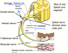
cutaneous and muscular nerves
|
|
|
What kinds of fibers will be found in a cutaneous nerve?
|
all types (sensory, and autonomic motor)
|
|
|
What kinds of fibers will be found in a muscular nerve?
|
Both. (motor for muscle and sensory for proprioception)
|
|
|
NOW WE TALK ABOUT DERMATONES!
|
DERMATASTIC!
|
|
|
breakdown the word dermatome
|
derma- skin
tome- to slice "a slice of skin" |
|
|
what is a dermatome?
|
It is the skin supplied by all the cutaneous branches of a pair of spinal nerves.
|
|
|
How many dermatomes are there? Why?
|
30 or 29. There is no C1 dermatome and you can argue there is no coccygeal dermatome either.
|
|
|
What dermatomes are on the face?
|
the 3 different branches of CN 5
|
|
|
Are the boundaries of dermatomes rigid?
|
No, they generally overlap
|
|
|
How much do they overlap?
|
They go into the adjacent (one over) dermatomes.
|
|
|
Would you be completely numb in one dermatome if you had a lesion there?
|
No because of the overlapping dermaterritories
|
|
|
NOW WE TALK ABOUT HISTOLOGY OF THE SPINAL CORD!
|
THIS IS SPASTIC!
|
|
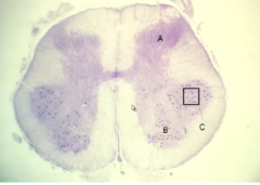
What kind of stain is this? What are you trying to see?
|
A nissl stain to see neuronal rER
|
|
|
Why can you only see dots in the anterior horn cells of the last slide?
|
Because they are cell bodies large enough to be seen in a low mag stain.
|
|
|
What are those cell bodies a part of?
|
They are part of large lower motor neurons
|
|
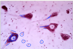
What are the big vs small spots here? WHat are the big ones shaped like and why?
|
Big- LMN cell bodies in a dendritic shape
Small- nissl substance of glial cells |
|
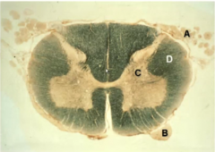
What kind of stain is this and what is it used for?
|
A myelin stain used to achieve contrast
|
|

Name what each letter is
|
A- dorsal root
B- ventral root C- grey matter D- white matter |
|
|
Would you see mixing of grey and white matter in a myelin stain? WHy?
|
Yes because axons have to come out of the grey matter and cell bodies may come out of the white matter.
|
|
|
NOW WE DO A SPINAL CORD REVIEW!!
|
YAY
|
|
|
How many spinal cord segments are there?
|
31
|
|
|
Show the cervical and lumbar enlargements
|
|
|
|
Why are there cervical and lumbar enlargements?
|
because you need extra large roots to innervate the limbs at these areas
|
|
|
Which segments have the cervical enlargement in them?
|
C5-T1 (brachial plexus)
|
|
|
Which segments have the lumbosacral enlargement in them?
|
L2-S3 (rami for the lower limbs)
|
|
|
Which spinal cord segmentscontain preganglionic sympathetic neurons?
|
T1-L2
|
|
|
Which spinal cord segmentscontain preganglionic parasympathetic neurons?
|
S2-S4
|
|
|
Where do the spinal cord vs the meninges end?
|
L2 vs S2
|
|
|
Which spinal cord segments are in the conus medullaris?
|
All the sacral segments.
|
|
|
What are the cauda equina structurally?
|
all spinal roots below L2
|
|
|
NOW WE TALK ABOUT THE ORGANIZATION OF GREY MATTER IN THE SPINAL CORD!
|
YAY THANKS
|
|
|
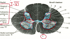
Who organized the grey matter of the spinal cord and how?
|
A scientist named Rexed did it and grouped similar types of neurons found there as laminae.
|
|
|
Show the spinal cord segment and divide it into the dorsal, intermediate, and anterior horns.
|
|
|
|
Which Rexed laminae are in the dorsal horn?
|
1-6
|
|
|
Which Rexed laminae are in the intermediate zone?
|
7
|
|
|
Which Rexed laminae are in the anterior horn?
|
8 and 9
|
|
|
Where are most of the cell bodies of the motor neurons going out to the ventral root going to be?
|
in the more lateral laminae 9
|
|
|
What does Rexed laminae 10 do?
|
No one knows, it just makes up the mysterior middle part
|
|
|
So what is found in Rexed 8 in the anterior horn?
|
the commissural nuclei
|
|
|
What types of nuclei are found in the intermediate zone?
|
the interomedial nuclei (for autonomic neurons)
Clarke's nucleus |
|
|
Will all segments have interomedial nuclei? Which ones?
|
No, only the ones with autonomic output. T1-L2, S2-S4.
|
|
|
What kind of info is carried in Clarke's nucleus and from which parts of the body?
|
proprioception from the lower limbs
|
|
|
What spinal segments have Clarke's nuclei? How do you remember this?
Mnemonic for Clarke's nucleus? |
T1-L2
same as sympathetic levels except they have nothing in common Lewis and Clarke were travelers by LEGS who liked to take the more SYMPATHETIC routes STRAIGHT ACROSS to travel America. |
|
|
NOW WE TALK ABOUT THE CLASSIFICATION OF MUSCULAR AND CUTANEOUS NERVES
|
YAY!
|
|
|
What criteria is used to rank the different types of muscular and cutaneous sensory nerves?
|
conduction velocity/fiber diameter
|
|
|
How do we name muscular nerves? Which has largest fiber diameter?
|
roman numerals I-!V
I is the fastest |
|
|
How do we name cutaneous nerves? Which has the fastest conduction velocity?
|
A-alpha, A-beta, A-delta, and C
A-alpha is the fastest |
|
|
so basically which ones are the fastest?
|
The ones earlier in line (I and A-alpha)
|
|
|
What are the thickest types of fibers going into the dorsal horn?
|
The ones coming from the muscles for proprioception
|
|
|
What are they called? Give them with their classifocations.
|
muscle spindle- Ia
golgi tendon organ- Ib |
|
|
How do you remember that muscle spindles are Ia?
|
They were taught first in the Najeeb lecture and they are the most famous proprioceptors
|
|
|
What force do the muscle spindles vs golgi tendons measure in the muscle? (what makes them fire more?)
|
muscle spindle- muscle stretch
GTO- muscle force |
|
|
What are the second thickest sensory fibers we have? (classification and modality)
|
A beta fibers (cutaneous)
For the touch modalities (touch, vibration, and pressure) |
|
|
What are the thinnest sensory fibers (think about what is slowest to reach us)?
(classification and modality) |
A-delta and C fibers for cutaneous (mostly) pain and temperature
|
|
|
Which fibers are responsible for fine touch?
|
A-beta (cutaneous) fibers
|
|
|
Which sensory fibers give collaterals to the dorsal column?
|
Ia, Ib (proprioception)
A-beta (fine touch) |
|
|
Show which types of fibers are "very interested" in synapsing in the dorsal horn and which aren't.
|

the pain and temperature fibers
|
|
|
What does this mean for our ability to locate where those sensations are coming from?
|
It is diminished (many entrances to one destination or vice versa)
|
|
|
Between A delta and C fibers, which one will have more synapses? Why?
|
C fibers because they are slower and more primitive and also cater to the viscera, creating dull, diffuse pain.
|
|
|
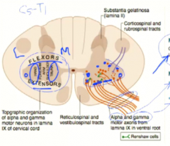
What are the two different type of efferent fibers coming out of the anterior horn?
|
alpha and gamma motor neurons
|
|
|
What will increasing gamma motor neuron firing do?
|
Increase muscle spindle contraction, making them MORE SENSITIVE TO STRETCH
|
|
|
What is added and where to the anterior horn in the spinal enlargements?
|
There will be more grey matter in the lateral aspect of the ventral horn.
|
|
|
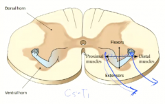
Show where the flexors vs extensor alpha motor neurons originate in the anterior horn.
|
|
|
|
Show a picture of an arm superimposed to the ventral horn to remember proximal/distal and flexor/extensor.
|
|
|
|
Do the dorsal horns expand in the spinal enlargements? How much?
|
Yes because they have more sensory info to take in, but not as much as the ventral horns.
|
|
|
NOW WE TALK ABOUT THE AUTONOMIC NERVOUS SYSTEM!! STARTING WITH THE SYMPATHETIC SYSTEM!
|
yay!
|
|
|
How can you tell if a neuron originated from neural tube or neural crest cells? (1 determinant)
|
whether or not their cell body is within the CNS.
|
|
|
What rexed laminae/zone will a preganglionic sympathetic neuron cell body come out of?
|
rexed laminae 7 or the intermediate zone
|
|
|
How do I use the cadaver to remember that preganglionic sympathetic neurons are short?
|
Remember the sympathetic chain that I saw?
|
|
|
What is the one sympathetic exception?
|
The adrenal medulla.
|
|
|
Where do sympathetic neurons go?
|
EVERYWHERE!
|
|
|
What effect does sympathetic stimulation have on our viscera?
|
1. inhibition digestion
2. cardiac stimulation 3. bronchodilation 4. release of glucose |
|
|
What effect does sympathetic stimulation have on our body wall?
|
1. constriction of blood vessel smooth muscle
2. increases sweat gland function |
|
|
What are the three types of sympathetic ganglia?
|
1. Prevertebral
2. Paravertebral 3. Adrenal |
|
|
Where can you find the prevertebral ganglia chain? What are they named for?
|
In front (PRE) of the spinal column inferior to the diaphragm and they are named for the blood vessel they run along.
|
|
|
Where can you find the paravertebral ganglia chain?
|
On either side of the spinal column
|
|
|
What structures does the prevertebral ganglia supply? How did you deduce this?
|
the viscera (you know this because they are below the diaphragm with the viscera)
|
|
|
What structures does the paravertebral ganglia supply? How did you deduce this?
|
The body wall (they are so nice and organized on the cadaver that it couldn't possibly be the viscera)
|
|
|
What is another name for the adrenal ganglia?
|
The cromaffin cells because they release catecholamines in response to sympathetic pregaglionic stimulation
|
|
|
What sympathetically controlled structures does the face have in common with the rest of the body walls?
|
vascular SM and sweat glands
|
|
|
What additional sympathetic structures does the face have? (think Horner's) (2)
|
2 smooth muscles of the orbit
superior tarsal muscle to raise the eyelids pupillary dilator muscle |
|
|
What spinal level gives sympathetic control to the face?
|
T1
|
|
|
What ganglion supplies sympathetic control to the whole face?
|
The superior cervical ganglion
|
|
|
Which chain of ganglia does the face sympathetic fibers travel up? How do you know this? (para/pre)
|
The prevertebral because this chain supplies body walls
|
|
|
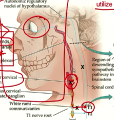
Can you show where this ganglion is?
|
|
|
|
What kind of tumor could compress the superior cervical ganglion? What disorder would this cause?
|
a neck tumor. This would cause ipsilateral horner's syndrome
|
|
|
Is the autonomic chain a 2 neuron pathway? How many are there?
|
NO! We haven't been told the whole truth. There are 3!
|
|
|
Where is the 3rd one from and where does it connect? What is it called?
|
Descending HYPOTHALAMIC tracts ending on preganglionic fibers.
|
|
|
Explain all the symptoms of horner's synrome and give it's sidedness.
|
ipsilateral:
Ptosis- droopy eyelid Anhydrosis- loss of sweating Miosis- constricted pupil |
|
|
What is so clinically significant about horner's syndrome?
|
It indicates a lesion anywhere from the sympathetics of the hypothalamus to the superior cervical ganglion.
(including the descending hypothalamic fibers, making it like a babinski's sign) |
|
|
NOW WE TALK ABOUT THE PARASYMPATHETIC SYSTEM!
|
LAST ONE OF THIS LECTURE!
|
|
|
If the sympathetics are thoracolumbar, what are the parasympathetics?
|
craniosacral
|
|
|
Which cranial and which sacral levels?
|
cranial- CN 3,7, 9, 10
sacral- S2-4 |
|
|
What parasympathetic function does each cranial nerve do?
|
CN 3- edinger westphal- constrict pupils to light on both sides
CN 7- salivation CN 9- same as 10 CN 10- visceral functions |
|
|
What parasympathetic function does the sacral nerves have?
|
bladder and bowel (hindgut) emptying
|
|
|
ANATOMY: Do the PS and S controlling neurons in the CNS run in a predictable bundle/tract?
|
sympathetics do, but we don't know about the PS's yet
|