![]()
![]()
![]()
Use LEFT and RIGHT arrow keys to navigate between flashcards;
Use UP and DOWN arrow keys to flip the card;
H to show hint;
A reads text to speech;
17 Cards in this Set
- Front
- Back
|
eye diagram |
|
|
|
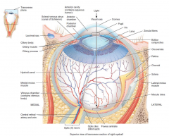
what are the three layers of the eye? |
fibrous tunic
vascular tunic
retina |
|
|
describe the fibrous tunic |
fibrous tunic
superficial layer consisting of the cornea (anterior) and sclera (posterior), the junction of these is called the scleral venous sinus, into which aqueous humor drains
cornea is a transparent epithelium covering the iris curved - helps focus light on the retina receives oxygen from outside air
sclera white of the eye, a layer of dense connective tissue
site of attachment for extrinsic eye muscles |
|
|
describe the vascular tunic |
vascular tunic
middle layer composed of choroid, ciliary body and iris
choroid posterior portion highly vascularised - supply the retina contains melanocytes producing melanin which absorbs stray light, keeping images on the retina sharp and clear
ciliary body extends from ora serrata (jagged anterior margin of the retina) to just posterior to the scleral venous sinus consists of ciliary processes and ciliary muscle
ciliary processes secrete aqueous humor from capillaries extend zonular fibres that attach to lens
ciliary muscle circular band of smooth muscle that contracts and relaxes to alter the shape of the lens for near/far vision
lens is transparent and avascular, behind the pupil and the iris, within the cavity of the eyeball |
|
|
describe the iris |
* suspended between the cornea and the lens, attached to ciliary processes |
|
|
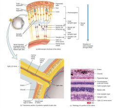
diagram and histological section of the retina |
|
|
|
describe the layers of the retina |
pigmented layer (outer)
sheet of melanin-containing epithelial cells - helps to absorb stray light
between choroid and the neural part
neural layer (inner) containing, from outer to inner…
photoreceptors - rods (black and white) and cones (colour - blue, green and red cones)
outer synaptic layer
bipolar cell layer - modifies signals from photoreceptors, contains bipolar cells, amacrine cells and horizontal cells
inner synaptic layer
ganglion cell layer - axons of ganglion cells extend posteriorly to the optic disc (‘blind spot’ - site where optic nerve, central retinal artery and central retinal vein exit the eyeball) to exit the eyeball as the optic nerve |
|
|
cavities and chambers f the eye |
the lens divides the interior of the eyeball into anterior cavity and vitreous chamber
anterior cavity
divided into anterior chamber (between cornea and iris) and posterior chamber (between iris and zonular fibres/lens)
filled with aqueous humor - transparent fluid that nourishes cornea and lens which is constantly supplied from ciliary processes of the ciliary body into the posterior chamber, and drains from the anterior chamber via the scleral sinus
vitreous chamber
between lens and the retina
contains vitreous body - jelly-like substance that holds the retina flush against the choroid giving the retina an even surface, with some phagocytes to keep it clear |
|
|
picture and description of rods and cones |

each rod and cone is divided into an outer segment, inner segment (with nuclear region) and a synaptic zone
saccules and discs in the outer segment contain photosensitive compounds that react to light to initiate APs in the the visual pathways
rods predominate outside the fovea
images of objects are focused on the retina, the light rays striking the retina generate APs in the rods and cones, these are conducted to the cerebral cortex where they produce the sensation of vision
|
|
|
explain how light is refracted by the eye
explain long and short sightedness too |
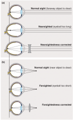
* light is refracted mostly at the anterior surface of the cornea, but also at the anterior and posterior surfaces of the lens * the retinal image is inverted
short-sightedness
eyeball is too long and light focuses in front of the retina
biconcave lens causes light rays to diverge before reaching the eye
long-sightedness
eyeball is too short and light focuses behind the retina
biconvex lens adds to the refractive power of the eye |
|
|
explain accommodation |
* the process by which the curvature of the lens is increased by contraction of the ciliary muscles in order to focus on close objects (less than 6m away) |
|
|
basic explanation of electrical response to light in the eye |
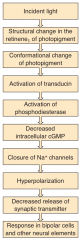
* when light is absorbed by the photosensitive compounds (made up of the protein opsin and retinene1) in the rods and cones their structure changes, triggering a series of events that initiate neural activity |
|
|
explain the ionic basis of photoreceptor potentials |
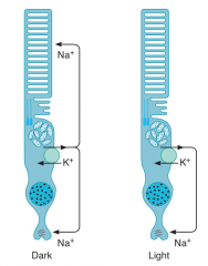
* Na+ channels in the outer segments of the rods and cones in the dark are held open by cGMP
* NaK pumps in the inner segment maintain ionic equilibrium * release of synaptic transmitter is steady in the dark * light leads to conversion of cGMP to 5’-GMP, and some of the channels close, produces hyper-polarisation of the synaptic terminal of the photoreceptor * this hyper-polarisation reduces the release of synaptic transmitter - generating a signal in the bipolar cells that ultimately leads to APs in the ganglion cells, which are transmitted to the brain |
|
|
explain photosensitive compounds |
made up of the protein opsin and retinene1
rods
the photosensitive pigment in the rods is called rhodopsin (contains retinene1 and an opsin called scotopsin), a G-protein linked receptor
light causes a conformational change in the rhodopsin, starting a G-protein linked pathway leads to the conversion of cGMP to 5’-GMP (causing some Na+channels to close)
cones
3 different kinds of cones with a different structured opsin for each
light causes a conformational change in the receptor, starting a G-protein linked pathway leads to the conversion of cGMP to 5’-GMP (causing some Na+channels to close)
melanopsin
a small number of photoreceptors contain melanopsin instead of rhodopsin or cone pigments
the axons of this neurons lead to the suprachiasmatic nuclei and the part of the lateral geniculate nucleus that controls pupillary response to light |
|
|
flowchart from light to neural response |
|
|
|
describe how the image changes throughout the different neural pathways of the eye |
* in a sense the processing of visual images in the retina involves three images |
|
|
psychological and social implication of blindness |
economic * unable to work - loss of employment and income* requires increased care - more likely to need residential care * health risks - falls and fractures * domestic - cooking, eating, dressing, etc., etc. * shopping * finance - bills, bank statements * navigation - safety * hearing impairment * non-verbal communication * social interactions * TV, film and media * isolation * anxiety * depression
|