Reading...
![]()
Play button
![]()
Play button
![]()
Use LEFT and RIGHT arrow keys to navigate between flashcards;
Use UP and DOWN arrow keys to flip the card;
H to show hint;
A reads text to speech;
21 Cards in this Set
- Front
- Back

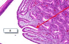
What is this? Identify what the arrows are pointing to.
|
Renal cortex
Top arrow-renal corpuscle middle arrow-convoluted tubules bottom arrow-medullary rays |
|
|
What are the two kinds of nephrons and what are they used for?
|
Cortical nephrons.
Juxtamedullar nephrons have loops of Henle that go into the medulla, seen in the inner stripe of the kidney. |
|
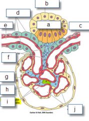
Identify the labels
|
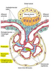
A-macula densa
b-distal tubule c-efferent arteriole d-juxtaglomerular cells e-afferent arteriole f-extraglomerular mesangial cells g-visceral layer-podocytes h-bowman's (urinary) space i-intraglomerular mesangial cells j-glomerular capillaries |
|
|
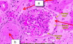
What type of epithelium constitutes the parietal layer of the bowman's capsule?
|
simple squamous
|
|
|
What kind of capillaries make up the glomerular capillaries
|
fenestrating capillaries, the basal lamina is fused with the basal lamina of the podocytes to make the filtration barrier
|
|
|
Explain the filtration barrier: in order of the capillary to the bowman's space
|
Endothelium-
basal lamina pedicels filtration slit |
|
|
What is a pedicel?
|
A pedicel is the secondayr process of a podocyte, it has a contractile diaphragm that is controlled by nephrin that filters out proteins selectively b/c of their negative charge
filtration slits are located between these pedicels |
|
|
What happens if you lose nephrin?
|
minimal change syndrome-nephrotic disease
|
|

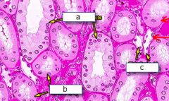
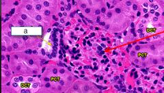
Where is this?
Identify the labels, how can you distinguish them |
Cortex,
A and B are PCT --the PCT has a brush border that has microvili C and D are DCT --are simple cuboidal |
|
Where is this? Identify the labeled structures?
What are the pink areas between the tubules? |

Renal Medulla
Vasa recta-from efferent arteriole A-Collecting duct (you can see the border between the cells) B-Thick Ascend/descend (simple cuboidal) C-thin ascending/descending (sqamous) |
|
|
Where does aldosterone act?
|
Cells of the DCT
|
|
|
Where does ADH act?
|
Cells of the Collecting Duct
|
|
What is this? What is the arrow pointing at?
|
Papillary ducts (ducts of bellini) (arrow) draining into the Minor Calyx
|
|

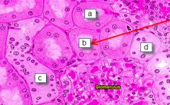
Where is this? What is A pointing to?
What is the red arrow pointing to? What about the light staining cells? |

Renal cortex,
A-macula densa -columnar cells-senses the volume of the blood Red arrow-juxtaglomerular cell? should be distinguished by its granular cytoplasm-secretes renin Light staining cells should be the extraglomerular mesangial cells |
|
|
What is this?
|
renal-vasa recta
|
|

Where is this? What is A pointing to?
What is the red arrow pointing to? What about the light staining cells? |
Renal cortex,
A-macula densa -columnar cells-senses the volume of the blood Red arrow-juxtaglomerular cell? should be distinguished by its granular cytoplasm-secretes renin Light staining/larger cells are the juxtaglomerular cells |
|
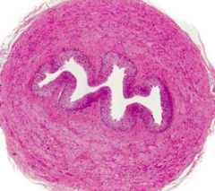
What is this and what are its layers?
|
Ureter:
Mucosa- --transitional epithelium --lamina propria--fibroelastic Muscular layer --inner longitudinal --outer circular ---lower third has an additional outer long layer Adventitia |
|
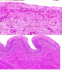
What is this?
Identify what is different about the pictures: What is special about its muscularis |

Urinary Bladder
Top is distended, Bottom is collapsed Transitional epithelium 3 layers of muscularis like the lower third of the ureter Only has adventitia instead of serosa where it touches the ureter |
|
|
What are the three parts of the urethra?
|
Prostatic, Membranous, penile
|
|
What is this? Identify the labels
|
A-distal tubule
b-arteriole |
|
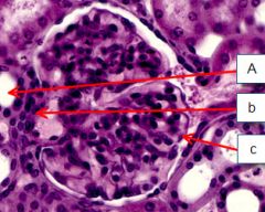
Identify the labels.
|

A-Macula densa in the DCT
b-juxtaglomerular cells C-podocytes |