![]()
![]()
![]()
Use LEFT and RIGHT arrow keys to navigate between flashcards;
Use UP and DOWN arrow keys to flip the card;
H to show hint;
A reads text to speech;
61 Cards in this Set
- Front
- Back
|
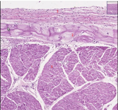
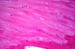
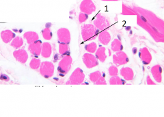
Heart (swine – 90)- 1 endocardium: 2 Purkinje fibers |
|
|
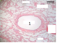
Aorta (slide 19) HE staining |
|
|
1. Haversian canals, 2.osteocytes
|
|
|
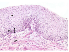
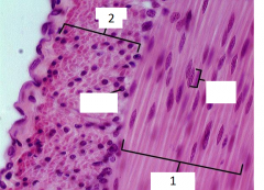
Esophagus (slide 40): Mucosa (1- nonkeratinized stratified squamous epithelium, 2-lamina propria, 3-muscularis mucosae), 4-Submucosa |
|

|
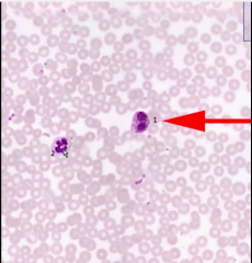
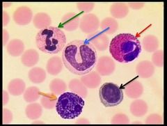
Peripheral blood -- 1-monocyte,3-lymphocyte, granulocytes (2-neutrophilic), 5-platelets (4 rbc)
|
|
|
7Peripheral blood -- granulocytes (1-eosinophilic,, 2-neutrophilic), |
|

|
Peripheral blood arrow: granulocytes ( basophilic)
|
|
|
Thymus - infant (slide 26) 2- Cortex, 3 medulla ej listed , 3 Hassal’s corpuscles - om pekar på en Hassal’s corpuscles = adult
|
|
|
. Thymus - infant (slide 26) |
|

|
Endochondral ossification - epiphysis : CHS:Resting cartilage - CHP: Proliferating cartilage: CHW calcifed cartilage bk:bone deposited onto trabeculae
|
|
|
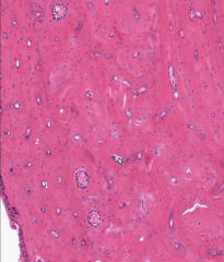
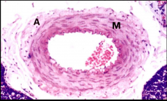
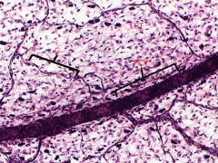
Muscular artery |
|
|
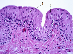
Esophagus 1.nonkeratinized stratified squamous epithelium |
|
|
Trachea 1- pseudostratified ciliated columnar epithelium 2 goblet cells,3 submucosal glands |
|
|
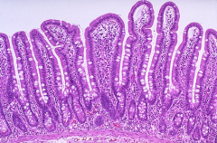
Large intestine (slide 45): 3 intestinal crypts, 2 goblet cells 1 simple columnar epithelium
|
|
|
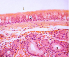
Urinary bladder (slide 53): 2 transitional epithelium, 1 umbrella cells |
|
|
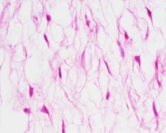
Mesenchymal tissue |
|

|
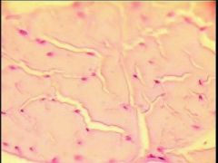
Loose connective tissue – mesothelium, intestine |
|
|
Regular fibrous connective tissue – tendon (Transverse section) |
|
|
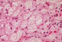
Brown adipose tissue |
|
|
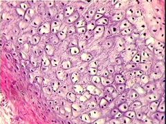
Elastic cartliage - chondrocytes, isogenic groups, perichondrium |
|
|
Fibrocartilage |
|

|
Compact bone (cross section) (slide 12): 1 Haversian canals, 2 osteocytes
|
|
|
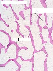
Spongy bone 1.trabeculae 2:Yellow bone marrow |
|
|
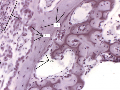
Intramembranous ossification (slide 14) 1. osteocytes, 2.osteoblasts, 3. bone matrix |
|
|
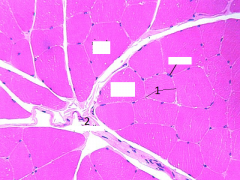
Striated skeletal muscle (cross section) 1.endomysium, 2.perimysium |
|
|
Striated skeletal muscle (cross section) 1.endomysium, 2.perimysium
|
|
|
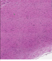
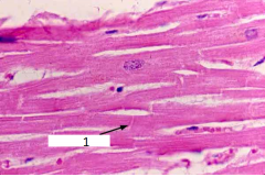
Cardiac muscle (longitudinal section) 1.intercalated disks
|
|
|
Smooth muscle – small intestine (1 longitudinal 2 cross section |
|
|
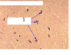
Spinal cord (slide 75): 1 central canal, 2 ependyma
|
|
|
Spinal cord -1 Multipolar (motor) neurons in anterior horns
|
|
|
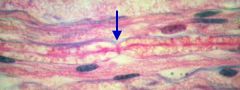
Myelinated nerve fibers Arrow: node of Ranvier |
|
|
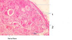
Peripheral nerve (cross section) 1 perineurium (2.Endo)
|
|

|
Peripheral nerve - cross section 1 epineurium, 2 perineurium
|
|
|
Dorsal root ganglion (slide 76): 1 pseudounipolar neurons, 2 satellite cells |
|
|
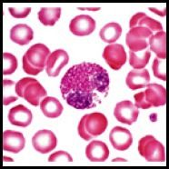
Peripheral blood neutrophilic(green)
|
|

|
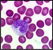
Peripheral blood granulocytes neutrophilic |
|
|
Peripheral blood granulocytes basophilic |
|
|
Peripheral blood granulocytes eosinophilic |
|
|
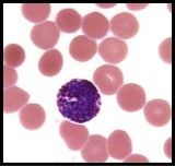
Peripheral blood monocyte |
|

|
Thymus - infant
|
|

|
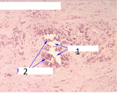
Thymus - adult Arrow: Hassal’s corpuscles
|
|

|
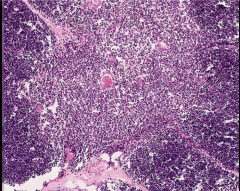
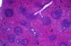
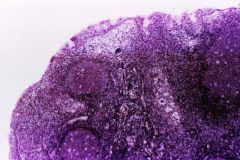
Lymph node 1 cortex, 2 paracortex, 3 medulla, 5 lymphoid follicles, 7 subcapsular, 8 cortical and 9 medullar sinuses, EJ 4 6
|
|
|
Spleen 1 white pulp (+lymphoid follicles) 2 red pulp (spleen sinuses) (ej 3+4) |
|
|
Spleen 4 trabecular artery, 2 white pulp (, 1lymphoid follicles), 3 red pulp |
|
|
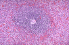
spleen 1 white pulp central arterioles |
|

|
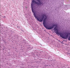
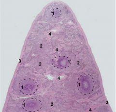
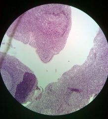
Palatine tonsil 2 lymphoid follicles, 1 stratified squamous nonkeratinized epithelium |
|
|
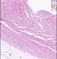
Heart (swine – 90)- 2 pericardium 1 endocardium |
|
|
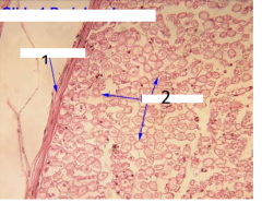
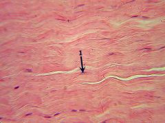
Regular fibrous connective tissue – tendon (longitudinal section) |
|
|
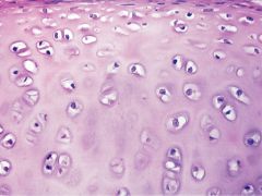
Hyaline cartilage |
|
|
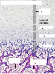
28 Endochondral ossification – epiphysis (slide 13): cartilaginous zones (1 quiescent cartilage, 2 proliferating cartilage, 3 hypertrophic cartilage, 4 calcareous cartilage, 5 trabeculae)
|
|
|
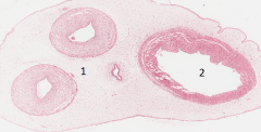
Umbilical cord: 1 muscular arteries 2 muscular vein |
|
|
PCV: postcapillaries |
|

|
Lymphatic vessels |
|
|
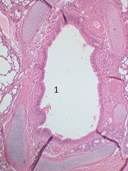
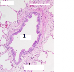
Lung bronchus |
|
|
Lung bronchus |
|
|
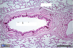
lung bronchiole |
|
|
lung bronchiole |
|
|
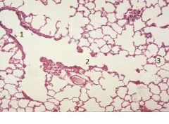
Lung 1 respiratory bronchiole, 2 alveolar duct, 3 alveolus |
|
|
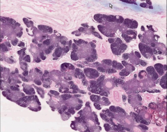
Pharyngeal tonsil |
|
|
Pharyngeal tonsil |
|

|
2 Precapillaries & 1 postcapillaries |