![]()
![]()
![]()
Use LEFT and RIGHT arrow keys to navigate between flashcards;
Use UP and DOWN arrow keys to flip the card;
H to show hint;
A reads text to speech;
45 Cards in this Set
- Front
- Back
|
How many layers of the neural retina are there?
|
10
|
|
|
What are the four types of cell in the retina that are associated with neuroligical function? |
Rods and cones (photoreceptor cells)
Bipolar cells (interneurons that bridge the rods and cones to the ganglion cells)
Horizontal cells and amacrine cells (help integrate signals from photoreceptos over a wide area of the retina)
Ganglion Cells: Dendrites connect with biopolar cells and axon to form layer of nerve fibes that cover the retinas inner surface and exit eye through eptic nerve
Ganglion cells |
|
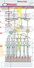
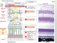
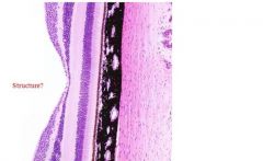
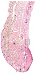
point out each relevant layer of the retina and name what cell types you would find there |
|
|
|
What are the first layer you would find after the pigmented epithelium? |
The rods and cones |
|
|
What are the supporting cells in the retina? |
Muller cells, horizontal cells and amacrine cells |
|
|
retina |
|
|
1. pigmented layer 2. Rod and cone cell layer 3. Outer nuclear layer 4. Inner nuclear layer 5. Ganglionic nuclear layer |
|
|
What makes up the layers of the retina? |
alternating layers of nuclei and processes |
|
|
What is the structure of a cone cell? What is it used for? |
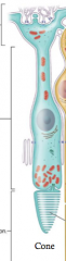
It has a cell body where you have your nucleus and then at the bottom it has a cone like extension where the outer segments are not enclosed
Plumper cells!
Color vision, bright light |
|
|
What is the structure of a rod |
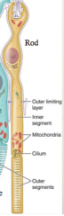
At the base the outer segements are enclosed
Longer cells
low levels of light |
|
|
What are the different visual pigments in the rods vs. cones |
Rods: Rhodopsin
Cones: iodopsin |
|
|
Where are rods found vs. cones? |
Rods: concentrated peripherally
Cones: concentrated in fovea centralis |
|
|
How does light travel in the eye? |

IT starts by going in through the ganglionic cells and then it hits the photerecptors last. The neural impulse then travels from the photoreceptor cells back out
|
|

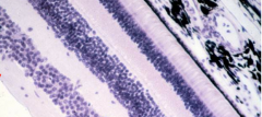
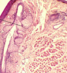
what is occurring here? |
The ora serrata
The point where you have the sensory layer meet the non sensory layer |
|
|
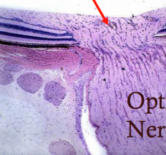
What is the optic disk? |

Where all of the nerves exit the back of the eye. It is a blind spot
Notice how there is no pigmented layer back there. |
|
|
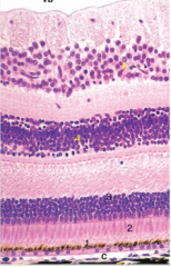
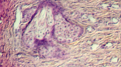
Fovea centralis |
Shallow pit along the temporal side that eptic disc that contains only cone cells
Site of most precise visual actity |
|
|
What surrounds the fovea centralis? |
macula lutea, this has an orange pigament that helps protect the cones |
|
|
Fovea centralis. Its so short because you lack those long rod cells |
|
|
Optic disk |
|

Name the parts of the eye! |
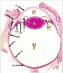
C= cornea AC= anterior cavity PC= posterior cavity CB= ciliary body CP= Ciliary process I = Iris L= lense S= sclera R= retina Ch = Chortoid V= virtous |
|
|
What are the accessory structures of the eye? |
Lacrimal apparatus Eyelids and eyelashes Conjunctiva |
|
|
Where is the lacrimal gland? What is its funciton |
The upper outer edge of your brow. tear production |
|
|
What are the components of the lacrimal apparatus? |
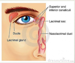
lacrimal gland, superior and inferior canaliculi, the lacrimal sac and the nasolacrimal duct |
|
|
What prevents tears from building up? |
suprerior and inferior canaliculi and they go into lacrimal sac and the nesolacrimal duct |
|
|
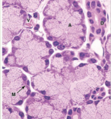
How do the lacrimal glands produce tears? |
merocrine serous glands
They produce tears through exocytosis |
|
|
lacrimal gland |
|
|
What are the chemicals found in tears |
Lactoferrin, lysozone, secrtoty immunoglobin A
A lot of baterial static agents |
|
|
What does the conjuctiva do? |
Covers the outer layer of eye and the inner layer of eye lid. Extremely thin |
|
|
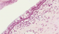
What is the histological appearance of conjuctiva |
Thin mucosa Rests on a loose vascular lamina propri |
|
|
What is the conjuctiva that lines your eyelids called? what is the histological appearance of those? |
palpebral conjuctiva.
It has stratified columnar epithelium, with numerous small cells resembling goblet cells
|
|
|
What is the conjuctiva that coats the anterior suface of eye |
nonkeratinized stratified squamous epithelium |
|
|
What is the structure of the eyelid? |
five layered skin fold.
Thin tskin on the surface lacks hair. Stratified squamous epithelium
This skin layer is underlain by muscular tissue that close the eyes.
Tarsal plate that is made up of dense fibroelastic connective tissue and contains glands |
|
|
What kins of glands are the found in the tarsal plate? |
Sebaceous tarsal glands
Small sweat glands of molle
Sebaceous glands of zeis |
|
|
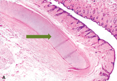
What do meibomian glands |
Secrete subum
Sebaceous glands, holocrine secretion method. Disengrate
|
|
What is that middle arrow pointing to? |
Meibomian glands |
|
|
What are the glands of zeis associated with? |
Sebaceous secreting glands
Surround follicles of eyelashes
Infection can lead to styes |
|
|
Gland of zeis |
|
|
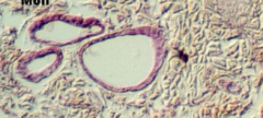
Glands of moll. Modified aprocrine sweat glands |
|
|
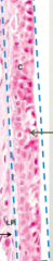
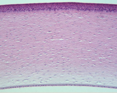
Cornea
Outermost edge is strafied epithelium, loose connective,
Membrane |
|
|
Conjuctiva
Goblet cells
|
|
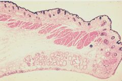
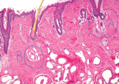
What is this and label |
Eyelid
Look at those tarsal gland |
|
|
What are the three parts of the external ear? |
1. Auricle, External auditory meatus
2. Auricle (pinna) |
|
|
1. What is the histological make up of the auricle?
|
* Review book "funnel like plate of elastic cartilaged sandwiched between two layers of skin"
* According to her there is keratinized stratified squamous epithelium |
|
|
Auricle |
|
|
What are the layers of the cornea |
1. Non keratinized
2. Contains many nerve endings 3. Surface of eye is extremely extremely sensitive 4. Thick basement membrane separating the surface epithelium and the stroma 5. Makes up majority of cornea's thickness 6. 60 layers of collagen bundles and fibroblasts. 7. Thick basement membrane of the corneal endothelium 8. Corneal endothelium. Simple squamous epithelium |