Reading...
![]()
Play button
![]()
Play button
![]()
Use LEFT and RIGHT arrow keys to navigate between flashcards;
Use UP and DOWN arrow keys to flip the card;
H to show hint;
A reads text to speech;
35 Cards in this Set
- Front
- Back
|
Where does the ligament of the head of the femur course between in the ruminant and horse?
|
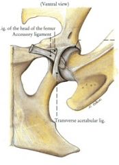
Horse – from the pubic groove near the acetabular notch to the fovea capitis femoris
Ruminant – from the acetabulum to the fovea capitis femoris |
|
|
Where does the accessory ligament of the coxofemoral joint course between? What is it's action? Is it present in horses or ruminants?
|

From the prepubic tendon, through the acetabular notch, to the fovea capitis femoris
Inserts caudal to the ligament of the head of the femur Tightens on abduction Prevents horse from cow-kicking Hip luxations are rare in the horses Not present in the ruminant |
|
|
Which ligament of the coxofemoral joint courses over the acetabular notch in the large animal?
|

Transverse acetabular ligament
|
|
|
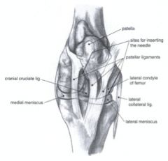
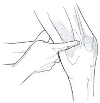
What are the boundaries of injection for the coxofemoral joint of the horse?
|
Difficult to inject - not commonly done (important to know)
Needle is inserted between the cranial and caudal projections of the greater trochanter, 1 cm dorsal to the caudal aspect of the cranial projection Needle is inserted to a depth of 11-12 cm |
|

What is 21 of the equine stifle? 21'?
|

Trochlea of the femur (21)
Medial trochlear ridge (21') – larger Lateral trochlear ridge Trochlear groove |
|
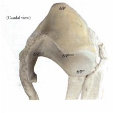
ID everything that goes along with the patella.
What muscle inserts here? |

Patella (69)
Base (69’) Apex (69’’) Cartilage process (69’’’) Parapatellar fibrocartilage (69’’’’) Articular surface (69’’’’’) Quadriceps femoris m. attaches here. Why a patellar fracture is more serious in large animals. |
|
|
What are the only sesamoid bones of the stifle joint in large animals?
|
The patella
|
|
|
What are the patellar ligaments of the equine stifle?
|
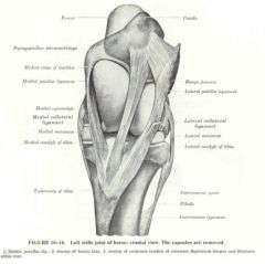
Medial patellar ligament
Courses from the parapatellar fibrocartilage to the tibial tuberosity Lateral patellar ligament Courses from the lateral part of the cranial surface of the patella to the lateral part of the tibial tuberosity Middle (intermediate) patellar ligament Courses from the cranial part of the apex of the patella to the distal part of the tibial tuberosity |
|

Where do the femoropatellar ligaments course between of the equine stifle?
Where do the collateral ligaments course between? |
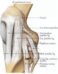
Medial and lateral femoropatellar ligaments
Course between the patella and the femoral epicondyles Prevent medial and lateral patellar luxations Collateral ligaments Medial collateral ligament – courses from the medial epicondyle of the femur to the tibia distal to the medial condyle Lateral collateral ligament – courses from the lateral epicondyle of the femur to the fibula |
|
|
What are the cruciate and meniscal ligaments of the equine stifle? What are the cruciate ligaments named in relation to?
|
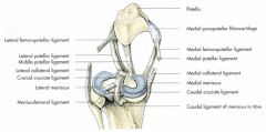
Cruciate ligaments – cranial and caudal (named by attachment to the tibia)
Menisci – medial and lateral Meniscotibial ligaments – cranial and caudal Meniscofemoral ligament -From the lateral meniscus to the femur |
|
|
What are the patellar locking mechanism in the horse?
|
The major part of the pelvic limb stay apparatus.
Components Medial patellar ligament, parapatellar fibrocartilage, patella, middle patellar ligament, trochlea of the femur - resting and gliding surfaces |
|
|
What are the patellar positions?
What muscle is actively involved in one of the positions? |
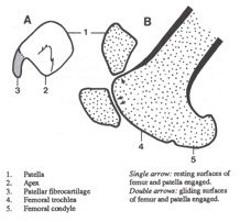
Gliding surface for progression
Resting surface for standing squarely Locking – patella rotates medially and medial patellar ligament locks in place - vastus medialis m. of quadriceps femoris m. contracts to hold the patella in this position (just doesn't require that much energy). Pulls it back just a bit to lock it. To unlock it, the horse needs to extend the limb a bit. |
|
|
What are the synovial joints of the equine stifle? Which communicate, if any?
|
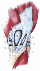
Femoropatellar joint (10)
Femorotibial joint Medial pouch (12) Usually communicates with the femoropatellar joint Lateral pouch (12’) May communicate with the femoropatellar joint |
|
|
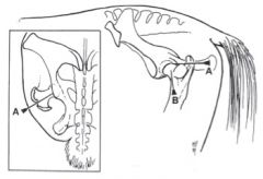
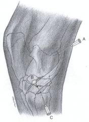
What are the stifle joint injection landmarks of the horse? What is the most straightforward injection?
|
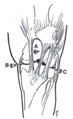
Femoropatellar joint (A) - most straightforward
Inject dorsal to the tibial crest between the middle and medial patellar ligaments Medial femorotibial pouch (B) Inject between the medial patellar ligament and medial collateral ligament approximately 4 cm dorsal to the proximal medial edge of the tibia Technically difficult Lateral femorotibial pouch (C) Inject between the lateral patellar ligament and lateral collateral ligament Less commonly performed |
|
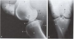
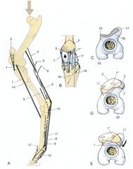
ID the structures of the equine stifle.
Lateral view and caudocranial view |
key
|
|
|
What are three conditions of the equine stifle?
|
Gonitis
Inflammation of the stifle Usually femoropatellar joint Upward fixation of the patella (pictured) Straight legged conformation To unlock the stifle – must extend the leg further Patellar luxation Usually lateral |
|
|
What are the ligaments of the patella in the bovine stifle? In the goat?
|
bovine -
Medial Lateral Middle (intermediate) caprine- 1 patellar ligament |
|
|
What is significant about the acetabular lip in the coxofemoral joint of the horse? What condition does this structure help prevent?
|

Greater depth to acetabular socket
Deeper in the horse - hip dysplasia is rare in the horse |
|
|
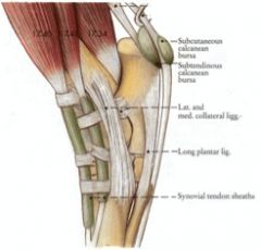
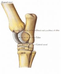
What are the collateral ligaments of the equine tarsus? How many parts do they have?
Which collateral ligament forms a canal for the lateral digital extensor tendon? |
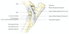
Medial collateral ligament
Long portion – courses from the caudal part of the medial malleolus to the talus, metatarsals II and III, and the distal tarsal bones Short portion – courses from the cranial part of the medial malleolus to the medial surface of the talus and the sustentaculum tali Lateral collateral ligament (pictured) Long portion – courses from the caudal portion of the lateral malleolus to the calcaneus, 4th tarsal bone, and metatarsals III and IV Forms a canal for the lateral digital extensor tendon Short portion – courses from the cranial part of the lateral malleolus to the lateral surface of the talus and adjacent surface of the calcaneus |
|
|
What ligament of the equine crus is involved in "curb"? Where does it course between? What is the clinical condition?
|
Long plantar ligament
Courses from the plantar surface of the calcaneus to the plantar surface of the 4th tarsal bone and metatarsal IV Counteracts the pull of the common calcaneal tendon Clinical condition – Curb Desmitis of the long plantar ligament Thickening of the plantar distal aspect of the tarsus Swelling causes the superficial digital flexor tendon to curve |
|
|
What are the joints and associated synovial pouches of the equine tarsus? Which ones communicate?
|
Tarsocrural (tibiotarsal, talocrural) joint
Greatest amount of mobility 4 pouches: Dorsomedial pouch Medioplantar pouch Dorsolateral pouch Lateroplantar pouch (all pouches here communicate) Proximal intertarsal joint Distal intertarsal joint Tarsometatarsal joint |
|
|
What is the difference between a joint capsule and a joint pouch? What are the clinical significances of the pouches?
|
A joint capsule not held down by a ligamentous or tendinous structure. Can see inflammation in the joint if the pouches are poofing. Allows for injection as well.
|
|
|
Which joints of the equine tarsal joint communicate?
|
Tarsocrural joint communicates with the proximal intertarsal joint
|
|
|
What are the boundaries of the dorsomedial pouch of the tarsocrural joint?
|
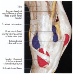
Dorsomedial surface of the hock
Bounded by the tendon of the peroneus tertius, medial collateral ligament, medial malleolus, and cunean tendon |
|
|
What are the boundaries of the medioplantar pouch of the tarsocrural joint of the horse?
|

Between the medial collateral ligament and the deep digital flexor tendon at the level of the medial malleolus
|
|
|
What are the boundaries of the dorsolateral pouch and lateroplantar pouch of the tarsocrural joint of the horse?
|
Dorsolateral pouch
Dorsal to the lateral collateral ligament, between the long and lateral digital extensor tendons and proximal to the short digital extensor muscle Lateroplantar pouch Caudal to the lateral collateral ligament, between the calcaneus and the lateral malleolus |
|
|
What are the boundaries of the tarsal joint injections in the horse? Which is the most important? What artery is crucial to be aware of in that injection?
|
A. Tarsocrural joint
***Dorsomedial pouch – distal and dorsal to the medial malleolus and plantar to the cranial branch of the medial saphenous vein B. Distal intertarsal joint Medial surface of the tarsus; between the palpable distal tubercle of the talus and the space between metatarsals II and III proximally C. Cunean bursa Medial surface of the tarsus; insert needle under the distal border of the cunean tendon Tarsometatarsal joint Plantarolateral aspect of tarsus Insert needle proximal to the head of metatarsal IV |
|
|
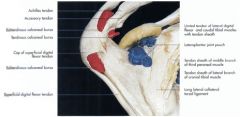
What is thoroughpin?
|
Tarsal sheath encloses the lateral and caudal heads of the deep digital flexor tendon
Tenosynovitis of the tarsal sheath is called thoroughpin Tenosynovitis – inflammation of the synovial membrane of the tendon sheath Fibrous layer of the tendon sheath is also usually involved Distension of the tendon sheath is due to synovial effusion |
|
|
What is spavin? Bog spavin? Bone spavin?
|
Condition of the equine tarsus
Osteoarthritis of the distal hock joints, specifically the distal intertarsal and tarsometatarsal joints Bone spavin – osteoarthritis Bog spavin – synovial joint effusion |
|
|
What is stringhalt? What is the treatment?
|
Condition of the equine tarsus
Hyperflexion of the joints of the pelvic limb Increased proprioception sensitivity to the stifle Treatment: Remove lateral digital extensor muscle insertion so that there is less input to the lateral collateral ligament. |
|
|
What is the stay apparatus of the pelvic limb?
|
Stifle joint
Locking mechanism of the stifle prevents flexion of the stifle joint Hock joint Superficial digital flexor tendon prevents flexion of the hock Below hock – similar to the thoracic limb Suspensory apparatus of the fetlock supports the fetlock joint Proximal check ligament is not present – SDF attaches to tuber calcanei and prevents overextension of the fetlock joint Distal check ligament weak and may not be present |
|
|
What is the stay apparatus of the equine pelvic limb specific to the stifle joint?
|

Locking mechanism of the stifle prevents flexion of the stifle joint
|
|
|
What is the stay apparatus of the equine pelvic limb specific to the hock joint?
|
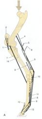
Superficial digital flexor tendon prevents flexion of the hock
|
|
|
What is the stay apparatus of the equine pelvic limb specific to the area below the hock?
|

Below hock – similar to the thoracic limb
Suspensory apparatus of the fetlock supports the fetlock joint Proximal check ligament is not present – SDF attaches to tuber calcanei and prevents overextension of the fetlock joint Distal check ligament weak and may not be present |
|
|
What take the place of the check ligaments in the equine pelvic limb stay apparatus?
|

SDF attaches to tuber calcanei so that there is a tendinous structure coursing down to the proximal end of the middle phalanx, distal end of the proximal phalanx. This takes the place of the check ligaments connecting bone to bone without muscle involved.
|