Reading...
![]()
Play button
![]()
Play button
![]()
Use LEFT and RIGHT arrow keys to navigate between flashcards;
Use UP and DOWN arrow keys to flip the card;
H to show hint;
A reads text to speech;
137 Cards in this Set
- Front
- Back
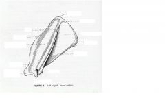
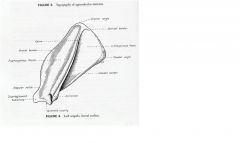
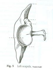
Identify these structures of the Scapula
|
|
|
|
Appendicular Skeleton
|
The bones of the thoracic girdle and forelimbs and the pelvic girdle and hind limbs
|
|
|
Axial Skeleton
|
The bones of the skull, vertebral column, ribs and sternum
|
|
|
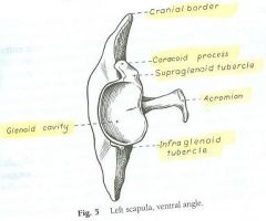
Glenoid Cavity
|
The angle parts of the scapula. It is formed by the distal or articular end. The constricted part that unites with the expanded blade is the neck.
|
|
|
Scapula
|
A flat, roughly triangular bone, possesses two surfaces, three borders and three angles.
|
|
|
Scapular Spine
|
The lateral surface of the scapula that is divided by nearly equal fossae which are the supraspinous and the infraspinous.
|
|
|
Acromion
|
Distal end of the Scapula which is a truncated process. Deltoideous muscle arises here.
|
|
|
Supraspinous Fossa
|
The entire surface cranial to the spine of the scapula.
The supraspinatus arises from all but the distal part of this fossa. |
|
|
Infraspinous Fossa
|
Caudal to the spine of the scapula. It is triangular with the apex at the neck. The infraspinatus arises from this fossa.
|
|
|
Serrated Face
|
A small proximal and cranial rectangular area of the scapula. It serves as insertion for the powerful subscapular fossa
|
|
|
Cranial Border
|
The thin portion of the scapula that is near the ventral angle and the border is concave as it enters the formation of the neck, It forms the scapular notch.
|
|
|
Scapular Notch
|
Notch formed by cranial border of scapula where border is concave.
|
|
|
Caudal Border
|
Just proximal to the ventral angle it bears the infraglenoid tubercle
|
|
|
Infraglenoid tubercle
|
on the caudal border of the scapula which arises the teres minor and the long head of the triceps.
|
|
|
Ventral angle
|
Forms the expanded distal end of the scapula. Clinically, the ventral angle is by far the most important part of the scapula because it enters the formation of the shoulder joint.
|
|
|
Supraglenoid tubercle
|
located at the cranial portion of the Glenoid cavity. This is were biceps brachii arise
|
|
|
Coracoid process
|
A small tubercle where the coracobrachialis arises from.
|
|
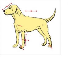
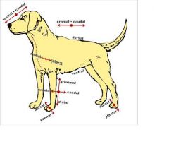
What are the names of the following directional terms?
|
|
|
|
What parts of the scapula can you palpate on a live dog?
|
Cranial, caudal and dorsal borders, the spine, acromion and supraglenoid tubercle.
|
|
|
Humerus
|
located in the arm (brachium). This bone enters into the formation of the shoulder joint and elbow joint. The shoulder joint is forrmed by the articulation of the scapula and humerous; the elbow joint by the articulation of the radius and ulna. The shoulder joint only undergoes flexion and extension.
|
|
|
Head of Humerus
|
The part that articulates with the scapula.
|
|
|
Intertubercular groove of humerus
|
Begins at the cranial end of the articular area. It lodges the tendon of origin of the biceps brachii and is deflected toward the median plane by the greater tubercle.
|
|
|
Greater Tubercle of Humerus
|
Forms the craniolateral part of the the proximal extremity. It is convex and in most breed it is higher than the head of the humerus. It receives insertions of the supraspinous and infraspinaus and part of the deep pectoral.
|
|
|
Lesser Tubercle of Humerus
|
lies medial on the medial side of the proximal extremity caudal to the intertubercular groove.
|
|
|
Neck of humerus
|
only caudally distinct, it is in line along which the head and parts of the tubercles have fused with the body.
|
|
|
Cranial Surface of the Humerus
|
found distinctly in the middle of the humeral body. It furnishes attachment for the brachiocephalicus and part of the pectorals.
|
|
|
What are the extrinsic muscles of the Thoracic Limb?
|
Superficial Pectoral
Deep Pectoral Brachiocephalicus Omotrasversarius Trapezius Rhomboideus Latissimus dorsi Serratus ventralis Sterocephalicus Sternohyoideus Sternothyroideus |
|
|
Superficial Pectoral Muscle (2 parts)
|
a) decending - 1st sternebrae that inserts at the crest of greater tubercle of the humerus. It is responsible for non weight bearing adduction.
b)Transverse - first 2 or 3 sternebrae. Inserts at a longer distance of the greater tubercle of the humerus. Responsible for abduction when weight bearing. |
|
|
Deep Pectoral
|
Ventral sternum, fibrous raphe, deep abdominal fascia near xiphoid cartilage. Inserts at lesser tubercle of the humerous.
Pulls the trunk cranially when limb is advanced, extend shoulder joint. Draw limb caudally, flex shoulder joint when non-weight bearing. Adduct limb. |
|
|
Brachiocephalicus
|
Clavicular tendon broken into cleidobrachialis (inserts at distal head of humerus) and cleidocephalicus. (inserts at cranial end of mid-dorsal fibrous raphe) Limb advancement, extend shoulder
joint, draw head & neck to the side |
|
|
Sternocephalicus
|
1st sternebra inserts at Mastoid part of temporal bone &
nuchal crest of the occipital bone. It is responsible for moving the head/neck side to side. |
|
|
Sternohyoideus
|
1st sternebra and costal cartilage. It inserts at basihyoid bone and is responsible for drawing the tongue and larynx caudally.
|
|
|
Sternothyroideus
|
1st costral cartilage. Inserts at the caudallateral surface of the thyroid cartilage. It is responsible for drawing the tongue and larynx caudally.
|
|
|
Omotransversarius
|
Distal spine of scapula to the
transverse spine of the atlas. In It is responsible for advancing the limb of or flex the neck laterally. |
|
|
Trapezius
|
Inserts at the spine of the scapula and is responsible for abducting and elevating the forelimb.
|
|
|
Rhomboideus (3 parts)
|
a) capitis-Nuchal crest of occiptial bone
b) cervics Median fibrous raphe of neck c) thoracis-Spinous process of T1-T7 Insert at cranial dorsal border (a) and dorsal border of scapula (b & c). They are responsible for Elevate forelimb, draw dorsal border of the scapula against the trunk (adduction) |
|
|
Latissimus dorsi
|
Attachment on last 2 ribs and inserts at teres major tuberosity of the humerus and tendon. It is responsible for moving limb caudally (digging) and flexes the shoulder joint
|
|
|
Serratus Ventralis
|
Transverse process of last C5 vertebra and 1st 7-8 ventral ribs. Its insertion is at the serrated face of the scapula. It is resonsible for depressing the scapula and supporting the truck between the limbs.
|
|
|
Cutaneous Trunci
|
It runs over the majority of the thorax and abdomen and is closely bound to the skin. It is the involuntary twitch muscle. It is innervated by the Lateral Thoracic.
|
|
|
Supination
|
Direction of an animals paw after limb rotation. A lateral rotation of the limb is a medial rotation of the paw. (cat cleaning itself)
|
|
|
Pronation
|
Direction of an animals paw after limb rotation. Medial rotation of the limb resulting in lateral rotation of the paw.
|
|
|
Brachium
|
Region of the forelimb between the shoulder and elbow
|
|
|
Antebrachium
|
Region of the forelimb between the elbow and the capus
|
|
|
Flexion
|
Taking the angle formed by the junction of two bones and making its angle decrease
|
|
|
Extension
|
Taking the angle formed by the junction of two bones and making its angle increase
|
|
|
Abduction
|
pulling the limbs farther away from your body
|
|
|
Adduction
|
bringing the limbs closer to your body
|
|
|
Summation on radiographs
|
When two structures are overlapping and the area on the x-ray where there is increased whiteness.
|
|
|
Silhouetting on radiographs
|
Two structures are close to each other but are not overlapping. They have a similar degree of whiteness in the x-ray and they are touching. The dimensions can not be determined fully due to their closeness and brightness.
|
|
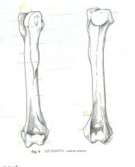
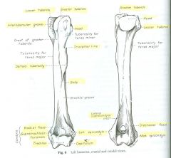
Identify the structures of the Humerus
|
|
|
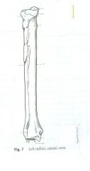
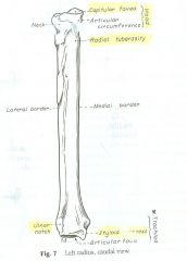
Identify the structures of the radius.
|
|
|
Identify the structures of the scapula on ventral angle
|
|
|
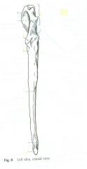
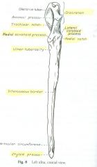
Identify the structures of the Ulna
|
|
|
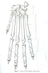
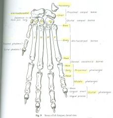
Identify the dorsal view of the paw
|
|
|
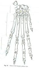
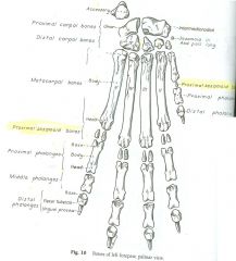
Identify the structures of the Palmar view of the paw
|
|
|
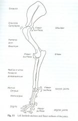
Identify the structures of the forelimb skeleton and flexor surfaces
|
|
|

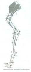
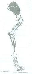
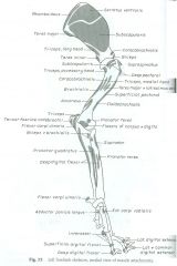
Identify the structures on the skeleton where muscles attach (lateral view)
|
|
|
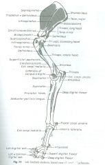
Identify the structures on the forelimb where muscles attach (medial view)
|
|
|
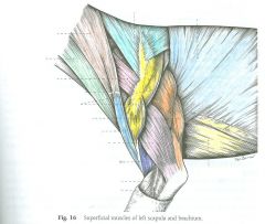
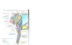
Identify the superfical structures of the left shoulder and arm
|
|
|
|
What are the intrinsic muscles of the lateral scapula and shoulder? (4)
|
Deltoideus
Infraspinatus Teres minor Supraspinatus |
|
|
What are the intrinsic muscles of the medial scapula and shoulder? (3)
|
Subscapularis
Teres Major Coracobrachialis |
|
|
What are the intrinsic muscles of the caudal arm? (3)
|
Tensor fasciae antebrachii
|
|
|
What are the intrinsic muscles of the cranial arm? (2)
|
Biceps Brachii
Brachialis |
|
|
What are the intrinsic muscles of the cranial and lateral forearm? (6)
|
Extensor carpi radialis
Common digital extensor Lateral digital extensor Ulnaris lateralis Supinator Abductor pollicus longus |
|
|
What are the intrinsic muscles of the caudal and medial forearm? (6)
|
Pronator teres
Flexor carpi radialis Superficial digital flexor Flexor carpi ulnaris Deep digital flexor Pronator quadratus |
|
|
Deltoideus muscle
|
Origin - the spine and acromial process of the scapula
Insertion - at the deltoid tuberosity. Action - flex the shoulder. |
|
|
Infraspinatus muscle
|
Origin – The infraspinous fossa
Insertion – A small, circumscribed area on the lateral side of the greater tubercle of the humerus Action- to extend or flex the joint (abduct shoulder and rotate arm laterally) |
|
|
Teres Minor muscle
|
Origin – The infraglenoid tubercle and distal third of the caudal border of the scapula
Insertion – The teres minor tuberosity of the humerus Action - to flex the shoulder and rotate the arm laterally |
|
|
Supraspinatus muscle
|
Origin – The supraspinous fossa
Insertion – The greater tubercle of the humerus, by a thick tendon Action - to extend and stabilize the shoulder joint |
|
|
Subscapularis
|
Origin – The subscapular fossa
Insertion – The lesser tubercle of the humerus Action - adduct, extend and stabilize the shoulder joint |
|
|
Teres Major Muscle
|
Origin – The caudal angle and adjacent caudal border of the scapula; the caudal surface of the subscapularis
Insertion – The teres major tuberosity of the humerus. Action - flexes the should and rotates the arm medially |
|
|
Coracobrachialis
|
Origin – The coracoids process of the scapula
Insertion – The crest of the lesser tubercle of the humerus proximal to the teres major tuberosity. Action - to adduct and extend the shoulder |
|
|
Tensor fasciae antebrachii
|
Origin – The fascia covering the lateral side of the latissimus dorsi
Insertion – The olecranon Action - extend the elbow |
|
|
Long head of Triceps
|
Origin – The caudal border of the scapula
Insertion – The olecranon tuber of the ulna Action - extend the elbow and flex the shoulder |
|
|
Lateral Head of the Triceps
|
Origin – The tricipital line of the humerus
Insertion – the olecranon tuber of the ulna Action – To extend the elbow |
|
|
Accessory head of the Triceps
|
Origin – The neck of the humerus
Insertion - The olecranon tuber of the ulna Action – To extend the elbow |
|
|
Medial Head of the Triceps
|
Origin –lesser tubercle of the humerus
Insertion – The olecranon of the ulna Action – Extend the elbow |
|
|
Anconeus
|
Origin – The lateral supracondylar crest and the lateral and medial epicondyles of the humerus
Insertion –The proximal end of the ulna. Action – To extend the elbow. |
|
|
Biceps brachii
. |
Origin – The supraglenoid tubercle
Insertion – The ulnar and radial tuberosities Action – To flex the elbow and extend the shoulder |
|
|
Brachialis
|
Origin – The proximal third of the humerus
Insertion – The ulnar and radial tuberosities Action – To flex the elbow |
|
|
Extensor carpi radialis
|
Origin – The lateral supracondylar crest
Insertion – The tuberosities of the bases of the metacarpals II and III Action – To extend the carpus |
|
|
Common digital extensor
|
Origin – The lateral epicondyles of the humerus
Insertion – The extensor processes of the distal phalanges of digits II, III, IV, and V Action – To extend the joints of the four principal digits and carpus. |
|
|
Lateral Digital Extensor
|
Origin – The lateral epicondyles of the humerus
Insertion – The proximal ends of all the phalanges of digits III, IV, and V, but mainly the extensor processes of the distal phalanges of these digits. Action – To extend the carpus and joints of digits III, IV, and V |
|
|
Ulnaris Lateralis (Extensor Carpi Ulnaris)
|
Origin – The lateral epicondyles of the humerus
Insertion – The lateral aspect of the proximal end of metacarpal V and the accessory carpal bone. Action –flexes carpal joint. |
|
|
Supinator
|
Origin – The lateral epicondyles of the humerus
Insertion – The cranial surface of the proximal fourth of the radius Action – Supinate the forearm; to flex the elbow |
|
|
Pronator teres
|
Origin – The medial epicondyles of the humerus
Insertion – The medial border of the radius Action – Pronate the forearm; flex the elbow |
|
|
Flexor carpi radialis
|
Origin – The medial epicondyles of the humerus and the medial border of the radius
Insertion – The palmar side of the base of metacarpals II and III Action - To flex the carpus |
|
|
Superficial digital flexor
|
Origin – The medial epicondyles of the humerus
Insertion – The middle phalanges of digits II, III, IV, and V Action - To flex the carpus, joints of digits II, III, IV, and V |
|
|
Flexor carpi ulnaris
|
Origin – Ulnar head –olecranon; humeral head – the medial epicondyles of the humerus.
Insertion – The accessory carpal bone. Action – To flex the carpus |
|
|
Deep digital flexor
|
Origin – Humeral Head – The medial epicondyles of the humerus; ulnar head –caudal border of the ulna; radial head –medial border of the radius.
Insertion –distal phalanx of each digit. Action - To flex the carpus, joints of the digits. |
|
|
Pronator quadratus
|
Attachments – The apposed surfaces of the radius and ulna
Action – To pronate the paw |
|
|
The crest of greater tubercle of the humerus.
|
The ridge that extends proximally in a craniomedial direction and is also the cranial border of the bone. This forms part of the area of insertion of the pectorals.
|
|
|
Lateral surface of the humerus
|
The ridge extending to caudal part of the greater tubercle. When this thickens distally it forms the deltoid tuberosity.
|
|
|
Deltoid tuberosity of the humerus
|
the distal thickened surface of the later surface of the humerus. This is where the deltoideus muscle inserts.
|
|
|
Tricipital line
|
From the deltoid tuberosity to the caudal part of the greater tubercle it is the most prominent ridge that is formed. The lateral head of the triceps arises here.
|
|
|
Tuberosity of the teres minor of the humerus
|
where the teres minor inserts and is adjacent to the promimal extremity of the tricpital line
|
|
|
Brachialis groove of humerus
|
located on the lateral surface of the body where the brachialis originates in the proximal part of the groove.
|
|
|
Lateral supracondylar crest of humerus
|
Distal to the brachialis groove where the extensor carpi radialis and part of the anconeus attach.
|
|
|
The caudal surface of the humerus
|
The smooth and rounded transversly and ends in the deep olecranon fossa
|
|
|
The crest of the lesser tubercle of the humerus (medial surface and teres major)
|
crosses the proximal end of the medial surface and ends distally at the teres major tuberosity. The teres major and latisimus dorsi insert on this tuberosity
|
|
|
Humeral condyle
|
located at the distal end of the humerus which includes the articular areas which are divided unevenly by a low ridge
|
|
|
Capitulum of the humerus
|
Small articular area lateral to the ridge which articulates only to the head of the radius
|
|
|
Lateral epicondyle of the humerus
|
occupies the enlarged distolateral end of the humerus proximal to the capitulum. (smaller than medial epicondyle)
|
|
|
Medial epicondyle of the humerus
|
The distomedial end of the humerus, its caudal projection deepens the olecranon fossa where the anconeus arises. Origin for intrinsic muscles of caudal and medial forearm.
|
|
|
Olecranon fossa of the humerus
|
a deep excavation of the caudal part of the humeral condyle. It receives the anconeal process of the ulna during extension of the elbow.
|
|
|
Radial fossa of the humerus
|
communicates with the olecranon fossa by an opening (the supratrochlear foramen) located on the cranial surface of the humeral condyle.
|
|
|
Supratrochlear foramen of the humerus
|
The opening in the olecranon fossa. No soft structures pass through here.
|
|
|
Radius
|
The shorter of the two bones of the antebrachium or forearm that articulates proximally with the humerus and distally with the carpus.
|
|
|
The head of the radius
|
widest medial to lateral and forms proximally an oval depressed articular surface, the fovea capitis
|
|
|
Fovea capitus of the radius
|
the depressed articular surface of the head of the radius that articulates with the capitulum of the humerus
|
|
|
Radial tuberosity of the radius
|
lies distal to the neck on the medial border of the bone. This is where the biceps brachii insert.
|
|
|
The body of the radius
|
possesses a cranial and caudal surface and a medial and lateral border. At the caudal surface there are ligamentous attachments to the ulna
|
|
|
Ulnar notch of the radius
|
On the lateral surface of the distal extremity of the radius where the ulna articulates.
|
|
|
Styloid process of the radius
|
The medial surface of the distal extremity of the radius which attaches the carpus by a the medial ligament.
|
|
|
Ulna
|
The longer of the two bones in the antebrachium and located caudally
|
|
|
Radial notch of the Ulna
|
Where the ulna is is medial to the radius and articulates with the humerus and with the articular circumference of the radius. It forms the elbow
|
|
|
Olecranon of the ulna
|
The proximal extremity which includes the olecranon tuber and the anconeal process. It serves as a lever for the extensor muscles of the elbow.
|
|
|
Olecanon tuber of the ulna
|
Where the triceps brachii, anconeus and tensor fasciae atebrachii attach to the caudal part of the olecranon. It is grooved cranially and enlarged and rounded caudally.
|
|
|
Anoneal process of the ulna
|
When the elbow is extended, it is what fits into the olecranon fossa
|
|
|
Medial and lateral coronoid process
|
at the distal end of the notch which articulate with the humerus and radius
|
|
|
Ulnar body
|
The body of the ulna is three sided in its middle third and proximal to this the bone is compressed laterally. It gradually loses its borders and becomes irregular
|
|
|
Ulnar Tuberosity
|
a small elongated eminence on the medial surface of the bone at its proximal end just distal to the medial coronoid process
|
|
|
Interosseous border of the ulna
|
the distinct, rough and irregular, especially at the junction of the proximal and middle thirds of the bone, where a large expansive but low eminence is found. (indicates the place of articulation with the radius by a heavy ligament)
|
|
|
Carpus
|
wrist, used to designate the part of the extremity between the forearm and the metacarpus
|
|
|
Intermedioradial carpal (radial carpal in dog)
|
Of the seven irregular bones it is the the largest of the the three proximal row bones. It is on the medial side and articulates proximally to the radius.
|
|
|
Ulnar carpal
|
The lateral member of the proximal row. Its palmar portion projects distally palmar and lateral to the fourth carpal
|
|
|
Accessory carpal
|
The palmer member is a short rod of bone that articulates with the styloid process of the ulna and the ulnar carpal bone and serves as a lever arm for some of the flexor muscles of the carpus.
|
|
|
Metacarpal
|
hand, contains 5 bones which are long bones in miniatures, possessing a slender body, or shaft and large extremities
Each metacarpal has a body base and head (shaft, proximal and distal extremity) |
|
|
Phalanges
|
Bones in the forepaw in which there are three bones for ever one main digit having a body, base and head.
|
|
|
Ungual crest
|
A thin sheath of bone that overlaps the claw.
|
|
|
Extensor process
|
the rounded dorsal part of the base which the common digital extensor tendon is inserted.
|
|
|
The ungual process
|
a curved conical extension of the distal phalanx into the claw.
|
|
|
Flexor tubercle
|
A small process on the palmer surface for insertion of the deep digital flexor tendon
|
|
|
Proximal Sesamoid bones
|
Located in the interosseos tendons on the palmer surface of each metacarpophalangeal joint
|
|
|
Dorsal sesamoid bones
|
embedded in the common digital extensor tendon as they pass over the metacarpophalangeal joints.
|
|
|
What are the joints of the thoracic limb?
|
Sin Sarcoidosis
Shoulder joint (glenohumeral) Elbow joint Antebrachiocarpal Joint (radiocarpal) Middle Carpal Joint Carpometacarpal Joint Metacarpophalangeal Joint Proximal Interphalangeal Joint Distal Interphalangeal Joint |
|
|
Cutaneous Trunci
|
A thin sheet of muscle that covers most of the dorsal, lateral and ventral walls of the thorax and abdomen. It is responsible for twitch from the lateral thoracic nerve.
|