![]()
![]()
![]()
Use LEFT and RIGHT arrow keys to navigate between flashcards;
Use UP and DOWN arrow keys to flip the card;
H to show hint;
A reads text to speech;
48 Cards in this Set
- Front
- Back
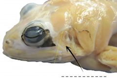
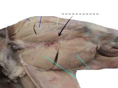
A broad muscle behind the tympanic ring that closes the mouth. |
Depressor mandibuli |
|
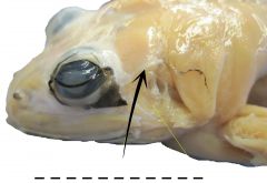
A small muscles that extends from the tip of the suprascapula to the region between the tympanic ring and the eye. |
Temporalis |
|
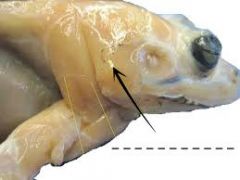
A small muscle located in the front of the tympanic ring that raises the lower jaw and closes the mouth. |
Masseter |
|
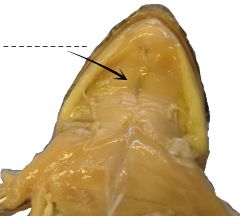
A very thin sheet of muscle at the ventral surface of the floor of the mouth. |
Mylohyoid |
|
|
The continuation of rectus abdominis forward to the head under the episternum. |
Sternohyoid |
|
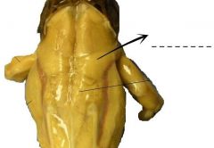
A small muscle overlapping the caudal portion of the suprascapula. |
Latissimus dorsi |
|
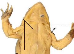
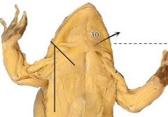
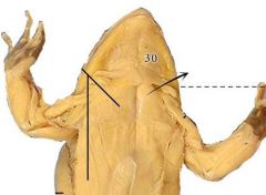
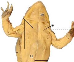
Muscles covering the outer surface of the suprascapula. |
Dorsalis scapulae |
|
A long muscle running close and along the vertebral column. |
Longissimus dorsi |
|
A narrow muscle located posteriorly to the longissimus dorsi and extends posterior in oblique position. |
Coccygeo-sacralis |
|
Running diagonally in the space between the ilia and the urostyle just caudal of the preceding muscle. It serves to hold the urostyle firmly. |
Coccygeo-iliacus |
|
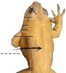
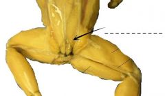
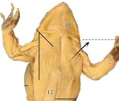
A broad, thin muscle covering the ventral side of the abdomen. The mid-ventral line is called the linea alba. The crosswise faint lines are called the ventral inscriptiones tendinae. |
Rectus abdominis |
|
A thin sheet of muscle, which forms the outer layer of the body wall at the side of the abdomen. |
External oblique |
|
Another sheet of oblique muscles internal to the external oblique. |
Internal oblique |
|
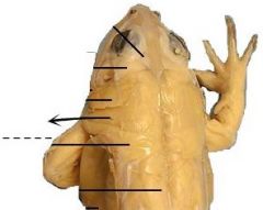
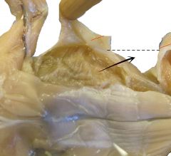
A triangularly-shaped muscle that lies at the anterior border of the shoulder. |
Deltoid |
|
Next to the preceding and arises in the midventral line passing outwards, piercing the distal portion of the deltoid. |
Sternordialis |
|
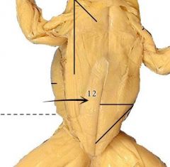
The anterior muscle of the chest just next to the sternordialis. |
Anterior pectoralis |
|
Next to the anterior pectoralis. |
Middle pectoralis |
|
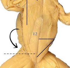
A paddle-shaped muscle, lateral and posterior to the middle pectoralis. |
Posterior pectoralis |
|
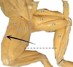
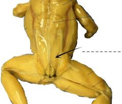
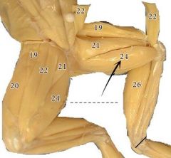
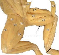
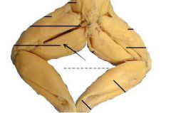
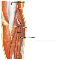
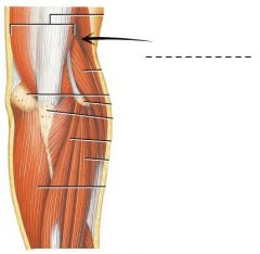
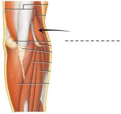
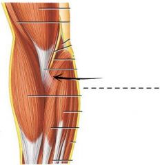
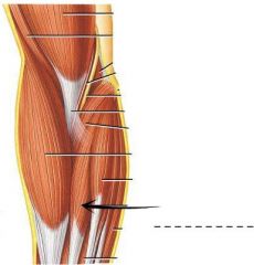
Large muscle covering the entire thigh from anterior part to the posterior margin. The proximal end is divided into three heads. |
Triceps extensor femoris |
|
The outer head of the triceps extensor femoris which can be seen at the dorsal surface of the thigh. |
Vastus externus |
|
The middle head of the triceps extensor femoris which can be seen in both dorsal and ventral surface of the thigh. |
Rectus femoris anticus |
|
The inner head of the triceps extensor femoris which can be seen at the ventral surface of the thigh. |
Vastus internus |
|
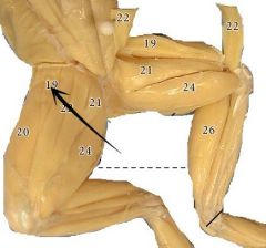
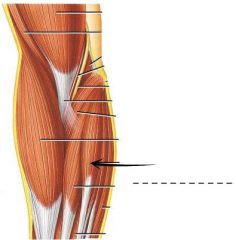
A thin slender muscle immediately below the vastus internus and partly covered by the sartorius, but a small portion is exposed along the preaxial side of the latter. |
Adductor longus |
|
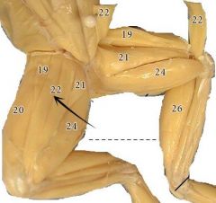
A thin flat muscle which traverse the thigh obliquely. |
Sartorius |
|
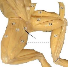
A stout triangular muscle lying behind the sartorius which crosses at its digital end. |
Adductor magnus |
|
A large muscle lying at the ventral side of the thigh. |
Gracilis major |
|
The most posterior, long slender muscle closely attached to the preceding, and run along the inner side of the thigh. |
Gracilis minor |
|
An internal muscle which could only be easily seen by separating the adductor magnus from the gracilis major. |
Semitendinosus |
|
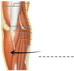
A long slender muscle immediately below the vastus externus and partly covered by it. |
Biceps femoris |
|
A large muscle lying on the posterior margin of the dorsal surface of the thigh. |
Semimembranosus |
|
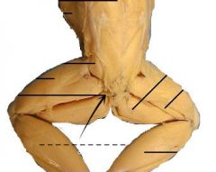
A small slender muscle extending ventrally and posteriorly from the tip of the urostyle and between the origin of the biceps femoris and the semimembranosus. |
Pyriformis |
|
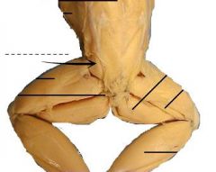
A short muscle lying between the rectus femoris anticus and vastus externus. |
Gluteus |
|
A narrow muscle lying at the most anterior side of the tibio-fibula and divided distally into two parts. |
Tibialis anticus |
|
Lies immediately posterior to the tibialis anticus. |
Peroneus |
|
A large muscle a the posterior side of the shank. It is covered with fascia. |
Gastrocnemius |
|
A long narrow muscle adhering closely behind the tibio-fibula. |
Tibialis posticus |
|
A small muscle lying on its proximal half of the tibio-fibula. |
Extensor cruris |
|
A very small muscle found between two distal ends of the tibialis anticus. |
Flexor tarsi anterior |
|
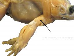
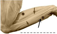
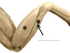
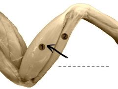
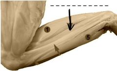
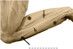
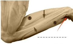
A big long muscle on the posterior side of the upper arm which composed of three heads, i.e the outer lateral head, the middle long head, and the medial head. |
Triceps brachii |
|
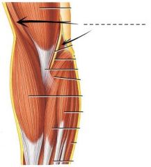
The next muscle anterior to the lateral head of the triceps brachii. |
Brachialis |
|
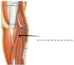
Found on the outer surface of the ulna. |
Extensor carpi ulnaris |
|
Two kinds; lateralis and communis. This is next and anterior to the extensor carpi ulnaris. |
Extensor digitorium |
|
Next o the extensor digitorium on the preaxial border of the forearm. |
Brachioradialis |
|
Next to the brachioradialis and lies along the inner surface of the forearm. |
Extensor carpi radialis |
|
Next to the extensor carpi radialis on the ventral and inner side. |
Pronator teres |
|
Lies next to the pronator teres and partly covered by it. |
Flexor carpi radialis |
|
Flat muscle next to the flexor carpi radialus and partly covering it. |
Palmaris longus |
|
Forms the ulnar border of the forearm next to the palmaris longus and immediately below the extensor carpi ulnaris. |
Flexor carpi ulnaris |