Reading...
![]()
Play button
![]()
Play button
![]()
Use LEFT and RIGHT arrow keys to navigate between flashcards;
Use UP and DOWN arrow keys to flip the card;
H to show hint;
A reads text to speech;
30 Cards in this Set
- Front
- Back
|
What are the precursor structures to the kidney?
|
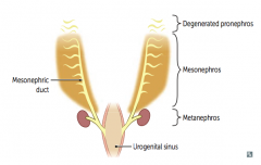
- Pronephros (until week 4)
- Mesonephros (1st trimester) - Metanephros (permanent) |
|
|
When is the fate of the Pronephros?
|

Renal precursor structure that is present until week 4 when it degenerates
|
|
|
When is the fate of the Mesonephros?
|

- Functions as interim kidney for 1st trimester
- Later contributes to male genital system |
|
|
When is the fate of the Metanephros?
|

- Permanent renal structures, first appears at 5th week of gestation
- Nephrogenesis continues through 32-36 weeks of gestation |
|
|
What structure is derived from the caudal end of the mesonephric duct? What does it become?
|

Ureteric Bud
- Gives rise to ureter, pelvises, calyces, and collecting ducts - Fully canalized by 10th week |
|
|
What structure interacts with the ureteric bud to induce differentiation and formation of the glomerulus through to the distal convoluted tubule?
|
Metanephric Mesenchyme
|
|
|
What structures form the permanent tubular system of the kidney? How?
|
- Metanephric Mesenchyme: forms glomerulus through distal convoluted tubule
- Ureteric Bud: forms collecting ducts, calyces, pelvises, and ureter - These form through interaction of the two structures |
|
|
What happens if there is aberrant interaction between the ureteric bud and metanephric mesenchyme?
|
May result in several congenital malformations of the kidney
|
|
|
What is the last part of the renal structures to canalize? Implications?
|
Ureteropelvic Junction - most common site of obstruction (hydronephrosis) in fetus
|
|
|
What is the most common site of obstruction, leading to hydronephrosis, in the fetus?
|
Ureteropelvic Junction
|
|
|
What syndrome occurs in babies who can't pee in utero?
|
Develop Potter Syndrome
|
|
|
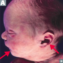
What syndrome is associated with Pulmonary hypoplasia, Oligohydramnios, Twisted face, Twisted skin, Extremity defects, and Renal failure (in utero)? Cause?
|

Potter Syndrome
- Causes: ARPKD, posterior urethral valves, bilateral renal agenesis - Leads to oligohydramnios → compression of developing fetus → limb deformities, facial anomalies (low set ears and retrognathia), and compression of chest → pulmonary hypoplasia (cause of death) |
|
|
What can cause Potter Syndrome?
|
Causes:
- ARPKD - Posterior Urethral Valves - Bilateral Renal Agenesis Leads to: - Oligohydramnios → compression of developing fetus → limb deformities, facial anomalies (low set ears and retrognathia), and compression of chest → pulmonary hypoplasia (cause of death) |
|
|
What are the consequences of ARPKD, posterior urethral valves, or bilateral renal agenesis?
|
Potter Syndrome:
- Pulmonary hypoplasia - Oligohydramnios - Twisted face - Twisted skin - Extremity defects - Renal failure (in utero) |
|
|
What facial abnormalities are associated with Potter Sequence?
|

- Low-set ears
- Retrognathia (posterior positioning of mandible) |
|
|
What happens in horseshoe kidney? Consequences?
|
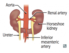
Inferior poles of both kidneys fuse
- As the kidneys ascend from the pelvis during fetal development, horseshoe kidneys get trapped under the inferior mesenteric artery - Remain low in abdomen |
|
|
How do kidneys that are fused at the inferior poles function?
|
Horseshoe kidneys function normally, but there is an increased risk of ureteropelvic junction obstruction, hydronephrosis, renal stones, and rarely renal cancer (Wilms tumor)
|
|
|
What is horseshoe kidney associated with?
|
Turner Syndrome
|
|
|
What kidney problem are patients with Turner Syndrome associated with having? Risk of?
|
Horseshoe kidney, increased risk of:
- Ureteropelvic junction obstruction - Hydronephrosis - Renal stones - Rarely renal cancer (Wilms tumor) |
|
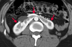
What does this axial CT of abdomen with contrast show?
|

Enhancing midline fused kidney (horseshoe kidney)
|
|
|
What kidney problem is often diagnosed prenatally via ultrasound with a unilateral cystic kidney and a contralateral hypertrophic kidney? Cause?
|
Multicystic Dysplastic Kidney - abnormal interaction between ureteric bud and metanephric mesenchyme → non-functional kidney
|
|
|
What is wrong if a patient has a Multicystic Dysplastic Kidney?
|
- Abnormal interaction between ureteric bud and metanephric mesenchyme
- Leads to a non-functional kidney consisting of cysts and connective tissue - If unilateral (most common), generally asymptomatic with compensatory hypertrophy of the contralateral kidney |
|
|
When is Multicystic Dysplastic Kidney typically diagnosed?
|
Prenatally with ultrasound
|
|
|
Which kidney is usually taken during living donor transplantation? Why?
|
Left kidney - longer renal vein
|
|
|
What is the branching pattern of the arteries in the kidney?
|
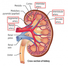
Renal artery → Segmental artery → Interlobar Artery → Interlobular Artery → Arcuate Artery
|
|
|
What does blood need to flow through to enter the kidney tubules?
|
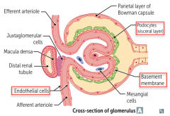
Glomerulus:
- Endothelial cells of blood vessel - Basement membrane - Podocytes (visceral layer) |
|
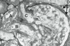
What are the labeled components of this glomerulus?
|

- A: Macula Densa
- B: Afferent arteriole - C: Efferent arteriole - D: Bowman Capsule |
|
|
Where is the Macula Densa?
|
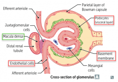
Distal Renal Tubule - adjacent to glomerulus and juxtaglomerular cells
|
|
|
What is the course of the ureters?
|

- Ureters pass UNDER uterine artery and UNDER ductus deferens (retroperitoneal)
- Water (ureters) are under the bridge (uterine artery and vas deferens) (But pass over the iliac arteries |
|
|
What can happen in gynecologic procedures involving ligation of the uterine vessels?
|
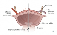
May damage the ureter → ureteral obstruction or ureteral leak
|