![]()
![]()
![]()
Use LEFT and RIGHT arrow keys to navigate between flashcards;
Use UP and DOWN arrow keys to flip the card;
H to show hint;
A reads text to speech;
67 Cards in this Set
- Front
- Back
- 3rd side (hint)
|
Enamel |
Most highly mineralized tissue in the body -acellular and avascular- unable to remodel or repair |
|
|
|
Enamel |
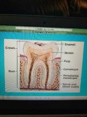
Thickest at cusp tips Thinnest near junction of crown and root |
|
|
|
Components of enamel |
96% inorganic- composed of mineral called hydroxyapatite (crystallized calcium phosphate) -4% water and organic matter (protein called enamelin |
|
|
|
Amelogenesis |
Enamel formation begins im late bell stage Dentin deposited first Then ameloblasts elongate and begin producing enamel |
|
|
|
Ameloblasts |
Columnar shaped with a secretory process at one end called a tome's process |
|
|
|
Tomes' process |
Shovel shaped & is responsible for the orientation of the enamel rods |
|
|
|
Ameloblasts |
Hexagonal in cross section |
|
|
|
2 stages of enamel formation |
1. Secretory stage 2. Resorbing stage |
|
|
|
Secretory stage |
Deposition of enamel matrix that contains both organic and inorganic material |
|
|
|
Resorbing stage |
Removal of most of the water and the organic matter |
|
|
|
Hydroxyapatite crystals |
Depositedd during the secretory stage Very thin and needle like As enamel matures, they grow in size |
|
|
|
Enamel crystals |
4 times larger than those in bone, dentin and cementum |
|
|
|
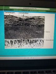
Enamel rods |
Enamel- composed of interlocking rods Help resist masticatory forces and prevent fracture between the rods |
|
|
|
Enamel rods |
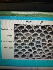
Extend from the DEJ to the outer enamel surface Run perpendicular to the incisal surface |
|
|
|
Shape of enamel rods |
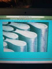
Key hole. One key hole shaped rod is formed by 4 ameloblasts Each ameloblasts (6 sided) contributed to 4 rods |
|
|
|
Enamel rods |
Appear wavy because of migratory path toward the periphery |
|
|
|
Hunter-schreger bands |
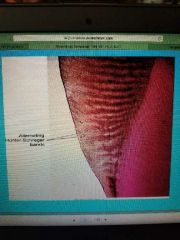
Alternating light and dark bands in the enamel due to configuration of the enamel rods Run perpendicular to the DEJ Extend about 2/3 of the way from the DEJ to the enamel surface |
|
|
|
Rod core |
Center of the enamel rod |
|
|
|
Rod sheath |
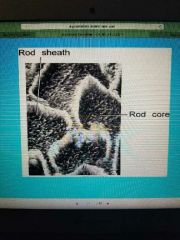
Around the periphery of the rod -especially noticeable in the head region of the rod Produced by a change in the angulation of the crystals formed as ameloblasts move This resists demineralization more than the rod core |
|
|
|
Rodless enamel |
A structureless layer of enamel about 30 microns Found on all deciduous teeth & 70% of permanent teeth Found most commonly at the cervical areas of the enamel Least commonly found over the cusp tips |
|
|
|
Gnarled enamel |
Enamel rods bend in exaggerated ways Begins near the DEJ under the areas of the cusp tips |
|
|
|
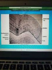
Incremental lines |
A result in rhythmic deposition of enamel End in small ridges on the tooth surface called perikymata |
|
|
|
Cross striations |
Daily apposition lines |
|
|
|
Striae of retzius |
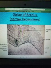
More prominent growth lines May reflect major interruptions in deposition of enamel |
|
|
|
Neonatal line |
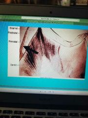
A significant striae if retzius Distinguishes prenatal enamel (fewer defects) from postnatal enamel |
|
|
|
Enamel spindles |
Single extension of odontoblast process into the enamel (shorter than tufts) |
|
|
|
Enamel tufts |
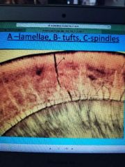
Hypomineralized inner ends of some enamel rods (defect in enamel) broader than spindles |
|
|
|
Enamel lamellae |
Cracks in the surface of the enamel visible to the eye Extend from the DEJ to the surface of the enamel Possible pathway for dental caries to spread |
|
|
|
Pits and fissures |
Form where ameloblasts become crowded between cusps -causes incomplete maturation of enamel More susceptible to caries due to hypocalcification |
|
|
|
Enamel etching |
Dilute acid is used to alter the surface of the enamel Acid attacks the mineral at the periphery of the enamel rods & leaves a rough surface Bonding material attached more firmly to roughened enamel area |
|
|
|
Enamel permeability |
Fluid, particles, and bacteria can pass through enamel by various pathways and can result in dental caries. These include: - lamellae - tufts - cracks - spindles - pits and fissures - spaces between crystals |
|
|
|
Tetracycline stain |
Appears as dark bands through enamel, especially near cervix of the tooth Tetracycline does not affect enamel Tetracycline binds to dentin and bone |
|
|
|
Darkened dentin |
Shows through the more translucent enamel, giving the tooth a darker appearance -does not usually get good results with bleaching bc the source of the problem is in the dentin |
|
|
|
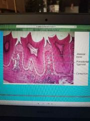
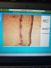
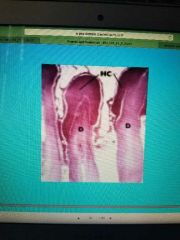
Cementum |
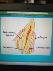
Mineralized connective tissue that covers roots of the teeth |
|
|
|
Cementum composed of |
Cells (cementoblasts, cementocytes) Fibers (collagen) Ground substance |
|
|
|
2 functions of cementum |
1. Seals the tubules of the dentin of the root 2. Serves as the attachment for the periodontal fibers that suspend the tooth in its socket |
|
|
|
Cementum |
Thinnest near the cervix of the tooth and thickest at the apex of the tooth Continues to form throughout life Less mineralized than enamel and dentin |
|
|
|
Cementum |
Lighter in color than dentin and softer than dentin Has no nerves therefore is not sensitive |
|
|
|
Bone |
A vascular tissue (haversian canal, volkmanns canal) cementum is avascular |
|
|
|
Cementum |
Does not have nerves, bone has nerves
Both have cells in Lacunae and canaliculi |
|
|
|
Cementum |
Less mineralized than bone Is more resistant to demineralization and resorption than bone Both have incremental lines |
|
|
|
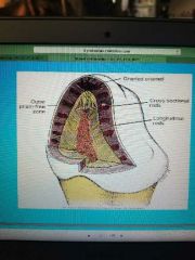
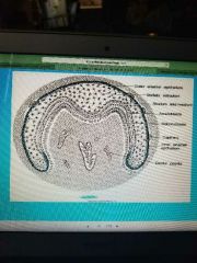
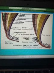
Cementogenesis |
After the crown forms, hertwig's epithelial root sheath is formed by joining of the OEE and the IEE The sheath forms the outline of the root and induces formation of root dentin |
|
|
|
Bell stage |
|
|
|
|
Hertwig's epithelial root sheath |
|
|
|
|
Epithelial rests of mallasez |
The sheath degenerates and the remnants are this |
|
|
|
Dental follicle |
Cells from here differentiate into cementoblasts They begin production of cementum on newly formed dentin |
|
|
|
3 types of cementum are formed |
1. Intermediate 2. Acellular 3. Cellular |
|
|
|
Intermediate cementum |
Thin, noncellular layer deposited by cells of inner epithelial root sheath More calcified than the dentin or the adjacent cellular cementum |
|
|
|
Acellular cementum |
Deposited on the surface if the intermediate cementum Formed from cementoblasts that originated as fibroblasts in the pdl (Derived from the dental follicle) |
|
|
|
Acellular cementum |
Covers the cervical 1/2 of the root No cells, does not increase after initial deposition Forms at a slower rate than cellular cementum |
|
|
|
Cellular cementum |
Covers apical portion of the tooth Much thicker layer Contains cells (cementocytes) |
|
|
|
Cellular cementum |
Deposited throughout life Deposited at intervals which produces arrest lines (highly calcified lines similar to those seen in bone) |
|
|
|
Cell types |
Cementoblasts, cementocytes, cementoclasts |
|
|
|
Cementoblasts |
Lay down cementoid (organic material) - becomes mineralized later They become embedded in mineralized matrix as more cementoid is produced They become enclosed in a Lacuna and become cementocytes |
|
|
|
Cementocytes |
Get their nutrients from the PDL via canaliculi -(oriented towards the PDL) |
|
|
|
Cementoclasts |
Cells that actively resorb cementum Associated with -primary teeth -trauma (caries, injury) |
|
|
|
Cementoenamel junction (CEJ) |
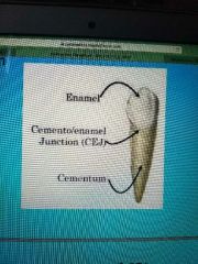
Point where cementum of root meets enamel of crown |
|
|
|
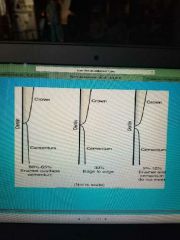
3 variations of CEJ |
1. O 2. M 3. G |
|
|
|
O |
Cementum overlaps the enamel slightly (60%) |
|
|
|
M |
Cementum meets the enamel (30%) |
|
|
|
G |
A small gap exists between the cementum and the enamel (10%) Results in exposed dentin and possibly root sensitivity and increased susceptibility to decay |
|
|
|
Cementoenamel junction |
|
|
|
|
Root resorption |
Can be a result of trauma For example: -traumatic occlusion -rapid orthodontic tooth movement -hypereruption due to loss of antagonist |
|
|
|
Cemental repair |
-new cementum can be deposited on top of an area with a defect Reveral line- the point where resorption stops & deposition begins |
|
|
|
Hypercementosis |
-abnormal thickening of the cementum -usually found in apical region -tooth may fuse to surrounding bone |
|
|
|
Cementicles |
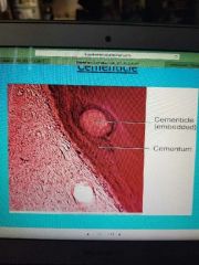
-calcified ovoid or round module -may be free in the PDL, attached, or embedded in the cementum -originate from epithelial cells |
|
|
|
Age related changes |
Increase in amount of cementum on the apical region usually occurs Cementum resorption is a characteristic of aging Root surface becomes more irregular with age due to continued resorption and deposition of cementum More cementicles Are found in the elderly |
|